Cyst in the Brain MRI: Preparation, Procedure and Results

MRI uses magnets and radio waves, not ionising radiation, to create detailed images of the brain. A brain cyst found on MRI is not automatically dangerous or cancerous; meaning depends on its type, size, location and effect on nearby structures.
Key Takeaways
- MRI uses magnets and radio waves, not ionising radiation, to create detailed images of the brain.
- A brain cyst found on MRI is not automatically dangerous or cancerous; meaning depends on its type, size, location and effect on nearby structures.
- Preparation usually includes completing a metal and implant safety questionnaire and following instructions about food, drink and medicines if contrast or sedation is planned.
- The scan commonly takes 30 to 60 minutes, and a radiologist interprets the images before the referring clinician discusses the results.
- Many brain cysts only need observation, while cysts causing symptoms, fluid blockage or pressure may require specialist treatment.
A cyst in the brain MRI can be identified in great detail, helping doctors determine whether it is a common harmless fluid-filled space or a finding that needs further assessment. Most brain MRI scans are painless and do not require special preparation, but safety screening for implants, metal and contrast allergy is essential.
Overview: what a cyst in the brain MRI can show
A cyst in the brain MRI refers to a fluid-filled area seen on detailed magnetic resonance imaging of the brain. MRI is especially useful because it shows soft tissues, fluid, nerves and brain structures clearly without using X-rays. The scan can help a doctor describe the cyst’s position, size, contents and relationship to nearby brain tissue and fluid spaces.
Not every cyst is a disease or requires treatment. Some, such as many arachnoid cysts, are congenital and discovered incidentally when a scan is performed for another reason. Others may be related to previous inflammation, infection, injury, surgery, a developmental variation or, less commonly, another underlying condition. The MRI appearance and a person’s symptoms guide the next steps.
A brain MRI may be requested for persistent headaches, seizures, changes in vision or balance, weakness, unusual neurological symptoms, or follow-up of a known finding. It is a diagnostic examination rather than a treatment. The images are interpreted in the context of the person’s medical history and neurological examination.
How to prepare before a brain MRI
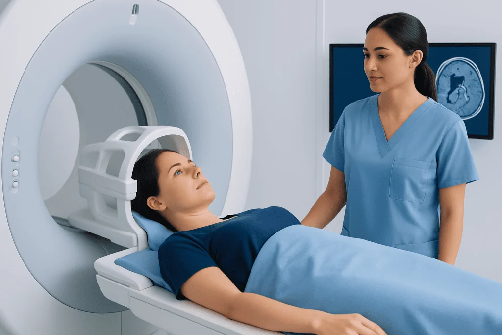
For most standard brain MRI examinations, a person can eat, drink and take usual prescribed medicines as normal. The imaging centre will provide personalised instructions in advance, particularly if contrast material, sedation or anaesthesia is planned. It is important to tell the team about pregnancy or a possible pregnancy, kidney disease, allergies and previous reactions to MRI contrast.
Before entering the scan area, patients complete a safety questionnaire. MRI uses a powerful magnet, so the team needs to know about pacemakers, implanted stimulators, cochlear implants, aneurysm clips, infusion pumps, metal fragments, surgical implants, joint replacements, dental devices and prior operations. Many modern implants are MRI-compatible under specific conditions, but this must be verified rather than assumed.
Patients should wear comfortable clothing without metal fasteners where possible, or change into a gown. Jewellery, watches, hairpins, belts, bank cards, hearing aids, removable dental work and electronic devices must be removed. If claustrophobia is a concern, it is helpful to discuss this when booking so the team can explain available support and whether medication or another arrangement is appropriate.
What should you not do before a brain MRI?
Patients should not enter the MRI room with metal objects, electronic devices or magnetic cards. This includes phones, keys, coins, jewellery, watches, credit cards and some cosmetics containing metallic particles. The imaging staff will check belongings carefully because the magnet remains active at all times.
People should not stop regular medicines unless their clinician specifically instructs them to do so. They should also not conceal information about implants, previous surgery, metal exposure at work, kidney problems, pregnancy, allergies or prior contrast reactions. Accurate safety information helps the MRI team choose the safest scanning approach.
If sedation is planned, the centre may advise avoiding food and drink for a defined period beforehand and arranging for a responsible adult to take the patient home. These instructions vary by patient and facility. Alcohol or recreational drugs should be avoided before an appointment because they can affect safety, consent and the ability to remain still during the examination.
How a brain MRI works and what happens during the procedure
MRI creates images by using a strong magnetic field and radiofrequency energy to detect signals from water molecules in the body. A computer converts these signals into detailed images in multiple planes. For a suspected brain cyst, different image sequences help distinguish simple fluid from blood products, protein-rich fluid, inflammation, solid tissue or other changes.
After checking in, the patient removes metal items and completes the safety review. They lie on a padded table, usually on their back, with a lightweight head coil placed around the head. Cushions or supports may be used to improve comfort and help keep the head still. Earplugs or headphones reduce the loud tapping and knocking sounds made during scanning.
The table moves slowly into the MRI scanner. The radiographer communicates through an intercom and can see and hear the patient throughout the examination. Remaining still is important because movement can blur images. A typical brain MRI takes about 30 to 60 minutes, although timing depends on the sequences required and whether contrast is used.
Sometimes gadolinium-based contrast is given through a vein to show blood vessels, inflammation or areas where the blood-brain barrier is altered. Contrast may help clarify the nature of a cyst-like finding, but it is not necessary for every scan. The radiologist and referring clinician decide whether it is appropriate based on the clinical question and safety information.
What does it mean if a cyst is found on a brain MRI?
A cyst-like finding on brain MRI means that an area containing fluid or fluid-like material has been detected. The most important questions are what type it is, where it is located, whether it changes nearby brain structures, and whether it matches the person’s symptoms. A radiology report may describe its size, signal characteristics, mass effect, surrounding tissue changes and whether there is any obstruction to normal cerebrospinal fluid flow.
Common examples include arachnoid cysts, pineal cysts, colloid cysts and cysts associated with developmental or prior inflammatory changes. Many remain stable and do not cause symptoms. In these situations, a doctor may recommend reassurance, clinical observation or a follow-up MRI to confirm stability.
Further assessment is more likely if a cyst is large, appears to be growing, has unusual imaging features, blocks fluid circulation, or is associated with seizures, progressive headache, visual changes, weakness, balance difficulty or other neurological symptoms. A neurologist or neurosurgeon may review the MRI alongside the patient’s examination and medical history. A related neurological finding may also be assessed through brain tumour evaluation when imaging features require specialist clarification.
How long after a brain MRI do you see results?
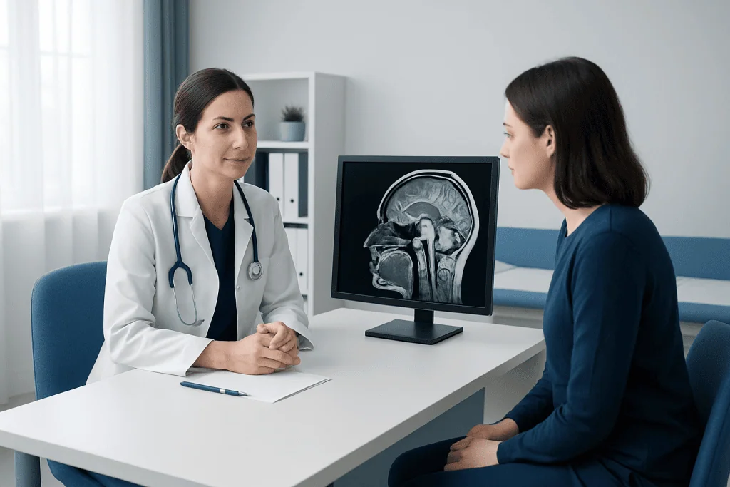
Images are available immediately after the scan, but the formal result requires interpretation by a radiologist. The radiologist reviews the sequences carefully, compares them with earlier imaging when available, and prepares a written report for the clinician who requested the MRI. Timing varies between facilities and according to how urgent the referral is.
For routine scans, results are often discussed within a few days, although this can take longer in some settings. Urgent findings are usually communicated promptly to the referring team. Patients should ask when and how they will receive their report, and whether a follow-up appointment has been arranged.
It is useful to remember that wording in a radiology report can be technical and may not indicate a serious problem. The clinician who knows the patient’s symptoms and history is best placed to explain what a described cyst means in practical terms. Patients may request a copy of the report and images for their personal health records or specialist consultation.
Cyst in the brain treatment, recovery and possible risks
Treatment for a brain cyst depends on its cause, type, location, size, symptoms and effect on surrounding structures. Observation is often the most appropriate plan for an incidental, stable cyst that is not causing symptoms. Follow-up may include repeat imaging at an interval recommended by the treating clinician.
When a cyst causes significant pressure, obstructs cerebrospinal fluid flow, contributes to neurological symptoms or has uncertain features, a neurosurgical opinion may be recommended. Cyst in the brain surgery can include endoscopic fenestration to create an opening for fluid drainage, microsurgical removal in selected cases, or placement of a shunt in specific circumstances. The approach is individualised; surgery is not needed simply because a cyst is visible on MRI.
Recovery after MRI itself is immediate. People can usually return to normal activities straight away unless they received sedation, in which case they need time to recover and should follow discharge instructions. Recovery after brain surgery varies considerably with the procedure and the individual’s health; the surgical team explains expected hospital stay, activity limits, follow-up imaging and rehabilitation needs.
MRI is generally very safe when screening procedures are followed. Possible limitations include anxiety in enclosed spaces, noise, discomfort from lying still and rare reactions to contrast. Surgical treatment has more substantial risks, such as bleeding, infection, seizures, neurological changes or fluid-related complications, which a neurosurgeon discusses in relation to the expected benefit. Acibadem International’s multidisciplinary specialists and JCI-accredited hospitals assess brain imaging findings and treatment needs for international patients.
When to seek medical care
A person should arrange medical review after a cyst is identified on MRI, even if they feel well, so the finding can be interpreted correctly and a follow-up plan can be made. They should seek earlier advice if headaches become persistent or markedly different, seizures occur, vision changes develop, or there are new problems with balance, coordination, speech, memory, strength or sensation.
Emergency care is appropriate for sudden severe headache, a first seizure, loss of consciousness, sudden weakness or numbness on one side of the body, new confusion, severe repeated vomiting, or an abrupt change in vision or speech. These symptoms can have many causes and need prompt assessment rather than self-diagnosis.
Patients can support their care by keeping records of symptoms, including when they occur, their duration and possible triggers. Bringing prior scan reports and image discs to appointments can help specialists compare findings over time and make well-informed recommendations.
Frequently asked questions
Is a cyst in the brain MRI always serious?
No. Many brain cysts are benign, stable and discovered incidentally during imaging for an unrelated reason. Their significance depends on the cyst type, location, size, imaging appearance and whether it is linked to symptoms or pressure on nearby structures.
Does a brain MRI use radiation?
No. MRI uses a powerful magnetic field and radio waves rather than ionising radiation such as X-rays or CT scans. This makes it valuable for detailed brain imaging, although MRI safety screening for metal and certain implants is essential.
Can a brain MRI tell whether a cyst is cancerous?
MRI can provide important clues about whether a lesion looks like a simple cyst or has features that need further investigation. However, imaging findings are interpreted with symptoms, examination results and sometimes follow-up imaging or additional tests; MRI alone does not always establish a final diagnosis.
Can I drive after a brain MRI?
Most people can drive and return to usual activities after a standard MRI. If sedation or medicine for anxiety was used, driving is not safe that day, and the patient should arrange transport home according to the facility’s instructions.
Do all brain cysts need surgery?
No. Many cysts need no treatment beyond observation or periodic imaging. Surgery is generally considered only when a cyst causes symptoms, blocks normal fluid flow, creates significant pressure, grows, or has features requiring a more definitive diagnosis.
What should I do if I am claustrophobic before a brain MRI?
Patients should tell the referring clinician or MRI centre before the appointment. The team can explain the process, offer communication and comfort measures, and discuss whether medication, sedation or another imaging arrangement may be suitable.
References
- Radiological Society of North America
- American College of Radiology
- National Institute of Neurological Disorders and Stroke
- National Health Service
- Mayo Clinic
This article is for general information only and is not a substitute for professional medical advice. Please consult a qualified doctor about your individual situation.
Oncology care in Turkey — second opinion and treatment plan
JCI-accredited · board-certified surgeons · reply within 24h
Add us as a Preferred Source to see more of our trusted health content across Google Search, AI Overviews and Discover.