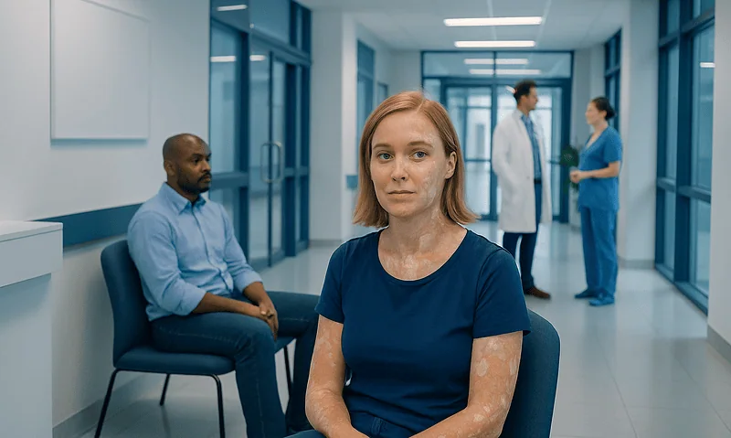
Diagnostic Mammogram and Ultrasound: Preparation, Procedure and Results

A diagnostic mammogram focuses on a specific breast concern or an area seen on screening imaging. Ultrasound uses sound waves and can help distinguish a fluid-filled cyst from a solid area.
Key Takeaways
- A diagnostic mammogram focuses on a specific breast concern or an area seen on screening imaging.
- Ultrasound uses sound waves and can help distinguish a fluid-filled cyst from a solid area.
- The tests do not usually require recovery time, and most people return to normal activities immediately.
- A radiologist may discuss preliminary findings on the day, but final reports and next steps can vary by center.
- Imaging can identify suspicious features, but a biopsy is needed to diagnose breast cancer with certainty.
A diagnostic mammogram and ultrasound are targeted breast imaging tests used to investigate a symptom, such as a lump or nipple discharge, or to clarify an abnormal screening mammogram. They provide complementary information and can often be completed during one visit, although further imaging or a biopsy may sometimes be needed.
Overview: why diagnostic mammogram and ultrasound are used
A diagnostic mammogram and ultrasound are breast imaging examinations performed when there is a specific question to answer. This may be a new lump, focal pain, skin or nipple change, nipple discharge, or an area that needs closer assessment after a screening mammogram. Unlike routine screening, diagnostic imaging is tailored to the person’s symptoms and previous images.
Mammography uses low-dose X-rays to show breast tissue, including calcifications and structural changes. Ultrasound uses sound waves to create real-time images and is particularly useful for examining a palpable area or determining whether a finding is more likely to be fluid-filled or solid. The tests are complementary; having both does not mean cancer is expected.
Breast imaging may also lead to reassurance, short-term follow-up imaging, or a recommendation for tissue sampling. If an imaging finding needs confirmation, a breast biopsy can provide a definitive diagnosis by examining cells or tissue in a laboratory.
Who may need these tests and how they work
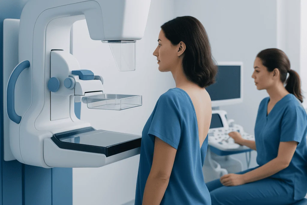
Diagnostic breast imaging may be appropriate for people with a new breast or underarm lump, persistent localized breast pain, a change in breast shape, dimpling of the skin, a newly inverted nipple, unusual nipple discharge, or a change noticed by a clinician. It may also be used to evaluate an abnormal or incomplete screening mammogram. The choice of tests depends on age, symptoms, breast density, pregnancy status, and prior imaging.
During mammography, the breast is briefly compressed between plates to spread the tissue and reduce motion. This improves image quality while using the lowest practical radiation dose. Diagnostic views may include additional angles, magnified views, or focused images of one area.
During ultrasound, a radiologist or trained sonographer applies gel to the breast and moves a handheld transducer over the skin. The sound waves do not use ionizing radiation. Ultrasound is commonly used alongside mammography, but it does not replace mammography for most diagnostic questions because each test can reveal findings the other may not show.
People who are pregnant, may be pregnant, have breast implants, have had breast surgery, or have had previous breast cancer should tell the imaging team. The team can adapt positioning and select the most appropriate imaging approach.
Preparation and step-by-step procedure
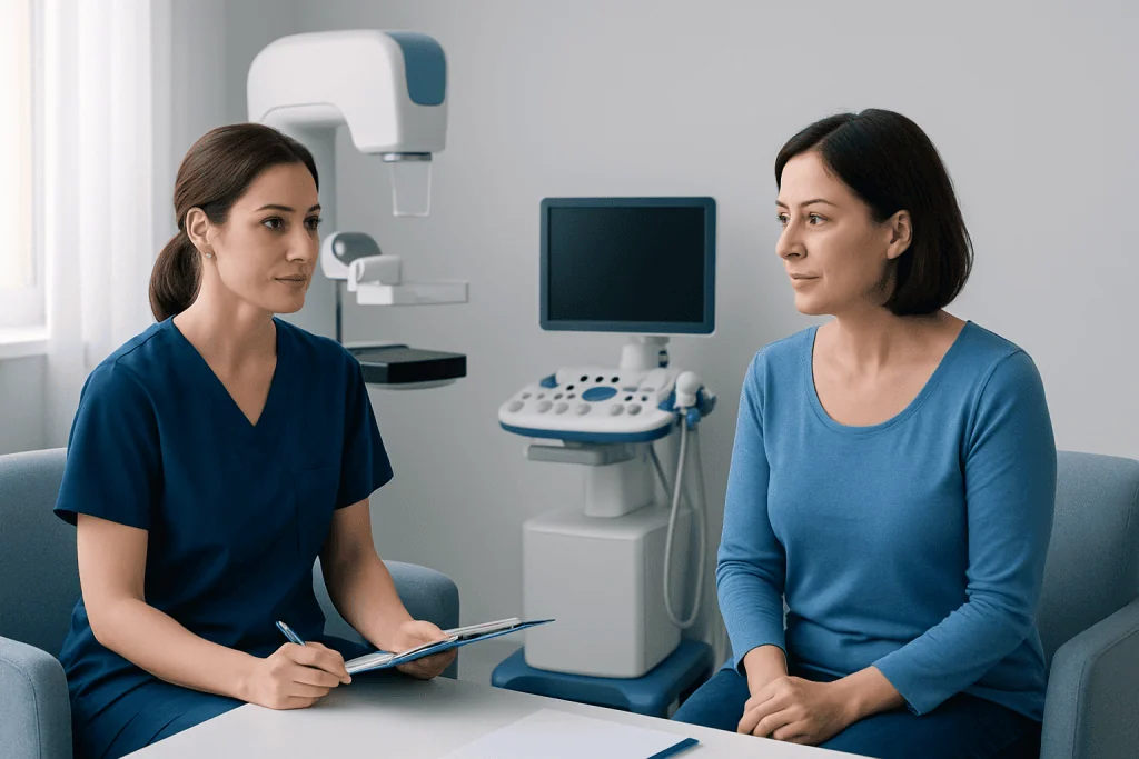
When booking, the imaging center may ask about symptoms, prior mammograms, breast surgery, implants, family history, and pregnancy possibility. Bringing previous breast images or ensuring they are transferred to the new center can help the radiologist compare changes over time. It is usually best to schedule mammography when breasts are least tender, if menstrual cycles make timing predictable.
On the day of the appointment, a person changes from the waist up and removes jewelry or deodorant from the chest and underarm area. Powders, lotions, creams, and deodorants can sometimes create image artifacts that resemble tiny calcifications. Comfortable two-piece clothing can make changing easier.
For the mammogram, the technologist positions one breast at a time and takes standard and, when needed, additional views. Pressure lasts only a few seconds for each image and can feel uncomfortable, particularly if the breasts are tender. For ultrasound, the person usually lies on their back or partly turned to one side while gel is applied and images are recorded. The examination may include the area of concern and nearby tissue.
A diagnostic mammogram and ultrasound appointment often lasts longer than a screening mammogram because the radiologist may review images while the person is still in the department and request extra views. The exact duration depends on the reason for imaging and whether further targeted assessment is required.
What should you not do before a breast ultrasound?
Most people do not need special preparation for a breast ultrasound. They can eat, drink, and take regular medicines unless their healthcare professional gives different instructions. There is no need to fast, and routine breast ultrasound does not involve injections, sedation, or radiation.
For an appointment that includes mammography, avoid applying deodorant, antiperspirant, powder, lotion, perfume, or cream on the breasts, chest, or underarms on the day. These products may leave particles on the skin that interfere with mammogram images. If they have already been applied, the imaging center can usually provide wipes.
People should not delay discussing a concerning breast change because of uncertainty about preparation. They should tell the imaging team about a new pregnancy, breastfeeding, breast implants, previous surgery, and any area that feels different so that the examination can be focused appropriately.
Results, benefits and possible next steps
A radiologist interprets the images and prepares a report for the referring clinician. The report may describe a normal result, a clearly benign finding such as a simple cyst, an area that should be monitored, or a finding that needs additional assessment. Results are often classified using a standardized breast imaging reporting system to guide follow-up.
The main benefit of diagnostic mammogram and ultrasound is that they provide a more detailed assessment than symptoms alone. They can identify many non-cancerous causes of a lump or breast change, including cysts, fibroadenomas, inflammation, scar tissue, and benign calcifications. They can also help locate a suspicious area accurately if a biopsy is advised.
Imaging has limitations. Dense breast tissue can make some changes more difficult to see on mammography, while ultrasound can identify findings that are not clinically important and may lead to follow-up tests. Mammography involves a small amount of radiation, but the exposure is carefully controlled and the clinical benefit generally outweighs this risk when diagnostic imaging is indicated.
If the findings appear suspicious, the radiologist may recommend image-guided biopsy, additional imaging, or referral to a breast specialist. This recommendation does not confirm cancer; it means more information is needed to reach an accurate diagnosis. Relevant care may involve breast radiology, pathology, breast surgery, and oncology specialists.
How long does it take to get results from a diagnostic mammogram and ultrasound?
Many imaging departments review diagnostic mammogram and ultrasound images during the visit, so a radiologist or another member of the care team may provide preliminary information before the person leaves. However, the timing of the official written report varies according to the center, the complexity of the findings, and whether prior images need to be obtained for comparison.
The referring clinician usually receives the final report and explains what it means, including whether follow-up is needed. People should ask the imaging center how and when results will be communicated and whom to contact if they have not heard back within the expected timeframe.
If a biopsy or additional test is recommended, the care team will explain the reason, how it is performed, and when to expect pathology results. Waiting for results can be stressful, but most diagnostic findings are not cancer, and a clear follow-up plan is an important part of safe breast care.
Can a radiologist tell if it is breast cancer on ultrasound?
Ultrasound can show features that make a breast finding appear benign or suspicious, including its shape, margins, internal appearance, and relationship to nearby tissue. A radiologist may be able to recognize a simple cyst or another typically benign pattern. However, ultrasound alone cannot confirm whether a solid mass is breast cancer.
When imaging features are concerning, the usual next step is a needle biopsy guided by ultrasound, mammography, or another imaging method. A pathologist examines the tissue sample, and this is the only way to diagnose breast cancer with certainty. Imaging and pathology results are interpreted together with the person’s symptoms and medical history.
Some breast cancers are more visible on mammography than ultrasound, particularly cancers that present as certain calcifications. Conversely, ultrasound may clarify an abnormality hidden by dense tissue or help assess a lump that is felt but not clearly seen on a mammogram. This is why combined assessment can be valuable.
Does breast ultrasound include armpits?
A breast ultrasound may include the underarm area, also called the axilla, when there is a lump, swelling, pain, an abnormal lymph node, a known breast diagnosis, or another clinical reason to examine it. The referral request and the radiologist’s assessment guide the area scanned. It is not automatically included in every routine breast ultrasound examination.
Axillary ultrasound can assess lymph nodes and other soft tissues in the underarm. Enlarged lymph nodes often have benign explanations, including recent infection, inflammation, vaccination, or skin conditions. If a node has features that need clarification, the radiologist may recommend follow-up or a biopsy.
There is no recovery period after mammography or ultrasound. Mild temporary breast tenderness or skin pressure marks can occur after mammography and usually settle quickly. Acibadem International’s multidisciplinary specialists and JCI-accredited hospitals can coordinate diagnostic breast imaging and subsequent care for international patients when needed.
When to seek medical care: A person should arrange prompt medical assessment for a new or persistent breast or underarm lump, bloody or spontaneous nipple discharge, skin dimpling, persistent redness or swelling, nipple changes, or a breast change that does not resolve. Urgent care is appropriate if breast redness and pain are accompanied by fever or feeling unwell, particularly during breastfeeding, as infection may need treatment.
Frequently asked questions
Is a diagnostic mammogram more painful than a screening mammogram?
The breast compression used for a diagnostic mammogram is similar to that used for screening mammography. Diagnostic imaging may include more views and therefore take longer, but each compression is brief. Letting the technologist know about pain or tenderness can help them position the breast as comfortably as possible.
Can I have a mammogram and ultrasound if I have breast implants?
Yes. People with breast implants can have diagnostic mammography and breast ultrasound. The imaging team uses additional positioning techniques and views when needed to see as much breast tissue as possible, so it is important to mention implants when booking and on arrival.
Can breast ultrasound find a lump that mammography misses?
Yes, ultrasound may help evaluate some lumps that are difficult to see on mammography, including findings in dense breast tissue. However, it can also miss certain abnormalities that mammography can detect, especially some calcifications. The tests are often used together because they provide different information.
What happens if the imaging result is abnormal?
An abnormal result does not necessarily mean cancer. Depending on the finding, the radiologist may recommend comparison with older images, additional focused imaging, a repeat scan after a defined interval, or a biopsy. The referring clinician or breast specialist will explain the recommended next step.
Can I drive after a diagnostic mammogram and breast ultrasound?
Yes. These tests do not normally involve sedation or medications that affect driving, and most people can return to work and usual activities immediately. If a separate procedure, such as a biopsy, is scheduled, its aftercare instructions may be different.
Should breast pain always be investigated with imaging?
Breast pain is common and is often not caused by cancer, particularly when it is diffuse or related to hormonal changes. Persistent pain in one specific area, pain accompanied by a lump or skin change, or pain that concerns the person should be assessed by a qualified clinician. The clinician can decide whether diagnostic imaging is appropriate.
References
- American College of Radiology
- American Cancer Society
- National Cancer Institute
- U.S. Food and Drug Administration
- World Health Organization
This article is for general information only and is not a substitute for professional medical advice. Please consult a qualified doctor about your individual situation.
Ultrasound in Turkey — costs, top hospitals & a free quote
JCI-accredited · board-certified surgeons · reply within 24h
Add us as a Preferred Source to see more of our trusted health content across Google Search, AI Overviews and Discover.

Check-up & Preventive Medicine
Comprehensive health screenings designed for early detection and prevention.
1 specialists in this unit