Does Lobular Breast Cancer Show Up on Ultrasound: Preparation, Procedure and Results

Ultrasound can detect some lobular breast cancers, but a normal ultrasound does not rule the condition out. Invasive lobular carcinoma often grows in single-file cells, which can make it less visible on mammography and ultrasound than other breast cancers.
Key Takeaways
- Ultrasound can detect some lobular breast cancers, but a normal ultrasound does not rule the condition out.
- Invasive lobular carcinoma often grows in single-file cells, which can make it less visible on mammography and ultrasound than other breast cancers.
- A biopsy is the only way to confirm whether an imaging abnormality is cancer.
- Breast MRI may be recommended to clarify the extent of confirmed or suspected lobular breast cancer.
- New breast changes, including thickening, a lump, nipple changes or unexplained swelling, should be assessed promptly.
Lobular breast cancer can show up on ultrasound, often as an area of shadowing or a subtle mass, but it may not have a distinct appearance and can sometimes be missed. Imaging findings are interpreted alongside a breast examination, mammogram and, when appropriate, breast MRI or biopsy.
Overview: Does Lobular Breast Cancer Show Up on Ultrasound?
Does lobular breast cancer show up on ultrasound? It can. Ultrasound may identify invasive lobular carcinoma as a mass, an area of altered tissue, or acoustic shadowing, but the cancer can be subtle and may not always be visible. A normal ultrasound therefore cannot, by itself, exclude lobular breast cancer when a person has concerning symptoms or another abnormal test result.
Lobular breast cancer usually refers to invasive lobular carcinoma (ILC), a cancer that begins in the milk-producing lobules and has moved into nearby breast tissue. Its cells often grow in a dispersed pattern rather than forming a firm, clearly outlined lump. This growth pattern helps explain why a combination of clinical examination, mammography, ultrasound, magnetic resonance imaging (MRI) and biopsy may be needed.
Breast ultrasound uses sound waves rather than radiation. It is commonly used to investigate a palpable change, assess an area seen on a mammogram or MRI, distinguish a fluid-filled cyst from a solid finding, and guide a needle biopsy. It is an important complementary test, not usually a replacement for routine mammography screening.
How Ultrasound Works and What It Can Show
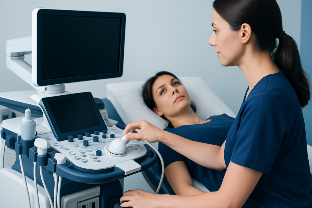
During a breast ultrasound, a trained sonographer or radiologist places gel on the skin and moves a small handheld device, called a transducer, over the breast and nearby underarm area. The transducer sends sound waves into the body and converts returning echoes into real-time images. Because the test does not use ionizing radiation, it can be used in many clinical situations, including pregnancy when imaging is clinically indicated.
On ultrasound, lobular cancer may appear as an irregular hypoechoic area, a distorted region of breast tissue, or an area that produces posterior acoustic shadowing. These descriptions are imaging terms, not a diagnosis. Benign conditions can also create unusual ultrasound appearances, while some cancers do not produce a clearly visible target.
Radiologists consider the ultrasound appearance together with symptoms, examination findings and other imaging. They report breast imaging using a standardized assessment approach and may recommend routine follow-up, short-interval repeat imaging, additional imaging, or tissue sampling depending on the level of concern.
- Benefit: Ultrasound can provide targeted, real-time evaluation of a specific area and can guide biopsies.
- Limitation: It can be less effective for detecting certain subtle cancers or widespread changes without a discrete mass.
- Important point: Results should be interpreted in context rather than viewed as a stand-alone answer.
Can Lobular Breast Cancer Be Missed on Ultrasound?
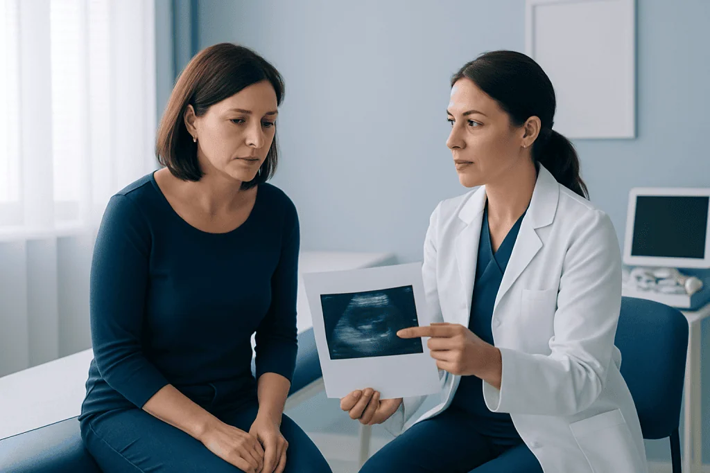
Yes. Lobular breast cancer can be missed on ultrasound, particularly when it does not form a defined mass or when its changes blend with normal breast tissue. Invasive lobular carcinoma may grow as thin strands of cells through the breast, causing only mild thickening, architectural distortion or no obvious change on a single imaging test.
Visibility can also depend on breast density, the size and location of the abnormality, the reason for the scan and whether there is a specific area to target. For example, ultrasound may be particularly useful when a clinician can direct the examination toward a palpable area. It may be less likely to detect disease that is diffuse or has no focal imaging correlate.
If symptoms or a physical examination remain concerning despite reassuring ultrasound findings, a doctor may recommend diagnostic mammography, breast MRI, specialist review or biopsy. Imaging-test agreement matters: when the clinical finding and imaging result do not match, further assessment is generally appropriate.
People should not assume that an ultrasound result explains a persistent breast change. Follow-up discussions with the radiology and breast-care teams help ensure that the next step reflects the overall clinical picture.
What Is the Best Test to Detect Lobular Breast Cancer?
There is no single best test for every person or every stage of assessment. Mammography remains a central screening and diagnostic tool, while ultrasound is often used to investigate specific findings. For suspected or confirmed invasive lobular carcinoma, breast MRI can be especially helpful because it may show the size and distribution of disease more clearly in selected situations.
A biopsy is the only test that can diagnose breast cancer with certainty. If imaging identifies a suspicious area, a radiologist may use ultrasound, mammography or MRI guidance to collect tissue samples. A pathologist examines these samples under a microscope and performs additional tests that help define the cancer type and guide treatment planning.
After a diagnosis, the breast team may use further imaging and pathology information to assess the cancer’s extent and biological features. The best investigation plan is individualized and may be influenced by symptoms, age, personal and family history, breast density, prior imaging and the initial test findings.
No imaging test should be interpreted as a guarantee. People with ongoing changes or questions about their results should ask their clinician whether the results are consistent with the examination and whether additional evaluation is needed.
Where Does Lobular Breast Cancer Usually Start?
Lobular breast cancer usually starts in the lobules, the glands that produce milk. The most common invasive form is invasive lobular carcinoma, in which abnormal cells have moved beyond the lobules into surrounding breast tissue. It may occur in one breast and, less commonly, can be associated with changes in the other breast as well.
Another condition, lobular carcinoma in situ (LCIS), involves abnormal cells within the lobules but is not invasive breast cancer. LCIS is generally considered a marker of increased future breast cancer risk rather than a cancer that has spread into surrounding tissue. Its management differs from that of invasive lobular carcinoma.
Invasive lobular carcinoma may be felt as a thickened area, fullness, firmness or a change in breast shape rather than a round, distinct lump. It can also cause skin or nipple changes, though many breast changes have non-cancerous causes. A new or persistent change merits professional assessment.
Procedure: Candidacy, Preparation and Step-by-Step Ultrasound
Breast ultrasound may be appropriate for someone with a new lump, focal pain, thickening, nipple discharge, skin change or an abnormal screening mammogram. It may also be used to examine breast symptoms during pregnancy or breastfeeding, when clinicians wish to avoid unnecessary radiation exposure. The referring clinician or breast-imaging team determines whether ultrasound, mammography, MRI or more than one test is appropriate.
Preparation is simple. There is usually no fasting, medication adjustment or recovery planning needed. It is helpful to avoid applying lotion, powder or deodorant to the chest and underarm area on the day of imaging, and to bring prior breast-imaging records when available. Wearing a two-piece outfit may make changing easier.
During the procedure, the person changes from the waist up and lies on an examination couch, often with one arm raised. Gel is applied to the skin, and the transducer is moved over the relevant breast and sometimes the underarm. The scan commonly takes around 15 to 30 minutes, although timing varies with the area being assessed and whether extra views are required.
There is no physical recovery time after a diagnostic ultrasound. The person can usually return to normal activities immediately. If a biopsy is recommended, the radiology team explains how it will be performed, what aftercare is needed and when results are expected.
Results, Benefits and Possible Next Steps
Ultrasound results may describe normal tissue, a benign-appearing finding such as a simple cyst, a finding that needs interval monitoring, or an abnormality that requires biopsy. A radiologist may also note whether lymph nodes in the underarm appear enlarged or unusual. These observations help guide care but do not establish a cancer diagnosis without tissue testing.
The principal benefits of ultrasound are that it is noninvasive, does not involve radiation and allows focused evaluation in real time. It can be particularly valuable for guiding a core needle biopsy when an abnormality is visible on the scan. Risks are minimal and usually limited to temporary discomfort from pressure by the transducer.
Ultrasound has no known long-term risks from the sound waves used in standard diagnostic examinations. Its main limitation is the possibility of false-negative or false-positive findings. A false-positive result can lead to additional imaging or biopsy, while a false-negative result can delay recognition if symptoms are not reassessed.
If invasive lobular carcinoma is diagnosed, treatment may include surgery, radiotherapy, systemic therapy such as endocrine therapy, and sometimes chemotherapy or targeted treatments, depending on the individual cancer features and stage. Care is planned by a multidisciplinary breast cancer team.
Which Is More Serious, Ductal or Lobular Breast Cancer?
Neither ductal nor lobular breast cancer is automatically more serious. Prognosis depends primarily on factors such as the cancer stage, tumor size, lymph-node involvement, grade, hormone-receptor and HER2 status, response to treatment, and overall health. Both invasive ductal carcinoma and invasive lobular carcinoma can be effectively treated, especially when identified at an earlier stage.
Invasive ductal carcinoma is more common and begins in milk ducts, whereas invasive lobular carcinoma begins in milk lobules. Lobular cancer’s diffuse growth pattern may make it harder to define on imaging and during surgical planning in some cases. This does not mean that every lobular cancer behaves more aggressively.
A pathology report provides essential information about the specific cancer. The breast-care team uses this information to discuss treatment options and likely outcomes in an individualized way. Comparing cancer types without the full clinical context can be misleading.
Acibadem International’s multidisciplinary specialists and JCI-accredited hospitals support international patients needing assessment and treatment planning for breast conditions and breast cancer.
When to Seek Medical Care
A person should arrange a medical assessment for a new breast lump, persistent thickening, a change in breast size or shape, dimpling of the skin, a new inverted nipple, unusual nipple discharge, persistent localized breast pain, or a swollen lymph node near the underarm or collarbone. Most breast symptoms are not caused by cancer, but timely evaluation helps clarify the cause.
It is also important to contact the care team if a breast change persists after a normal ultrasound or mammogram, or if a person feels that the imaging result does not match what they notice in their body. Clinicians can review prior studies, perform a repeat examination and decide whether another imaging approach or biopsy is appropriate.
People should follow local breast-screening recommendations and attend scheduled follow-up appointments. Those with a strong family history of breast or ovarian cancer, known inherited cancer risk, previous chest radiotherapy or prior high-risk breast findings may need a personalized screening plan.
Frequently asked questions
Does a normal breast ultrasound rule out lobular breast cancer?
No. A normal ultrasound can be reassuring, but it does not completely rule out invasive lobular carcinoma because this cancer may be subtle or diffuse. Persistent symptoms, an abnormal examination or a concerning mammogram may require further imaging or biopsy.
Is ultrasound better than mammography for lobular breast cancer?
Ultrasound and mammography provide different information, so one is not universally better than the other. Ultrasound is useful for assessing a targeted area and guiding biopsy, while mammography remains an important screening and diagnostic examination. Breast MRI may be useful in selected cases of suspected or confirmed lobular cancer.
What does lobular cancer look like on ultrasound?
Lobular cancer may appear as an irregular dark area, tissue distortion or an area with shadowing behind it. However, there is no single ultrasound pattern that confirms lobular cancer. A biopsy is needed to establish a diagnosis.
Can breast ultrasound detect cancer in dense breasts?
Ultrasound can find some abnormalities that are harder to see on mammography in dense breast tissue. However, it can also identify benign findings that require further assessment, and it may still miss some cancers. The most suitable imaging plan should be discussed with a clinician.
How is invasive lobular carcinoma confirmed?
Invasive lobular carcinoma is confirmed with a tissue biopsy reviewed by a pathologist. Imaging helps locate the area for biopsy and assess the extent of disease, but imaging alone cannot make the diagnosis.
Does a breast ultrasound hurt?
Breast ultrasound is usually painless. Some people may feel mild pressure from the transducer, especially if an area is already tender. There is typically no recovery period and normal activities can resume immediately.
References
- American Cancer Society
- National Cancer Institute
- American College of Radiology
- Breast Cancer Now
- World Health Organization
This article is for general information only and is not a substitute for professional medical advice. Please consult a qualified doctor about your individual situation.
Breast Cancer in Turkey — costs, top hospitals & a free quote
JCI-accredited · board-certified surgeons · reply within 24h
Add us as a Preferred Source to see more of our trusted health content across Google Search, AI Overviews and Discover.

Medical Oncology Department
Medical treatment of cancer with chemotherapy, immunotherapy and targeted therapies under a multidisciplinary tumor board.
60 specialists in this unit