Early stage blood clot in foot pictures: Symptoms, Causes, and Treatment — Complete Patient Guide

Pictures may hint at a blood clot, but they cannot confirm or rule one out. Common warning signs include one-sided swelling, pain, warmth, redness, and difficulty walking.
Key Takeaways
- Pictures may hint at a blood clot, but they cannot confirm or rule one out.
- Common warning signs include one-sided swelling, pain, warmth, redness, and difficulty walking.
- A blood clot may form after immobility, surgery, injury, pregnancy, or with certain medical conditions.
- Ultrasound and a clinical exam are the main ways doctors diagnose a suspected clot.
- Urgent care is important if symptoms are sudden, worsening, or occur with chest pain or shortness of breath.
Early stage blood clot in foot pictures may show swelling, color change, or visible surface veins, but images cannot reliably diagnose a clot. A true blood clot in the foot or leg needs medical assessment because symptoms can overlap with injury, infection, arthritis, and circulation problems.
Overview: What early stage blood clot in foot pictures can and cannot show
People often search for early stage blood clot in foot pictures to compare what they are seeing at home with online images. In some cases, a clot affecting the veins of the foot or lower leg may be associated with visible swelling, a patch of redness or bluish discoloration, skin that looks tighter than usual, or veins that appear more prominent. These changes may be subtle in the early stage, especially in people with darker skin tones or in those whose main symptom is pain rather than obvious color change.
However, pictures alone are not a safe way to diagnose a blood clot. Many other conditions can look similar, including a sprain, cellulitis, gout, venous insufficiency, tendon inflammation, and arthritis. A clot may also be present without dramatic surface changes, which is why doctors rely on symptoms, risk factors, and imaging tests rather than appearance alone.
Most concern centers on venous clots, especially deep vein thrombosis, or DVT, which usually occurs in the deep veins of the calf or leg but can cause symptoms felt in the foot. When a clot is suspected, the goal is not only to ease pain and swelling but also to prevent complications such as clot extension or movement to the lungs. For broader context on this condition, readers may find deep vein thrombosis helpful.
What symptoms may appear in the early stage

Early symptoms of a blood clot in the foot area can vary. Some people notice aching, cramping, throbbing, or tenderness that is different from ordinary muscle soreness. Others notice swelling on one side, warmth, or a change in skin color. Pain may become more noticeable when standing or walking, and shoes may suddenly feel tighter on one foot.
A clot does not always produce all symptoms at once. In the early stage, there may be only mild swelling or a vague heavy sensation in the foot and ankle. Because of this, it can be mistaken for overuse, a minor injury, or fluid retention. Symptoms that are limited to one foot or one leg are generally more concerning than symptoms affecting both sides equally.
Signs that may raise suspicion include:
- Swelling in one foot, ankle, or lower leg
- Pain or tenderness not clearly explained by injury
- Warmth over the painful area
- Red, purple, or bluish skin color change
- Prominent surface veins
- Discomfort that worsens with walking or standing
If chest pain, sudden shortness of breath, coughing blood, fainting, or a racing heartbeat occurs along with leg or foot symptoms, emergency care is needed right away because these can be signs of a clot that has traveled to the lungs.
What a blood clot in the foot may look like compared with other problems
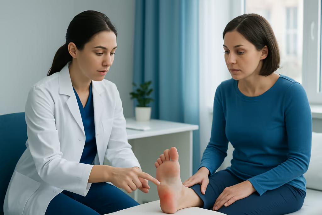
When people search for pictures, they are often trying to answer a practical question: does this look like a clot or something else? A blood clot may cause one-sided swelling, stretched-looking skin, redness or dusky discoloration, and an area that feels warmer than the surrounding skin. The foot may look fuller over the top or around the ankle, and the difference may be easier to spot by comparing both feet side by side.
Still, there is no single “classic” picture. A sprain may also cause swelling and pain, but it usually follows a twist, fall, or clear injury and often causes bruising around a joint. Infection may cause warmth, redness, swelling, and tenderness, but it is more likely to come with fever, spreading redness, or a wound. Gout often affects the big toe suddenly and can make the joint extremely painful, red, and swollen.
Because appearance overlaps so much, doctors ask about the timing of symptoms, recent travel, surgeries, medications, pregnancy, cancer history, and whether there is calf pain or swelling higher in the leg. The physical exam helps narrow the possibilities, but imaging is usually needed to confirm whether a clot is present.
Causes and risk factors
Blood clots form when blood flow slows, the blood becomes more likely to clot, or the lining of a vein is affected. This can happen during long periods of immobility, such as bed rest, recovery after surgery, or long-distance travel. Injury to the leg or foot, fractures, and casting can also increase risk by reducing movement and affecting nearby veins.
Medical and hormonal factors matter as well. Pregnancy, the postpartum period, estrogen-containing birth control, hormone therapy, certain cancers, smoking, obesity, and inherited clotting disorders can all increase the chance of a clot. Risk also rises with age and with a personal or family history of deep vein thrombosis or pulmonary embolism.
Not every painful or swollen foot is caused by a clot, and many people with risk factors will never develop one. But when new one-sided swelling or unexplained pain appears in someone with these risks, doctors tend to investigate promptly. Related circulation problems can sometimes overlap with or mimic clot symptoms, and readers may also want to understand peripheral artery disease.
How doctors diagnose a suspected clot
Diagnosis begins with a medical history and physical examination. A doctor will ask when symptoms started, whether there was recent travel, surgery, injury, pregnancy, illness, or reduced mobility, and whether there is any history of clots. They will also assess whether symptoms are confined to the foot or extend into the calf or leg, since many clinically significant clots are located above the foot even when the foot is where symptoms are first noticed.
The main test for a suspected clot in the leg is usually a venous duplex ultrasound. This imaging test is painless, does not use radiation, and helps show whether blood is flowing normally through the veins. In some cases, blood testing such as a D-dimer may be used to help decide how likely a clot is, although it cannot diagnose a clot on its own.
If the diagnosis remains uncertain, doctors may repeat imaging or use additional tests depending on the situation. Because circulation problems can involve different vessel types, some patients may need further vascular evaluation and treatment when symptoms suggest a broader blood-flow issue rather than a simple strain or local injury.
Treatment options and what recovery may involve
Treatment depends on where the clot is, how extensive it is, the person’s overall health, and the risk of bleeding. Many venous clots are treated with anticoagulant medicines, often called blood thinners, which do not dissolve the clot instantly but help stop it from growing and reduce the chance of new clots forming. Over time, the body gradually breaks down the clot.
Some people may need compression therapy, closer monitoring, or treatment in the hospital, especially if symptoms are severe, the clot is extensive, or there is concern about spread to the lungs. Doctors also advise walking and movement plans carefully, since complete rest is not always recommended once treatment has started. Pain control and elevation may help relieve swelling and discomfort.
In selected cases, a specialist may consider more advanced procedures, particularly when there is a large clot, impaired circulation, or a high risk of complications. This can include image-guided care through interventional radiology or specialist management by cardiovascular surgery teams when appropriate. Treatment should always be individualized by a qualified clinician.
Near the end of the care pathway, some international patients choose centers with coordinated vascular, radiology, and internal medicine support. Acibadem International’s multidisciplinary specialists and JCI-accredited hospitals diagnose and treat clot-related conditions for international patients when further evaluation is needed.
Prevention and self-care
Prevention focuses on keeping blood moving and reducing known risks where possible. During travel or long periods of sitting, it helps to stand up regularly, flex the ankles, stretch the calves, and stay well hydrated. After surgery or illness, following the medical team’s advice about early movement, compression, and preventive medicines can be important.
General health measures also support vein health. Maintaining a healthy weight, not smoking, managing chronic conditions, and discussing hormone-related clot risks with a doctor may lower overall risk. People with a history of clots should let healthcare professionals know before surgeries, long flights, or pregnancy-related care so that prevention can be planned in advance.
At home, a person should not massage a painful swollen foot if a clot is suspected, and they should not rely on online pictures as reassurance. Gentle movement may be reasonable, but unexplained one-sided swelling or pain should be assessed by a clinician rather than treated as a routine sprain without evaluation.
When to seek medical care
Medical care should be sought promptly for new one-sided foot or leg swelling, unexplained pain, warmth, redness, or a sudden change in walking comfort, especially after travel, surgery, injury, or prolonged immobility. The same applies to people who are pregnant, recently postpartum, taking estrogen-containing medications, or who have a personal or family history of blood clots.
Emergency care is needed immediately if symptoms occur together with chest pain, shortness of breath, fainting, coughing blood, or sudden severe weakness. These symptoms may signal a pulmonary embolism, which is a medical emergency.
If symptoms are mild but persistent, it is still wise to arrange a prompt medical review. Early assessment can identify whether the cause is a clot or another problem such as infection, injury, arthritis, or poor circulation, and it allows timely treatment before complications develop.
Frequently asked questions
Can a blood clot in the foot be diagnosed from pictures alone?
No. Pictures may show swelling, redness, or color change, but they cannot confirm whether a clot is present. A medical history, physical exam, and usually an ultrasound are needed for a reliable diagnosis.
What does an early stage blood clot in the foot usually feel like?
It may feel like aching, throbbing, tenderness, or a sense of heaviness in one foot or lower leg. Some people also notice warmth, swelling, or pain when walking or standing.
Is a blood clot in the foot always visible?
Not always. Some clots cause obvious swelling or skin color changes, while others produce very little visible change. This is one reason symptoms should not be ignored even if the foot looks nearly normal.
How is a suspected blood clot in the foot or leg tested?
Doctors usually start with a clinical assessment and often order a venous ultrasound. In some situations, a D-dimer blood test may help estimate the likelihood of a clot, but it is not enough by itself to make the diagnosis.
What conditions can look like a blood clot in the foot?
Common look-alikes include sprains, fractures, cellulitis, gout, arthritis, tendon problems, and chronic vein disease. Because these problems can overlap in appearance, self-diagnosis from online images is not dependable.
When should someone go to the emergency room?
Immediate emergency care is needed for chest pain, sudden shortness of breath, coughing blood, fainting, or a rapid heartbeat, especially if there is also leg or foot swelling or pain. These symptoms can suggest a clot has traveled to the lungs.
References
- Centers for Disease Control and Prevention
- National Heart, Lung, and Blood Institute
- American Heart Association
- Society for Vascular Surgery
- National Health Service
This article is for general information only and is not a substitute for professional medical advice. Please consult a qualified doctor about your individual situation.
Joint and spine care in Turkey — expert assessment & treatment
JCI-accredited · board-certified surgeons · reply within 24h
Add us as a Preferred Source to see more of our trusted health content across Google Search, AI Overviews and Discover.

More from the Health Library



Related Specialists

Dr. Mehmet Bilhan Hayırlıoğlu
Anesthesiology
Prof. Dr. İsmet Tamer
Family Medicine
Fzt. Dilek Küçükvardar
Physical Medicine & Rehabilitation
Dr. Aslı Saran İkizler
Radiation Oncology