Endoscopic Ultrasound: High-Detail Imaging and Biopsy From Inside the Body
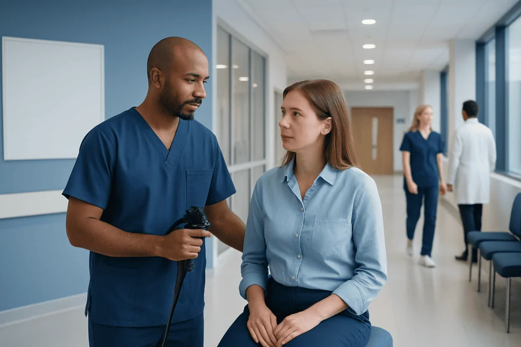
Endoscopic ultrasound combines a flexible endoscope with ultrasound imaging to show detailed views of the digestive tract, pancreas, bile ducts, lymph nodes and nearby structures. EUS may help doctors investigate unexplained abdominal symptoms, evaluate tumors, stage certain cancers, and assess cysts or lesions found on other scans.
Key Takeaways
- Endoscopic ultrasound combines a flexible endoscope with ultrasound imaging to show detailed views of the digestive tract, pancreas, bile ducts, lymph nodes and nearby structures.
- EUS may help doctors investigate unexplained abdominal symptoms, evaluate tumors, stage certain cancers, and assess cysts or lesions found on other scans.
- When necessary, EUS can guide a fine needle aspiration or biopsy, allowing tissue or fluid sampling through the wall of the digestive tract.
- Most procedures are performed with sedation, and patients usually return home the same day after a short recovery period.
- Preparation, risks and aftercare depend on the reason for the test and whether a biopsy or therapeutic step is performed.
Endoscopic ultrasound, often called EUS, is an advanced procedure that creates detailed images of the digestive tract and nearby organs from inside the body. It can also guide a thin needle to collect tissue or fluid samples when a biopsy is needed.
Overview: What Is Endoscopic Ultrasound?
Endoscopic ultrasound, or EUS, is a minimally invasive imaging procedure that combines two technologies: endoscopy and ultrasound. A doctor passes a thin, flexible tube called an endoscope through the mouth or, less commonly, through the rectum. At the tip of the endoscope is a small ultrasound probe that produces sound waves and converts them into detailed images of the tissues and organs close to the digestive tract.
Because the ultrasound probe is placed inside the body, very close to the area being examined, EUS can show fine details that may not be visible on standard abdominal ultrasound. It is especially useful for evaluating the esophagus, stomach, duodenum, rectum, pancreas, bile ducts, gallbladder, liver, adrenal glands and nearby lymph nodes.
EUS is both a diagnostic and, in selected cases, a guiding procedure. If a suspicious area, cyst or lymph node is seen, the doctor may use EUS to guide a very thin needle into the target to collect cells, tissue or fluid. This is known as EUS-guided fine needle aspiration or biopsy, and it can help clarify a diagnosis without the need for open surgery.
How EUS Works and What It Can Show
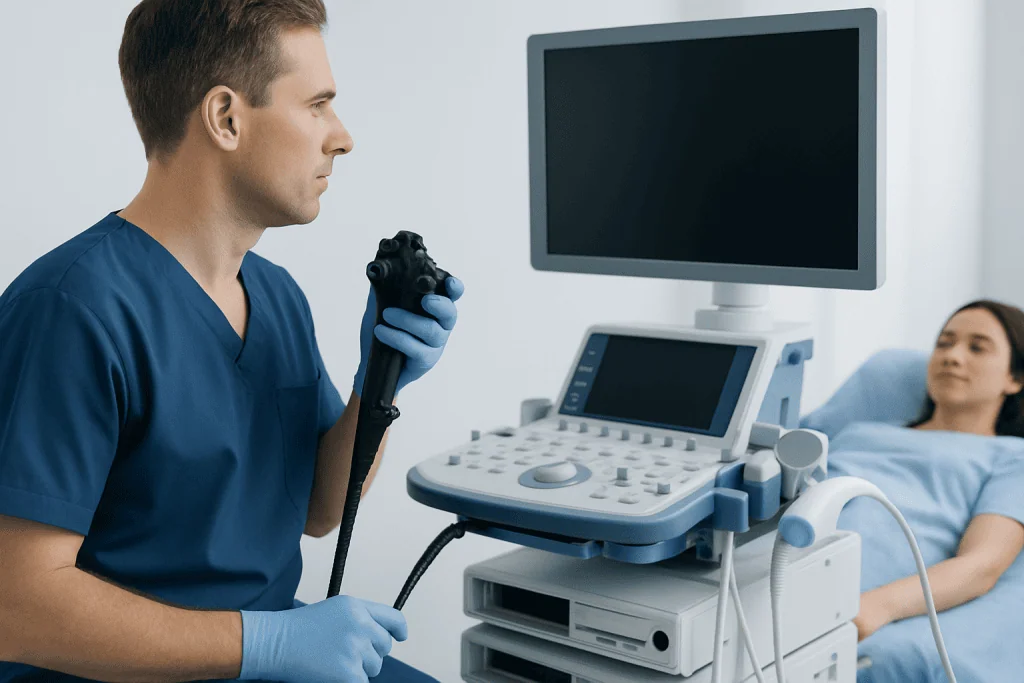
Ultrasound uses sound waves rather than radiation. During EUS, sound waves travel from the probe into nearby tissues and return as echoes. A computer transforms these echoes into images that show the layers of the digestive wall and structures just outside it. The closeness of the probe to the organ being examined is what gives EUS its high-detail view.
Doctors may recommend EUS to investigate findings seen on CT, MRI, standard ultrasound, endoscopy or blood tests. It can help assess whether a growth starts in the lining of the digestive tract or deeper layers, whether nearby lymph nodes look abnormal, and whether a pancreatic cyst or mass has features that need further evaluation. EUS can also help examine bile duct stones or blockages when other tests are unclear.
Common reasons for EUS include evaluation of pancreatic cysts or tumors, unexplained pancreatitis, suspected bile duct disease, submucosal lesions in the stomach or esophagus, rectal or esophageal cancer staging, and enlarged lymph nodes near the digestive tract. In some centers, EUS is also used to guide selected treatments, such as drainage of certain fluid collections, when clinically appropriate.
EUS-Guided Biopsy: Sampling Tissue From Inside the Body
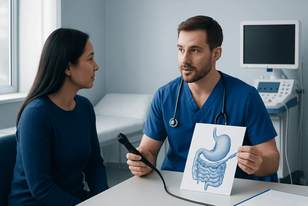
One of the important advantages of endoscopic ultrasound is that it can guide a needle with real-time imaging. If the doctor sees an area that requires sampling, a fine needle can be passed through a channel in the endoscope and directed into the target. The ultrasound image helps the doctor avoid blood vessels and reach the area accurately.
There are two main types of sampling. Fine needle aspiration collects cells or fluid, while fine needle biopsy is designed to obtain a small core of tissue. The choice depends on the location of the abnormality, the suspected condition and what the laboratory needs to examine. Samples may be reviewed by a pathology team to look for inflammation, infection, cyst characteristics or cancer cells.
EUS-guided biopsy is often used for pancreatic masses, pancreatic cysts, enlarged lymph nodes, lesions under the lining of the digestive tract and selected abnormalities near the stomach or rectum. Although it is generally well tolerated, it is still a medical procedure. The doctor will weigh the expected benefit of obtaining a sample against possible risks, such as bleeding, infection, pancreatitis or a reaction to sedation.
Preparing for an Endoscopic Ultrasound
Preparation depends on whether the examination is performed through the mouth, through the rectum, and whether biopsy or treatment is planned. For upper EUS, patients are usually asked not to eat or drink for several hours before the procedure so the stomach is empty. For lower EUS, bowel preparation may be needed to clear the rectum or colon, depending on the area being examined.
Patients should tell their doctor about all medications, allergies and medical conditions before the procedure. This is especially important for blood-thinning medicines, diabetes medications, heart or lung disease, kidney disease, pregnancy, previous reactions to anesthesia, and implanted devices. The medical team will explain whether any medication should be adjusted, paused or continued.
Because sedation is commonly used, patients generally need a responsible adult to take them home afterward. It is not usually safe to drive, operate machinery, sign important documents or drink alcohol on the day of sedation. The care team will provide individualized instructions, including when to stop eating and drinking and when to restart regular medicines.
What Happens During the Procedure?
Before EUS begins, the care team checks vital signs, confirms the procedure plan and places an intravenous line if sedation is used. For an upper EUS, a mouthguard may be placed to protect the teeth and endoscope. The patient is usually positioned on the left side. Sedation helps most people feel relaxed and comfortable, and many remember little or nothing about the procedure.
The doctor gently advances the endoscope to the area of interest. The ultrasound probe is then used to create images from inside the digestive tract. Water may be added through the endoscope to improve contact between the probe and the tissue. If a biopsy is needed, the needle is passed through the endoscope and guided by the ultrasound image to collect a sample.
The length of the procedure varies. A straightforward diagnostic EUS may be relatively brief, while a biopsy or more complex evaluation may take longer. Afterward, patients are monitored in a recovery area until the sedation wears off. Mild bloating, a sore throat or temporary drowsiness can occur and usually improves within a short time.
Benefits, Limitations and Possible Risks
EUS provides very detailed imaging of structures that can be difficult to assess from outside the body, especially the pancreas and the layers of the digestive wall. It may help doctors make a more accurate diagnosis, choose the most appropriate treatment plan, and avoid more invasive procedures in selected situations. The ability to combine imaging and biopsy in one session is a major benefit.
Like all medical tests, EUS has limitations. It may not be the best test for every patient or every organ, and results sometimes need to be interpreted together with CT, MRI, blood tests, standard endoscopy or surgery. A biopsy sample can occasionally be insufficient or inconclusive, and repeat testing or another diagnostic method may be recommended.
Complications are uncommon, but they can happen. Possible risks include sore throat, bleeding, infection, perforation of the digestive tract, pancreatitis after certain pancreatic procedures, aspiration, and side effects from sedation or anesthesia. The risk level depends on the patient’s health, the area examined and whether biopsy or therapeutic intervention is performed. The medical team should discuss the expected benefits and risks before consent is given.
Aftercare, Results and When to See a Doctor
After EUS, patients usually rest until they are alert and stable. Eating and drinking may resume when the medical team says it is safe, often after the throat numbness has worn off for upper procedures. If a biopsy was taken, the doctor may give specific instructions about diet, activity and medications. Most people can return to normal activities the next day, but this varies.
Some findings may be explained immediately, especially if the doctor could clearly see the area of concern. Biopsy or fluid analysis results take longer because the sample must be processed and examined by a laboratory. The final report should be discussed with the treating doctor, who can explain what the findings mean and whether follow-up imaging, medication, surgery, oncology care or simple monitoring is recommended.
Patients should contact a doctor promptly if they develop severe or worsening abdominal pain, persistent vomiting, fever, black stools, chest pain, shortness of breath, difficulty swallowing, or signs of bleeding after the procedure. These symptoms do not mean a serious complication has occurred, but they should be assessed without delay. Acibadem International’s multidisciplinary specialists and JCI-accredited hospitals provide diagnostic and therapeutic EUS services for international patients, with care plans tailored to each person’s medical needs.
Frequently asked questions
Is endoscopic ultrasound painful?
Most patients do not describe EUS as painful because sedation is commonly used. Some may notice a mild sore throat, bloating or drowsiness afterward. Discomfort is usually temporary, but any severe or persistent pain should be reported to a doctor.
How is EUS different from a regular ultrasound?
A regular ultrasound is performed from outside the body, through the skin. EUS places a small ultrasound probe inside the digestive tract, much closer to organs such as the pancreas, bile ducts and nearby lymph nodes. This close position can provide more detailed images of selected areas.
Why might a doctor recommend an EUS biopsy?
A doctor may recommend EUS-guided biopsy when imaging shows a mass, cyst, enlarged lymph node or abnormal tissue that needs further evaluation. The biopsy can provide cells, tissue or fluid for laboratory analysis. This helps guide diagnosis and treatment planning.
How long does it take to recover after EUS?
Many patients go home the same day after a short observation period. Because sedation can affect coordination and judgment, patients are usually advised not to drive or make important decisions for the rest of the day. Recovery may take longer if a complex biopsy or therapeutic procedure is performed.
Are there risks with endoscopic ultrasound?
EUS is generally considered safe when performed by trained specialists, but no procedure is risk-free. Possible complications include bleeding, infection, perforation, pancreatitis in selected cases, and reactions to sedation. The doctor will explain the individual risk based on the planned procedure and the patient’s health.
When will biopsy results be available?
Timing varies by laboratory and by the type of tests needed on the sample. Some preliminary information may be available sooner, while final pathology results can take several days or longer. The treating doctor should review the results and explain the next steps.
References
- American Society for Gastrointestinal Endoscopy
- European Society of Gastrointestinal Endoscopy
- National Institute of Diabetes and Digestive and Kidney Diseases
- Mayo Clinic
- National Comprehensive Cancer Network
This article is for general information only and is not a substitute for professional medical advice. Please consult a qualified doctor about your individual situation.
Ultrasound in Turkey — costs, top hospitals & a free quote
JCI-accredited · board-certified surgeons · reply within 24h
Add us as a Preferred Source to see more of our trusted health content across Google Search, AI Overviews and Discover.

Check-up & Preventive Medicine
Comprehensive health screenings designed for early detection and prevention.
3 specialists in this unit