Enthesophyte: A Complete Medical Overview

An enthesophyte is a bone growth at the attachment point of a tendon, ligament, or capsule. Many enthesophytes are found incidentally on imaging and do not always need treatment.
Key Takeaways
- An enthesophyte is a bone growth at the attachment point of a tendon, ligament, or capsule.
- Many enthesophytes are found incidentally on imaging and do not always need treatment.
- Symptoms depend on the location and the underlying cause, such as overuse, aging-related wear, or inflammatory disease.
- Treatment usually focuses on the source of pain with rest, physical therapy, footwear changes, medicines, or selected procedures.
- Medical evaluation is important if pain persists, mobility worsens, or inflammatory arthritis is suspected.
An enthesophyte is a bony outgrowth that forms at an enthesis, the point where a tendon, ligament, or joint capsule attaches to bone. It may cause no symptoms at all, but when it is linked to inflammation, overuse, or arthritis, it can contribute to pain, stiffness, and reduced movement.
What Is an Enthesophyte?
An enthesophyte is a bony projection that develops where a tendon, ligament, or joint capsule attaches to a bone. This attachment site is called the enthesis. In simple terms, an enthesophyte is a form of bone response at a stress point, and it is sometimes described as a type of bone spur.
Enthesophytes can appear in different parts of the body, including the heel, knee, hip, spine, elbow, and pelvis. They are often seen on X-rays or other scans done for pain or stiffness, but they may also be found accidentally in people who have no symptoms. Whether an enthesophyte is important depends less on its appearance alone and more on the person’s symptoms, physical examination, and overall joint or tendon health.
These growths are not all the same. Some form mainly from long-term mechanical stress and age-related tissue change, while others are associated with inflammation at the enthesis, a process called enthesitis. For this reason, an enthesophyte is best understood as a sign that the body has been adapting to repeated strain, degeneration, or inflammation over time.
How an Enthesophyte Differs From Other Bone Spurs
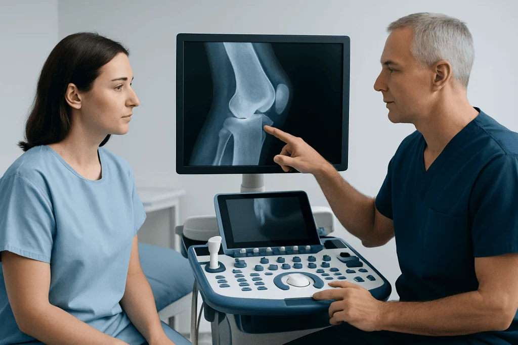
Although the terms are sometimes used loosely, an enthesophyte is not exactly the same as every other bone spur. The key difference is location. An enthesophyte forms specifically at the site where soft tissue attaches to bone. By contrast, an osteophyte usually develops along the edge of a joint and is more commonly associated with cartilage wear and osteoarthritis.
This distinction matters because it helps guide the medical evaluation. For example, a heel enthesophyte may be linked to traction and stress on the plantar fascia or Achilles tendon, while joint-edge spurs may reflect arthritis within the joint itself. In some people, both patterns can exist together.
Doctors also consider whether the finding is degenerative, inflammatory, or both. Inflammatory conditions that affect the entheses can contribute to enthesophyte formation over time. Mechanical stress, altered foot or joint mechanics, obesity, repetitive sports activity, and long-term tissue wear can also play a role.
Because the imaging appearance does not always explain pain by itself, clinicians interpret an enthesophyte together with symptoms, movement, tenderness, and any signs of a broader musculoskeletal condition such as rheumatoid arthritis or other inflammatory disorders.
Symptoms and Common Locations

Many enthesophytes do not cause symptoms. When symptoms do occur, they are usually related to irritation of the nearby tendon, ligament, or surrounding soft tissues rather than the bone growth alone. Pain may be worse with first steps after rest, with exercise, or after repeated loading of the affected area.
Common symptoms can include localized pain, tenderness at the attachment site, stiffness, reduced flexibility, swelling around the area, or discomfort during walking, climbing stairs, gripping, or bending depending on the location. Some people notice symptoms only during activity, while others feel persistent morning stiffness or pain after sitting still.
Frequent sites include:
- Heel: at the Achilles tendon insertion or plantar fascia attachment, sometimes called a calcaneal enthesophyte.
- Elbow: around tendon attachment sites involved in repetitive arm use.
- Knee: at the quadriceps tendon or patellar tendon attachment.
- Hip and pelvis: where strong muscle groups attach.
- Spine: where ligaments and soft tissues anchor along the vertebrae.
If the pain is centered in the heel, the person may also be evaluated for overlapping conditions such as heel spur or plantar fascia irritation, since several causes of heel pain can exist at the same time.
Causes and Risk Factors
Enthesophytes develop over time, usually in response to repeated pulling, stress, or inflammation at the enthesis. The body may lay down extra bone where soft tissue repeatedly tugs on the attachment site. This process can be part of normal aging, but it is more likely when additional risk factors are present.
Mechanical causes are common. These include repetitive sports, prolonged standing, running on hard surfaces, poor footwear, reduced flexibility, muscle imbalance, obesity, and jobs that place repeated strain on certain joints or tendons. Prior injury can also change tissue mechanics and make an enthesis more vulnerable to chronic irritation.
Degenerative joint conditions can contribute as well. For example, people with joint wear may develop altered movement patterns that increase stress at tendon and ligament insertions. In some cases, enthesophytes are seen alongside osteoarthritis, especially in areas already exposed to long-term loading.
Inflammatory diseases are another important group of causes. Certain forms of arthritis, especially the spondyloarthritis family, can inflame the entheses. Over time, repeated inflammation may leave behind structural change, including enthesophyte formation. That is why doctors may ask about back pain, psoriasis, bowel symptoms, eye inflammation, or a family history of inflammatory joint disease when evaluating this finding.
How Doctors Diagnose an Enthesophyte
Diagnosis begins with a medical history and physical examination. The doctor asks where the pain is felt, what activities trigger it, how long it has been present, and whether there are signs of inflammation such as prolonged morning stiffness, swelling, or pain in multiple sites. Examining range of motion, tenderness, gait, and nearby tendons helps identify whether the enthesophyte is likely to be clinically relevant.
Imaging often confirms the finding. Standard X-rays can show mature bony outgrowths clearly, especially in the heel, knee, elbow, and spine. Ultrasound can be helpful for looking at soft tissue changes around the tendon or fascia and may show inflammation at the enthesis. MRI may be used when the diagnosis is uncertain, when a deeper structure is involved, or when inflammatory disease is suspected.
Blood tests are not used to diagnose an enthesophyte itself, but they can be useful when there is concern about an inflammatory arthritis or another systemic condition. The main question is not simply whether the growth exists, but what process caused it and whether it explains the patient’s symptoms.
When symptoms are complex or persistent, assessment by an orthopedic specialist, rheumatologist, or rehabilitation physician may be recommended. In advanced cases, detailed imaging can also help guide procedures such as MRI evaluation or personalized rehabilitation planning.
Treatment Options
Treatment depends on symptoms, location, and the underlying cause. If an enthesophyte is found incidentally and causes no discomfort, no immediate treatment may be needed. When pain is present, the goal is usually to reduce stress on the attachment site, calm surrounding inflammation, and improve function.
Conservative treatment is the first step for most people. This may include activity modification, short-term rest from aggravating movements, ice after overuse, supportive footwear, heel lifts or orthotics when appropriate, and structured stretching or strengthening exercises. Physical therapy can be particularly valuable because it addresses flexibility, muscle balance, walking mechanics, and load management rather than focusing only on the image finding.
Doctors may also recommend pain-relieving or anti-inflammatory medicines when suitable for the individual, as well as targeted treatment for the specific tendon or fascia involved. In selected cases, guided injections or other non-surgical procedures may be considered, especially if soft tissue inflammation is a major source of pain. Persistent symptoms related to biomechanics may benefit from physical therapy and rehabilitation.
Surgery is not needed for most enthesophytes. It may be considered only when symptoms remain significant despite appropriate conservative care, or when the bony prominence is clearly contributing to mechanical irritation and loss of function. Surgical decisions are based on the whole clinical picture, not the X-ray alone, and may be part of broader orthopedic care for chronic tendon, foot, or joint problems.
Self-Care, Prevention, and Daily Management
Not every enthesophyte can be prevented, especially when age-related tissue change is involved, but many people can reduce symptoms and lower the chance of ongoing irritation. The most helpful approach is usually steady load management rather than complete inactivity. Sudden increases in exercise intensity are more likely to aggravate an already stressed enthesis.
Daily measures may include wearing supportive shoes, warming up before activity, improving calf or hamstring flexibility if advised by a clinician, and strengthening the muscles that support the affected area. Maintaining a healthy body weight can also reduce stress on weight-bearing attachment sites such as the heel, knee, and hip.
If work or sport contributes to repeated overload, it may help to review movement patterns, surfaces, training schedules, and recovery habits. Rest periods are important, but long-term immobilization is usually not ideal unless specifically advised. Gradual return to activity is generally safer than pushing through pain.
For people with inflammatory conditions, prevention also means good control of the underlying disease. Treating inflammation early can help limit pain and protect long-term mobility. In international centers such as Acibadem International, multidisciplinary specialists in JCI-accredited hospitals evaluate these causes and coordinate treatment for patients who need further assessment.
When to Seek Medical Care
Medical review is appropriate if pain lasts more than a few weeks, returns repeatedly, affects walking or daily tasks, or does not improve with basic self-care. An assessment is also important if there is marked swelling, warmth, night pain, or a sudden drop in function after injury.
People should seek medical attention sooner if symptoms occur in multiple sites, if there is significant morning stiffness, or if there are clues to inflammatory disease such as back pain that improves with movement, psoriasis, eye inflammation, or bowel symptoms. These features suggest that the enthesophyte may be part of a broader condition rather than a simple overuse problem.
Urgent evaluation is needed for severe pain after trauma, inability to bear weight, fever with joint pain, numbness, or rapidly progressive swelling. These symptoms can point to causes other than a routine enthesophyte and should not be managed at home alone.
Frequently asked questions
Is an enthesophyte the same as a bone spur?
An enthesophyte is a type of bony growth, so it can be described broadly as a bone spur. More specifically, it forms at the point where a tendon, ligament, or capsule attaches to bone. That location helps distinguish it from other bone spurs that form mainly around joint edges.
Can an enthesophyte go away on its own?
The bony growth itself usually does not disappear completely. However, symptoms can improve greatly when the underlying irritation, inflammation, or mechanical strain is treated. Many people feel better with conservative care even though the imaging finding remains.
Does every enthesophyte cause pain?
No. Many enthesophytes are found by chance during imaging and do not cause symptoms. Pain is more likely when nearby soft tissues are inflamed, overused, or compressed, or when the growth is part of a larger tendon or joint problem.
What is the most common place for an enthesophyte?
The heel is one of the most common locations, especially where the plantar fascia or Achilles tendon attaches to the calcaneus. Enthesophytes can also occur in the knee, elbow, pelvis, hip, and spine. The significance depends on symptoms and the cause.
How is an enthesophyte treated?
Treatment usually targets the source of pain rather than the bone growth alone. Common options include activity changes, physical therapy, stretching, supportive footwear, orthotics, and medications when appropriate. Surgery is reserved for selected cases when symptoms persist despite well-planned conservative treatment.
Can enthesophytes be related to arthritis?
Yes. Enthesophytes may appear with wear-related conditions such as osteoarthritis, and they can also be associated with inflammatory disorders that affect the entheses. If symptoms involve multiple sites or come with prolonged stiffness and swelling, a doctor may look for an underlying arthritis.
This article is for general information only and is not a substitute for professional medical advice. Please consult a qualified doctor about your individual situation.
Joint and spine care in Turkey — expert assessment & treatment
JCI-accredited · board-certified surgeons · reply within 24h
Add us as a Preferred Source to see more of our trusted health content across Google Search, AI Overviews and Discover.

More from the Health Library

Boostrix: What Patients Need to Know

Bv in Men: An Evidence-Based Guide for Patients

Pulmicort Inhaler: What Patients Need to Know

Patent Ductus Arteriosus: An Evidence-Based Guide for Patients

Nightshade Vegetables: An Evidence-Based Guide for Patients