

Eye Pain With Vision Changes: Could It Be an Optic Nerve Problem?

Eye pain with vision changes can sometimes be linked to optic nerve inflammation, compression, or reduced blood flow. Warning signs include blurred vision, dim vision, color desaturation, pain with eye movement, and blind spots.
Key Takeaways
- Eye pain with vision changes can sometimes be linked to optic nerve inflammation, compression, or reduced blood flow.
- Warning signs include blurred vision, dim vision, color desaturation, pain with eye movement, and blind spots.
- A neuroophthalmic evaluation may include an eye exam, visual field testing, optical coherence tomography, blood tests, and MRI.
- Treatment depends on the cause and may involve medication, managing an underlying disease, or urgent specialist care.
- Sudden vision loss, severe pain, or neurological symptoms need prompt medical attention.
Eye pain accompanied by blurred vision, reduced color vision, or visual field changes may sometimes point to a problem affecting the optic nerve. Because the optic nerve carries visual information from the eye to the brain, prompt medical assessment is important to identify the cause and protect vision.
Overview
Eye pain with vision changes is a symptom combination that should not be ignored. In some cases, it may be caused by a problem affecting the optic nerve, the structure that carries visual signals from the eye to the brain. When the optic nerve becomes inflamed, compressed, injured, or does not receive enough blood flow, vision may change quickly or gradually, and pain may occur at the same time.
Not every painful eye problem involves the optic nerve. Dry eye, migraine, infection, glaucoma, and corneal disease can also cause discomfort and visual symptoms. However, certain patterns, such as pain with eye movement, reduced color brightness, a central blurry area, or vision dimming in one eye, can raise concern for an optic nerve disorder and deserve specialist assessment.
Neuroophthalmology focuses on conditions that affect both the eyes and the nervous system. This includes optic nerve disorders such as optic neuritis, compressive lesions, and other neurological causes of visual loss. Early evaluation helps determine whether the issue is in the eye itself, the optic nerve, or the brain pathways involved in vision.
Symptoms That May Suggest an Optic Nerve Problem
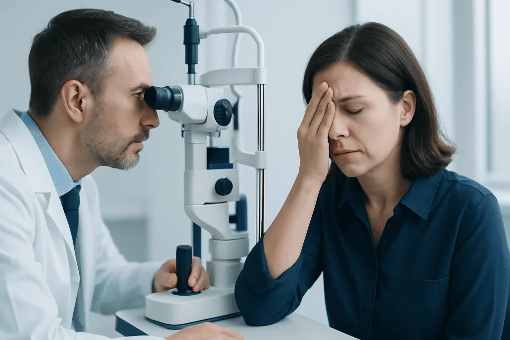
Symptoms can vary depending on the underlying cause, but many people notice that vision in one eye becomes blurry, dim, or less sharp over hours to days. Some describe colors, especially red, as looking washed out or less vivid. Others notice a dark spot in the center of vision, missing areas to the side, or a general sense that one eye is not seeing as clearly as the other.
Pain is often felt behind the eye or deep in the orbit and may be more noticeable when moving the eye. This pattern is common in inflammatory optic nerve conditions, though it is not present in every case. Some people also develop headaches, sensitivity to light, or a difference in how bright a room appears between the two eyes.
Symptoms that can occur with optic nerve disorders include:
- Blurred or dim vision in one or both eyes
- Pain with eye movement
- Reduced color vision or color intensity
- Blind spots or patchy areas of missing vision
- Decreased contrast sensitivity
- Flashing lights or visual distortion in some cases
Because these symptoms can overlap with other eye and neurological conditions, self-diagnosis is not reliable. A prompt professional examination is the safest way to clarify the cause.
Causes and Risk Factors

One important cause is optic neuritis, an inflammation of the optic nerve. It may occur on its own or in association with autoimmune or demyelinating conditions that affect the central nervous system. Inflammation can interfere with normal signal transmission and lead to pain, blurred vision, and reduced color perception.
Other optic nerve problems include compression by nearby structures, reduced blood flow to the nerve, infections, toxic exposures, nutritional deficiencies, and raised pressure within the skull. Rarely, trauma can injure the nerve directly. In older adults, sudden painless or mildly painful vision loss may raise concern for ischemic optic neuropathy, which happens when the nerve’s blood supply is impaired.
Risk factors depend on the specific diagnosis but may include autoimmune disease, recent infection, smoking, vascular risk factors such as diabetes or high blood pressure, certain medications or toxins, and a history of neurological symptoms. A compressive optic neuropathy may be related to masses near the optic pathways, sinus disease, or structural problems around the eye socket or brain.
Because the causes range from temporary inflammation to urgent neurological disease, the goal of assessment is not simply to treat the pain, but to identify why the optic nerve may be affected. In selected cases, doctors may investigate related neurological conditions such as multiple sclerosis or other inflammatory disorders.
How Doctors Diagnose the Cause
Diagnosis begins with a careful history. The doctor will ask when symptoms started, whether one or both eyes are affected, whether pain worsens with movement, and whether there are associated symptoms such as headache, weakness, numbness, fever, or jaw pain. A history of autoimmune disease, infection, trauma, or previous vision episodes can also provide important clues.
The eye examination usually includes checking visual acuity, color vision, pupil reactions, eye pressure, eye movements, and the appearance of the optic nerve at the back of the eye. A relative afferent pupillary defect, changes in the optic disc, or impaired color vision may suggest optic nerve dysfunction. Visual field testing helps map areas of missing vision that patients may not fully notice on their own.
Imaging and other tests are often needed. Doctors may use MRI scanning of the brain and orbits to look for optic nerve inflammation, demyelination, compression, or other neurological causes. Optical coherence tomography can measure the retinal nerve fiber layer and help assess optic nerve damage. Blood tests may be recommended to evaluate inflammation, infection, autoimmune conditions, or vitamin deficiency.
In some situations, diagnosis involves a wider neurological workup, especially when symptoms suggest a disorder that affects both vision and the nervous system. When a tumor or structural lesion is suspected, further review by specialists may be needed, including evaluation through a neurology assessment or other multidisciplinary care.
Treatment Options
Treatment depends entirely on the cause. For inflammatory optic neuritis, doctors may recommend corticosteroid treatment in selected cases, especially when vision loss is significant or when MRI findings suggest broader central nervous system involvement. Steroids are used under medical supervision and are not appropriate for every patient or every type of optic neuropathy.
If the optic nerve is compressed, treatment focuses on relieving the pressure and addressing the underlying lesion. This may involve medication, sinus treatment, or surgery depending on the source and urgency. When reduced blood flow is responsible, management often centers on identifying vascular risk factors and treating the broader medical condition to reduce future harm.
Infectious, autoimmune, toxic, or nutritional causes are managed by treating the underlying problem. This can include antimicrobial therapy, immune-directed treatment, stopping a harmful medication or substance, or correcting a deficiency. Some people may benefit from visual rehabilitation strategies if there is lasting impairment.
Because the causes may involve both the eye and the brain, coordinated care is often important. Neuroophthalmologists, neurologists, radiologists, and other specialists may work together, and in selected cases further evaluation may involve surgical treatment for compressive brain lesions when imaging identifies a structural cause.
Prevention and Self-Care
There is no single way to prevent all optic nerve problems, but some general steps can support eye and neurological health. Managing blood pressure, blood sugar, and cholesterol can help reduce vascular risk. Avoiding smoking and limiting exposure to toxic substances may also lower the chance of certain optic neuropathies.
Routine eye exams are useful, especially for people with chronic health conditions or a family history of eye disease. People with autoimmune disorders or neurological conditions should report new visual symptoms early, even if they seem mild at first. Fast evaluation is especially important if symptoms affect only one eye or progress over a short period.
Self-care should focus on safety rather than home treatment. It is reasonable to rest the eyes, avoid driving if vision is impaired, and keep a brief note of when symptoms began and how they are changing. However, new eye pain with reduced vision should not be treated only with over-the-counter drops or pain relievers without a medical assessment.
If ongoing care is needed, patients may benefit from a structured follow-up plan with eye and nerve specialists. Near the end of the care pathway, centers such as Acibadem International may provide multidisciplinary diagnosis and treatment for international patients through JCI-accredited hospitals when neuroophthalmic expertise is required.
When to See a Doctor Urgently
Urgent assessment is recommended when eye pain occurs together with sudden or worsening vision changes. The same is true if there is loss of color vision, a new blind spot, double vision, or symptoms in only one eye that were not present before. While some causes are treatable and may improve, delays can increase the risk of permanent visual loss in certain conditions.
Emergency care is especially important if visual symptoms are accompanied by weakness, numbness, difficulty speaking, severe headache, fever, confusion, or loss of balance. These features may suggest a broader neurological or systemic problem that needs immediate attention. Severe redness, nausea, halos around lights, or intense eye pain can also point to an eye emergency unrelated to the optic nerve.
People over 50 should seek prompt care for sudden vision changes with scalp tenderness, jaw pain while chewing, or unexplained fatigue, because these can be signs of giant cell arteritis, a condition that can threaten vision. Even when symptoms seem to improve, follow-up remains important because the underlying condition may still require investigation and treatment.
Frequently asked questions
Can eye pain with vision changes really come from the optic nerve?
Yes. When the optic nerve is inflamed, compressed, or not receiving enough blood flow, a person may develop eye pain along with blurred or dim vision. This pattern is not the only cause of painful vision changes, but it is one that should be evaluated promptly.
What does optic nerve pain feel like?
People often describe it as a deep ache behind the eye rather than surface irritation. It may become more noticeable when moving the eye, especially in inflammatory conditions such as optic neuritis.
Is optic neuritis always linked to multiple sclerosis?
No. Optic neuritis can occur on its own, after infection, or in connection with several inflammatory or autoimmune disorders. Because it can sometimes be associated with multiple sclerosis, doctors may recommend MRI or other tests to understand the broader picture.
Will vision return after an optic nerve problem?
Recovery depends on the cause, severity, and how quickly treatment begins. Some inflammatory cases improve substantially over time, while other causes may leave some lasting visual changes. Regular follow-up helps monitor healing and guide rehabilitation if needed.
What tests are usually done for suspected optic nerve disease?
Doctors usually perform an eye examination, color vision testing, pupil testing, and visual field testing. Depending on the findings, they may also order optical coherence tomography, blood tests, and MRI of the brain and orbits.
When should someone go to the emergency department?
Immediate care is important for sudden vision loss, rapidly worsening symptoms, severe eye pain, or visual changes with neurological symptoms such as weakness or trouble speaking. Emergency evaluation is also needed if there is severe redness, nausea, or halos around lights, since these can signal another urgent eye condition.
References
- American Academy of Ophthalmology
- National Eye Institute
- American Academy of Neurology
- National Institute of Neurological Disorders and Stroke
- Mayo Clinic
This article is for general information only and is not a substitute for professional medical advice. Please consult a qualified doctor about your individual situation.
Explore treatments in Turkey — costs, top hospitals & a free quote
JCI-accredited · board-certified surgeons · reply within 24h
Add us as a Preferred Source to see more of our trusted health content across Google Search, AI Overviews and Discover.
Check your numbers in seconds
BMI, calories, due date, blood pressure and 30+ more clinical calculators — free, instant, doctor-reviewed ranges.

More from the Health Library



Related Specialists

Damla Tuncay
Physical Medicine & Rehabilitation
Dr. Muhammed Taha Taş
Emergency Service
Prof. Dr. İbrahim Kaya
Orthopedic Surgery & Traumatology
Prof. Dr. Mehmet Uğur Özbaydar
Orthopedic Surgery & Traumatology