Fibroglandular Density — Explained by Medical Evidence, Not Myths

Fibroglandular density is an imaging description, not a disease. Dense breast tissue is common and can change with age, hormones, and body composition.
Key Takeaways
- Fibroglandular density is an imaging description, not a disease.
- Dense breast tissue is common and can change with age, hormones, and body composition.
- Higher breast density can make abnormalities harder to see on a mammogram.
- Breast density may be associated with a modestly increased breast cancer risk.
- Follow-up depends on the full imaging report, symptoms, age, and individual risk factors.
Fibroglandular density refers to the amount of glandular and fibrous tissue seen in the breast on imaging, especially mammograms. It is a common and often normal finding, but it can make breast images harder to interpret and may slightly affect breast cancer risk.
Overview: what fibroglandular density means
Fibroglandular density means the breast contains a certain proportion of fibrous tissue and glandular tissue compared with fatty tissue. This is a description used by radiologists when reviewing breast imaging, especially mammograms. In simple terms, it tells how the breast looks on the image; it does not mean a person is ill, and it is not a diagnosis by itself.
Breasts are made up of several normal components: milk-producing glands, ducts that carry milk, supportive fibrous tissue, and fat. On a mammogram, fatty tissue usually appears darker, while fibrous and glandular tissue appears whiter. Because many breast changes, including some cancers, can also appear white, denser tissue can make images more difficult to read.
The phrase “scattered fibroglandular density” often appears on imaging reports and usually means there are some areas of dense tissue mixed with fat. Other reports may describe the breasts as heterogeneously dense or extremely dense. These terms are part of a standard system used to classify breast density and help guide imaging interpretation.
How breast density is classified
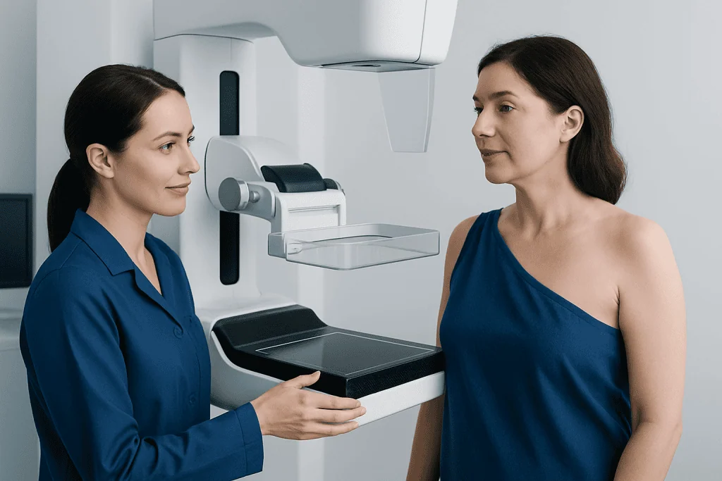
Radiologists commonly describe breast density using categories developed for breast imaging reports. These categories range from mostly fatty breasts to extremely dense breasts. The categories help standardize reporting, but they do not measure density in the same way a blood test measures a laboratory value.
The main categories are:
- Almost entirely fatty: most of the breast is fatty tissue.
- Scattered areas of fibroglandular density: there are some dense areas, but much of the breast is still fatty.
- Heterogeneously dense: more of the breast is dense, which may obscure small findings.
- Extremely dense: most of the breast tissue is dense, making mammogram interpretation more challenging.
Many people hear the term “dense breasts” after a mammogram and understandably worry. In reality, dense tissue is common and often reflects normal biology. What matters most is how the imaging finding fits with the person’s age, family history, symptoms, prior mammograms, and overall breast cancer risk.
Why fibroglandular density matters

Fibroglandular density matters for two main reasons. First, dense tissue can reduce the sensitivity of mammography because both dense tissue and some abnormal findings appear white on the image. This can make small lesions harder to detect, particularly in people with heterogeneously dense or extremely dense breasts.
Second, breast density is considered one factor linked to breast cancer risk. It is not as strong a risk factor as certain inherited genetic mutations or some personal cancer histories, but it is part of the overall picture. Density alone does not mean cancer is present, and many people with dense breasts never develop breast cancer.
This is why breast density should be interpreted alongside other information rather than in isolation. A doctor may combine mammogram findings with clinical breast examination, family history, risk assessment tools, and sometimes additional imaging. If an abnormality is suspected, the next steps may include breast cancer evaluation or targeted imaging to clarify what the mammogram shows.
What influences breast density
Breast density is shaped by normal biological factors and may change over time. Younger people often have denser breasts because they generally have more active glandular tissue and less fat. With age, especially after menopause, density often decreases as more fatty tissue replaces glandular tissue, although this pattern is not the same for everyone.
Hormonal influences can also play a role. Pregnancy, breastfeeding, menstrual cycle changes, and hormone-based medications may affect how breast tissue appears on imaging. Body composition matters as well, since people with lower overall body fat may appear to have denser breasts on mammograms.
Family patterns can contribute, but breast density does not necessarily follow a simple inheritance pattern. Having dense breasts does not automatically mean a person is at high risk, and having mostly fatty breasts does not guarantee that no problems will develop. It is best understood as one piece of a broader health assessment.
How fibroglandular density is evaluated and what tests may be used
Fibroglandular density is usually identified on routine screening mammography. The radiologist reviews the images, compares them with prior studies if available, and assigns a density category. Comparison with previous mammograms is helpful because stability over time often supports a benign explanation for many findings.
If the report shows dense tissue but no suspicious abnormality, routine follow-up may be all that is needed. In some cases, especially when a mammogram is difficult to interpret or a person has additional risk factors, a doctor may discuss supplemental imaging. Depending on the situation, this can include mammography follow-up views, breast ultrasound, or breast MRI.
If there is a specific area of concern, targeted breast ultrasound may help determine whether the finding is solid, fluid-filled, or likely benign. Sometimes additional tests are recommended not because density is dangerous on its own, but because the goal is to see through overlapping tissue more clearly and avoid missing an important abnormality.
Treatment, follow-up, and common report findings
Fibroglandular density itself does not need treatment. Because it is a description of tissue composition rather than a disease, management usually focuses on appropriate screening and follow-up rather than medication or surgery. The most important question is whether the imaging shows only normal density or whether there is a separate finding that needs more evaluation.
People are sometimes told they have “scattered fibroglandular densities” and assume this means a problem was found. In most cases, that wording simply describes the background appearance of the breast. If the report also states there is no suspicious mass, calcification, or architectural distortion, it usually means no cancer-specific sign was seen on that study.
If a mammogram detects a mass, distortion, grouped calcifications, or another area that cannot be confidently classified as benign, the radiologist may recommend short-term follow-up imaging or tissue sampling. When needed, breast biopsy can provide a definite diagnosis. Decisions are based on the full report and clinical context, not on density alone.
Self-care, screening habits, and reducing confusion around myths
A common myth is that fibroglandular density means cancer is already present. Another is that dense breasts always require extensive testing. Medical evidence does not support either idea. Most dense breast tissue is normal, and many people need only routine screening based on age and personal risk.
Good self-care starts with understanding the imaging report and keeping regular screening appointments. People should know their family history of breast and ovarian cancer, inform their doctor about any hormone use, and mention new symptoms promptly. Comparing new images with prior mammograms can also improve accuracy, so it helps to keep records accessible when changing clinics.
General health habits may support breast health overall, even though they do not directly “treat” density. These include maintaining a healthy weight when possible, staying physically active, limiting alcohol intake, and following medical advice on age-appropriate screening. For international patients, Acibadem International’s multidisciplinary specialists in JCI-accredited hospitals diagnose and manage breast imaging concerns with individualized follow-up plans.
When to seek medical care
Medical advice is important if a person notices a new breast lump, persistent focal pain, skin thickening, nipple inversion, nipple discharge that is bloody or occurs without squeezing, or swelling in one breast or underarm. These symptoms do not always mean cancer, but they should be assessed by a qualified clinician.
People should also contact a doctor if they receive a mammogram report they do not understand, are told they have dense breasts and have additional risk factors, or are called back for extra imaging. A callback after screening is common and often leads to benign results, but it should still be completed without unnecessary delay.
Those with a strong family history, known genetic risk, prior chest radiation, or previous high-risk breast lesions may need a more personalized screening plan. A breast specialist or radiologist can explain whether standard mammography is enough or whether supplemental imaging is reasonable in that specific case.
Frequently asked questions
Is fibroglandular density normal?
Yes. Fibroglandular density is a common and normal imaging description of breast tissue composition. It refers to how much fibrous and glandular tissue is present compared with fat, not to a disease.
Does fibroglandular density mean breast cancer?
No. Fibroglandular density does not mean a person has breast cancer. It may make mammograms harder to interpret, which is why doctors sometimes recommend additional imaging in selected cases.
What does scattered fibroglandular density mean on a mammogram?
This usually means there are some dense areas in the breast, but much of the tissue is fatty. It is one of the standard density descriptions used in mammography reports and is often a routine finding.
Can breast density change over time?
Yes. Breast density can change with age, menopause, pregnancy, breastfeeding, hormone exposure, and body composition. Many people become less dense over time, although this varies from person to person.
Do dense breasts always require ultrasound or MRI?
Not always. Whether extra imaging is useful depends on the mammogram findings, symptoms, and overall breast cancer risk. A doctor or radiologist can explain if supplemental imaging would likely add value in that situation.
Should a person worry if called back after a mammogram because of dense tissue?
A callback can be stressful, but it is common and does not automatically mean something serious was found. Often, the purpose is simply to get clearer images or focus on an area that was difficult to evaluate on the first study.
References
- American College of Radiology
- Breast Imaging Reporting and Data System (BI-RADS)
- National Cancer Institute
- American Cancer Society
- World Health Organization
This article is for general information only and is not a substitute for professional medical advice. Please consult a qualified doctor about your individual situation.
Explore treatments in Turkey — costs, top hospitals & a free quote
JCI-accredited · board-certified surgeons · reply within 24h
Add us as a Preferred Source to see more of our trusted health content across Google Search, AI Overviews and Discover.
Check your numbers in seconds
BMI, calories, due date, blood pressure and 30+ more clinical calculators — free, instant, doctor-reviewed ranges.

More from the Health Library

Catatonic Schizophrenia: What Patients Need to Know

Cause of ulcers on the tongue: A Complete Medical Guide for Patients

Ventricular Extrasystole Bigeminy: A Complete Medical Overview

Terconazole: A Complete Medical Overview

Zone of Proximal Development: An Evidence-Based Guide for Patients