Flow Diversion for Brain Aneurysms: Candidates, Procedure, and Follow-Up

Flow diversion is usually used for unruptured or carefully selected brain aneurysms, particularly wide-neck, large, or complex aneurysms. The procedure is performed through the blood vessels, most often from the groin or wrist, without opening the skull.
Key Takeaways
- Flow diversion is usually used for unruptured or carefully selected brain aneurysms, particularly wide-neck, large, or complex aneurysms.
- The procedure is performed through the blood vessels, most often from the groin or wrist, without opening the skull.
- Patients typically need antiplatelet medicines before and after treatment to reduce the risk of clot formation on the device.
- The aneurysm often closes gradually over months, so follow-up imaging is an essential part of care.
- Treatment decisions depend on aneurysm size, shape, location, rupture status, patient health, and the expertise of a neurovascular team.
Flow diversion is a minimally invasive endovascular treatment for selected brain aneurysms, especially wide-neck or complex aneurysms that may be difficult to treat with traditional coiling or surgery. It works by placing a fine mesh device across the aneurysm opening to redirect blood flow and encourage the aneurysm to seal over time.
Overview
Flow diversion is an endovascular treatment for certain brain aneurysms. A brain aneurysm is a weak, bulging area in the wall of an artery inside the skull. Some aneurysms remain stable for years, while others may enlarge or, less commonly, rupture and cause bleeding around the brain. The goal of treatment is to reduce the risk of future rupture while keeping nearby arteries and brain tissue safe.
A flow diverter is a flexible, stent-like device made of a fine metal mesh. During the procedure, a neurointerventional specialist places the device inside the parent artery, across the neck of the aneurysm. Instead of filling the aneurysm directly, as in coiling, the device redirects blood along the normal path of the artery. Over time, reduced flow inside the aneurysm promotes clotting within the aneurysm sac and healing of the artery lining over the device.
This approach is particularly useful for aneurysms that have a wide neck, a large or giant size, a fusiform shape, or an anatomy that makes other treatments more challenging. Flow diversion is not the right choice for every aneurysm. A careful assessment by a multidisciplinary team helps determine whether observation, coiling, microsurgical clipping, flow diversion, or another strategy offers the safest balance of benefits and risks.
Who May Be a Candidate for Flow Diversion
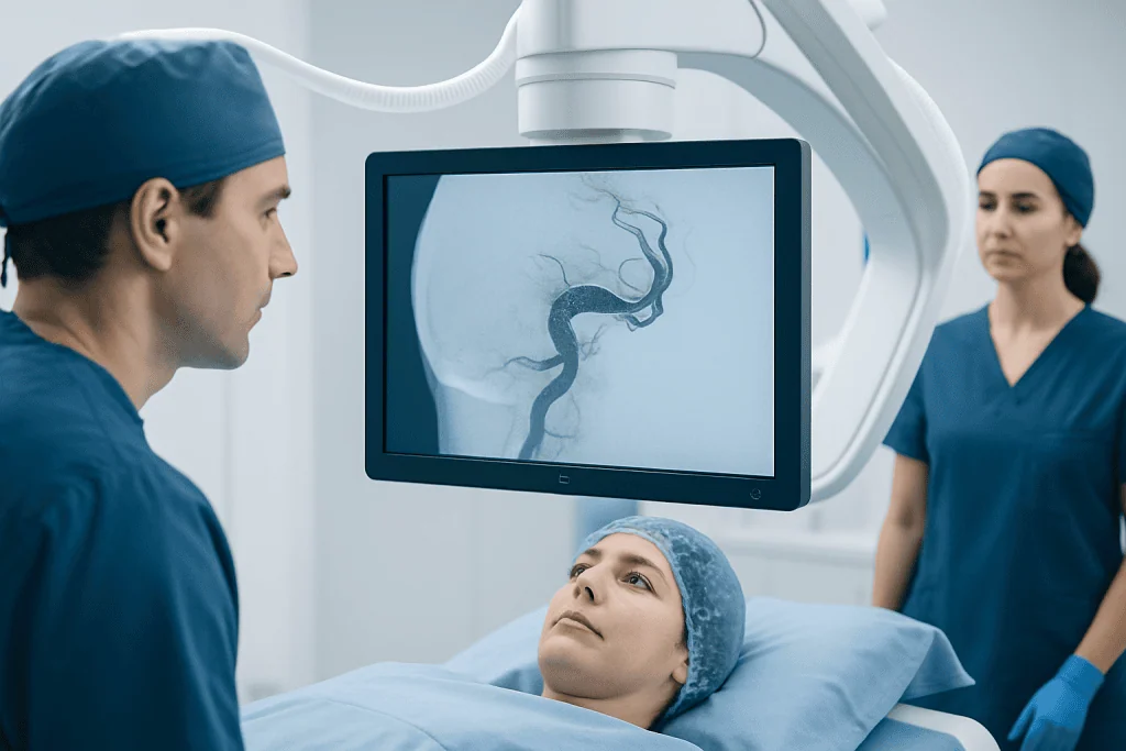
Good candidates are often people with unruptured brain aneurysms that are considered suitable based on size, shape, location, and risk profile. Flow diversion is commonly considered for aneurysms along the internal carotid artery and other selected arteries where the device can be placed safely. It may be helpful when the aneurysm has a broad opening, when coils might not stay securely inside the aneurysm, or when previous treatment has not fully closed the aneurysm.
Doctors also consider the person as a whole. Age, general health, kidney function, bleeding risk, allergies to contrast dye or medications, and the ability to take antiplatelet medicines all matter. Because flow diverters are metal devices placed inside an artery, patients usually need medicines that reduce platelet activity before and after the procedure. This requirement can affect timing and suitability, especially if recent bleeding, planned surgery, or certain medical conditions are present.
Factors that may support consideration of flow diversion include:
- A wide-neck aneurysm that is difficult to treat with coils alone.
- A large, giant, or complex aneurysm in a suitable vessel.
- A recurrent aneurysm after previous coiling or other treatment.
- An aneurysm shape that involves a long segment of the artery, such as selected fusiform aneurysms.
- A clinical situation in which the expected long-term benefit outweighs the procedure and medication risks.
Flow diversion is used more cautiously for recently ruptured aneurysms, aneurysms with important branch arteries coming out of the aneurysm sac, or aneurysms in very small or tortuous vessels. In these situations, other treatments may be safer or more appropriate.
Benefits and Possible Risks

The main advantage of flow diversion is that it treats the diseased segment of the artery rather than only packing the aneurysm. For selected aneurysms, this can lead to durable closure and reconstruction of the parent vessel. Because the treatment is performed through the blood vessels, it avoids a craniotomy, which is an open skull operation. Many patients recover more quickly than they would after open surgery, although individual recovery varies.
Like all brain aneurysm treatments, flow diversion has possible risks. These include stroke from clot formation or vessel blockage, bleeding, injury to the artery, device movement or incomplete opening, contrast-related kidney or allergic reactions, and complications at the catheter entry site. There is also a small risk that the aneurysm may not close completely or may require additional treatment. The treating team reviews these risks in the context of the aneurysm’s natural history and the patient’s personal health factors.
Antiplatelet therapy is an important part of risk reduction. These medicines help prevent clots from forming on the flow diverter while the artery lining heals over it. However, antiplatelet medicines can increase bleeding tendency. For this reason, patients should not stop, start, or change these medicines without direct medical advice from the treating neurovascular team.
How the Procedure Is Performed
Before treatment, patients typically undergo detailed imaging such as CT angiography, MR angiography, or digital subtraction angiography. Blood tests and medication planning are also performed. The medical team may check how the patient responds to antiplatelet medicines, depending on local practice and individual risk. The procedure is usually performed in a specialized angiography suite by an interventional neuroradiologist, endovascular neurosurgeon, or similarly trained neurovascular specialist.
Flow diversion is commonly done under general anesthesia, although some centers may use deep sedation for selected cases. A small puncture is made in an artery, usually in the groin or sometimes the wrist. Through this access point, the doctor guides thin tubes called catheters through the blood vessels to the artery that contains the aneurysm. Continuous X-ray imaging and contrast dye help the team see the vessels clearly.
Once the catheter is positioned, the flow diverter is carefully deployed across the aneurysm neck. The specialist checks that the device is well expanded, properly positioned, and covering the target area. In some cases, coils or additional devices may be used along with the flow diverter, but many aneurysms are treated with the flow diverter alone. After final imaging confirms blood flow through the artery, the catheters are removed and the puncture site is closed.
The hospital stay varies by case and by institutional practice. Some patients are monitored in a high-dependency or intensive care setting for a short period after the procedure. Nurses and doctors check neurological status, blood pressure, the access site, and comfort. Most recovery instructions focus on medication adherence, puncture site care, hydration, and avoiding strenuous activity for a short time.
Recovery and Follow-Up Imaging
Flow diversion works gradually. Unlike surgical clipping, which can close an aneurysm immediately, a flow diverter changes blood flow so the aneurysm can thrombose and the artery can heal over weeks to months. This means follow-up is not optional; it is a key part of treatment. Imaging helps doctors confirm whether the aneurysm is shrinking or closing and whether the parent artery remains open.
Follow-up schedules differ according to the aneurysm, device type, and the treating center’s protocol. Patients may have CT angiography, MR angiography, or catheter angiography at planned intervals. Catheter angiography provides very detailed images and may be recommended when precise assessment is needed. The care team will explain which imaging test is preferred and when it should be done.
During recovery, patients are usually advised to take prescribed antiplatelet medicines exactly as directed and attend all appointments. Mild fatigue, bruising at the puncture site, or temporary discomfort can occur, but new neurological symptoms require urgent evaluation. Patients should seek immediate medical help for sudden severe headache, weakness, numbness, speech difficulty, vision changes, fainting, chest pain, or significant bleeding.
Preparing for Treatment and Self-Care
Preparation begins with a clear conversation about why treatment is recommended, what alternatives exist, and what follow-up will involve. Patients should provide a full list of medicines, supplements, allergies, previous reactions to contrast dye, and any history of bleeding or clotting problems. It is also important to mention pregnancy, kidney disease, stomach ulcers, recent surgery, or planned dental or surgical procedures.
Medication instructions must be followed carefully. The team may prescribe antiplatelet medicines before the procedure and continue them afterward for a defined period. Patients should ask what to do if a dose is missed, whether any over-the-counter pain relievers should be avoided, and when routine activities can resume. Alcohol use, smoking, and uncontrolled blood pressure can affect vascular health, so individualized lifestyle advice may be part of the care plan.
After discharge, helpful self-care steps include keeping the puncture site clean and dry as instructed, drinking fluids if permitted, avoiding heavy lifting until cleared, and arranging transportation for follow-up visits. Long-term vascular health also matters. Managing blood pressure, avoiding tobacco, controlling cholesterol and diabetes when present, and maintaining regular medical care can support overall brain and blood vessel health.
When to See a Specialist
A person diagnosed with a brain aneurysm should be evaluated by a clinician experienced in cerebrovascular disease, even if the aneurysm is small and observation is being considered. The decision to treat depends on aneurysm features, symptoms, age, family history, medical conditions, and patient preferences. A second opinion can be useful when the aneurysm is complex or when several treatment options are possible.
Specialist assessment is especially important for aneurysms that are growing, causing symptoms, large or wide-necked, located in a higher-risk area, or previously treated but not fully closed. Symptoms such as a sudden severe headache, neurological weakness, speech problems, or vision changes should be treated as urgent and evaluated immediately. These symptoms do not always mean an aneurysm has ruptured, but they require prompt medical attention.
For international patients seeking coordinated care, Acibadem International’s multidisciplinary specialists and JCI-accredited hospitals diagnose and treat brain aneurysms, including cases where flow diversion may be considered. The most appropriate plan should always be based on a direct review of imaging, medical history, and an individualized discussion of risks and benefits.
Frequently asked questions
Is flow diversion the same as a brain stent?
A flow diverter is a stent-like device, but it is designed specifically to change blood flow near an aneurysm. Its fine mesh reduces blood entering the aneurysm while maintaining flow through the parent artery. Doctors may use the term stent when explaining it, but its purpose differs from many conventional vascular stents.
Does flow diversion cure a brain aneurysm immediately?
Not usually. Flow diversion encourages the aneurysm to close gradually as blood flow into the aneurysm decreases and the artery lining heals over the device. Follow-up imaging is needed to confirm how well the aneurysm has sealed.
Will every patient need blood-thinning medication?
Most patients need antiplatelet medicines before and after flow diversion to reduce the risk of clot formation on the device. The exact medicine plan and duration depend on the patient’s condition and the treating team’s protocol. These medicines should never be stopped without medical guidance.
How long does recovery take after flow diversion?
Many patients are able to return to light activities within days, but recovery varies based on the aneurysm, overall health, anesthesia, and any complications. The doctor will provide specific instructions about work, exercise, driving, and medication use. Follow-up imaging continues for months or longer.
Can a ruptured aneurysm be treated with flow diversion?
Flow diversion may be considered in selected ruptured aneurysms, but it is used cautiously because antiplatelet medicines can increase bleeding risk. Many ruptured aneurysms are treated with coiling, clipping, or other urgent methods. The safest choice depends on the aneurysm anatomy and the patient’s emergency condition.
What happens if the aneurysm is not fully closed on follow-up?
Incomplete closure does not always mean treatment has failed, because healing can continue over time. The specialist may recommend continued observation, additional imaging, medication adjustment, or another procedure depending on the findings. Decisions are individualized and based on the aneurysm’s behavior and patient safety.
References
- American Heart Association and American Stroke Association
- Society of NeuroInterventional Surgery
- European Stroke Organisation
- National Institute of Neurological Disorders and Stroke
- Mayo Clinic
This article is for general information only and is not a substitute for professional medical advice. Please consult a qualified doctor about your individual situation.
Explore treatments in Turkey — costs, top hospitals & a free quote
JCI-accredited · board-certified surgeons · reply within 24h
Add us as a Preferred Source to see more of our trusted health content across Google Search, AI Overviews and Discover.