Foot Bones — Explained by Medical Evidence, Not Myths
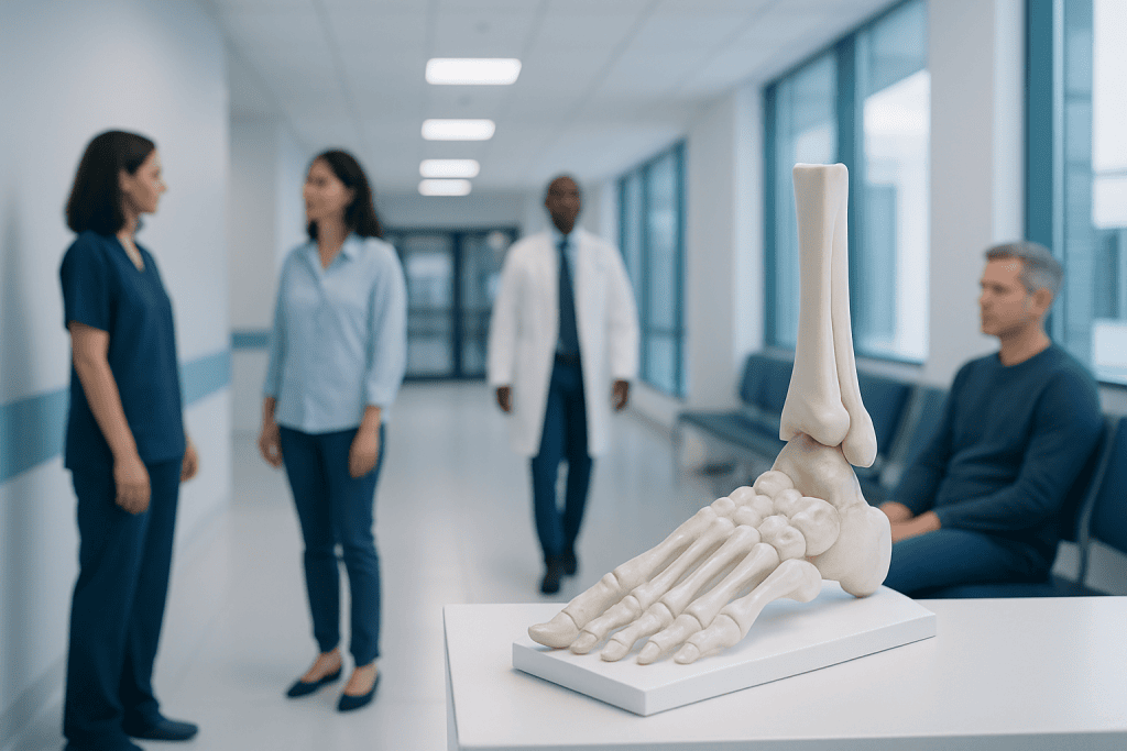
The adult foot usually has 26 bones organized into tarsals, metatarsals, and phalanges. Foot bones work together with joints, ligaments, muscles, and tendons to support movement and balance.
Key Takeaways
- The adult foot usually has 26 bones organized into tarsals, metatarsals, and phalanges.
- Foot bones work together with joints, ligaments, muscles, and tendons to support movement and balance.
- Pain in the foot may come from a fracture, overuse injury, arthritis, deformity, or soft-tissue problem rather than from the bone alone.
- Diagnosis often involves a physical examination and imaging such as X-rays, with MRI or CT used in selected cases.
- Many foot bone problems improve with rest, footwear changes, physical therapy, or bracing, while some need specialist treatment or surgery.
Foot bones are the 26 bones that form the framework of the foot, helping the body stand, walk, balance, and absorb impact. Understanding how these bones are arranged can make common symptoms, injuries, and treatment options easier to recognize and discuss with a doctor.
Overview: what foot bones are and why they matter
Foot bones are the 26 bones in each adult foot that create a strong but flexible structure for standing, walking, running, and balance. They do not work alone: joints, cartilage, ligaments, tendons, muscles, nerves, and blood vessels all help the foot function as one coordinated unit. When people ask about “foot bones,” they are often trying to understand not only anatomy, but also why foot pain, swelling, deformity, or difficulty walking happens.
The foot is designed to do several demanding jobs at once. It supports body weight, adapts to uneven ground, absorbs shock with each step, and helps push the body forward. Because of this constant workload, foot bones can be affected by injury, overuse, age-related wear, inflammatory disease, or changes in alignment. A clear understanding of the basic structure can make symptoms easier to interpret and can guide safer self-care decisions.
Unlike oversimplified descriptions, medical evidence shows that foot problems rarely come from a single bone in isolation. Bone alignment, arch shape, joint motion, footwear, activity level, and underlying health conditions all influence symptoms. This is why an accurate diagnosis is more useful than relying on common myths, such as assuming all forefoot pain is a “spur” or that every bunion needs surgery.
How the 26 foot bones are arranged
The bones of the foot are grouped into three main regions: the tarsals, the metatarsals, and the phalanges. The seven tarsal bones form the rearfoot and midfoot. They include the calcaneus, or heel bone; the talus, which connects the foot to the ankle; the navicular; the cuboid; and three cuneiform bones. These bones create the foundation for weight transfer and help form the arches of the foot.
The five metatarsal bones are the long bones in the middle of the foot. They connect the tarsal bones to the toes and play an important role in balance and push-off during walking. The heads of the metatarsals, located near the ball of the foot, bear substantial pressure, especially during running or prolonged standing.
The toes contain 14 phalanges. Each lesser toe has three phalanges, while the big toe has two. Although small, these bones are important for stability and gait. The big toe in particular contributes significantly to forward propulsion. Beneath the big toe are also two small sesamoid bones in many people; these help the tendons move smoothly and help distribute pressure.
The foot’s shape depends not just on the bones themselves but also on how they line up to form the arches. The medial longitudinal arch, lateral arch, and transverse arch help spread body weight and absorb force. Changes in these arches may contribute to pain, altered walking mechanics, or conditions such as flatfoot.
What foot bones do during daily movement
Foot bones provide structure, but their function is dynamic rather than rigid. At heel strike, the rearfoot helps absorb impact. As body weight moves forward, the midfoot adapts to the surface and stabilizes the arch. During push-off, the forefoot and toes become firmer, allowing efficient forward motion. This sequence happens with every step and depends on coordinated motion between bones and joints.
The talus and calcaneus help manage forces coming from the leg and ground. The midfoot bones contribute to flexibility and stability, allowing the foot to adjust while still protecting the body from excessive motion. The metatarsals and toe bones help distribute pressure and support balance, especially when changing direction, climbing stairs, or standing on one foot.
Because the foot performs these repeated tasks throughout the day, even small changes in alignment can matter. A fracture, arthritis, deformity, or chronic ligament strain may shift the way forces move through the foot bones. Over time, this can lead to pain in areas that are not the original source of the problem. For example, altered pressure under the metatarsals may cause forefoot pain, calluses, or difficulty wearing shoes comfortably.
Common problems that affect foot bones
Foot bone problems range from minor injuries to chronic structural conditions. Fractures may occur suddenly after a twist, fall, direct impact, or sports injury. Stress fractures are different: they develop gradually from repeated loading, often in runners, dancers, or people who increase activity too quickly. A person may notice localized pain that worsens with weight-bearing and improves with rest.
Joint and alignment problems can also involve the foot bones. Bunions affect the alignment at the base of the big toe and may gradually change the shape of the forefoot. Flatfoot can alter the position of several bones and joints, sometimes causing arch pain or fatigue. Arthritis may affect the toe joints, midfoot, or hindfoot, leading to stiffness, swelling, and reduced mobility. Some people evaluated for chronic pain may also need assessment for arthritis.
Children and adults can experience conditions related to the heel or growth areas, while older adults may be more vulnerable to fractures linked to reduced bone strength. Inflammation of nearby structures can also mimic a bone problem. For instance, heel pain may come from the plantar fascia rather than the calcaneus itself, and some patients with persistent heel symptoms are eventually diagnosed with heel spur changes or related soft-tissue irritation.
Not all visible changes are dangerous, but they should be interpreted correctly. A bump, widening of the forefoot, or toe deviation may reflect mechanical stress over time. Pain, redness, progressive deformity, or sudden inability to bear weight deserves medical review rather than self-diagnosis.
Symptoms, warning signs, and how doctors diagnose foot bone conditions
Symptoms linked to foot bones can include pain, tenderness, swelling, bruising, stiffness, difficulty bearing weight, limping, or a visible change in shape. Some people notice pain only during activity, while others feel it even at rest. Pain in one specific spot on the top, side, heel, or ball of the foot can help a clinician narrow down the likely cause, but symptoms alone do not always identify the exact structure involved.
Diagnosis begins with a careful history and physical examination. A doctor may ask when symptoms started, whether there was an injury, what activities make the pain better or worse, and whether there are other conditions such as diabetes, inflammatory disease, or osteoporosis. The foot is examined for swelling, tenderness, alignment, skin changes, range of motion, and walking pattern.
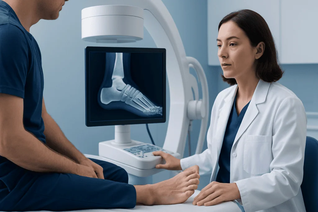
Imaging is often important when a bone injury or structural problem is suspected. Plain X-rays are usually the first test because they can show fractures, dislocations, joint narrowing, bone spurs, and alignment changes. If X-rays are normal but symptoms strongly suggest a stress fracture or soft-tissue injury, the doctor may consider MRI to look in greater detail. In complex injuries or for surgical planning, CT scanning may be helpful because it shows bone structure very clearly.
In some cases, blood tests or additional evaluation may be recommended to look for infection, gout, inflammatory arthritis, or metabolic bone disease. Accurate diagnosis matters because treatment for a stress fracture, bunion, arthritis, and tendon problem can differ significantly even when the pain feels similar.
Treatment options for foot bone injuries and structural problems
Treatment depends on the specific diagnosis, symptom severity, and the person’s daily needs. Many foot bone problems improve with conservative care. This may include temporary rest from painful activities, ice for swelling, supportive footwear, shoe inserts or orthotics, taping, and medication recommended by a doctor for pain or inflammation. Physical therapy may help restore mobility, improve strength, and reduce abnormal loading across the foot.
Fractures may require immobilization with a stiff-soled shoe, walking boot, cast, or limited weight-bearing while the bone heals. Stress fractures typically need activity modification and a gradual return to exercise. Conditions related to poor alignment, such as bunions or flatfoot, are often managed first with footwear changes, pressure relief, strengthening, and custom support. Some patients with severe deformity or persistent symptoms may be evaluated for hallux valgus surgery when the problem is centered on a painful bunion.
When arthritis affects the foot bones and joints, treatment may combine lifestyle adjustments, supportive devices, physical therapy, and targeted medical care. Surgical treatment is usually considered only if symptoms remain limiting despite appropriate non-surgical care, or when there is significant instability, deformity, or a displaced fracture. Depending on the problem, surgery may repair, realign, fuse, or remove damaged tissue around the involved bones.
Recovery times vary widely. A simple toe fracture may heal with basic protection, while a complex midfoot injury can require a longer period of rehabilitation. Follow-up is important because returning to full activity too early can delay healing or increase the risk of re-injury.
Prevention and self-care for healthy foot bones
Good foot care begins with reducing avoidable stress on the bones and surrounding tissues. Shoes should fit well, support the arch appropriately, and leave enough room for the toes. Footwear that is too tight can increase pressure on the forefoot and contribute to deformity or pain, while worn-out soles may reduce shock absorption and stability. Activity should be increased gradually, especially when starting a new exercise routine.
Strength and flexibility also matter. Gentle exercises for the foot and ankle may improve support and balance, and calf flexibility can reduce strain through the foot during walking. Maintaining a healthy body weight can decrease repeated load across the heel, midfoot, and forefoot. People with osteoporosis risk factors should discuss bone health with a clinician, since stronger bones are less vulnerable to fracture.
Self-care has limits. Persistent pain should not simply be pushed through, especially if there is swelling, bruising, or pain in a very specific bony area. People with diabetes, circulation problems, or reduced sensation in the feet should be especially cautious, because injuries may be less obvious and complications can develop more easily.
For international patients seeking specialist evaluation, Acibadem International’s multidisciplinary teams in JCI-accredited hospitals diagnose and treat foot and ankle conditions with orthopedic, imaging, and rehabilitation support when needed.
When to seek medical care
Medical care is important if foot pain follows a fall, twist, sports injury, or direct blow and walking becomes difficult. A doctor should also evaluate pain that does not improve after several days of rest, swelling that keeps returning, or a visible change in the shape of the foot or toes. These signs may point to a fracture, joint injury, or structural problem that benefits from early treatment.
Urgent assessment is needed if the foot looks severely deformed, if a person cannot bear weight at all, or if there is increasing redness, warmth, fever, or an open wound. Numbness, color change, or sudden coldness in the foot also deserves prompt medical attention. In children, older adults, and people with diabetes or poor circulation, it is sensible to seek help early rather than wait for symptoms to worsen.
Early diagnosis can prevent prolonged pain and protect long-term function. Because many foot problems overlap, a qualified clinician is the best person to determine whether the issue involves a bone, joint, tendon, ligament, nerve, or a combination of these structures.
Frequently asked questions
How many foot bones are there in one foot?
An adult foot usually has 26 bones. These are divided into 7 tarsal bones, 5 metatarsal bones, and 14 phalanges in the toes. Some people also have small sesamoid bones near the big toe that help tendons move smoothly.
What are the main groups of foot bones?
The main groups are the tarsals, metatarsals, and phalanges. Tarsals form the rearfoot and midfoot, metatarsals are the long bones in the middle of the foot, and phalanges make up the toes. Together they help support the arches and movement of the foot.
Can foot bone pain happen without a fracture?
Yes. Foot pain can come from arthritis, tendon strain, ligament injury, bunions, nerve irritation, plantar fascia problems, or inflammation around a joint. That is why a medical examination is often needed when pain persists or affects walking.
What is the difference between a stress fracture and a regular fracture?
A regular fracture often happens after a clear injury such as a fall or direct impact. A stress fracture usually develops more gradually from repeated strain, often after increased training or prolonged standing. Stress fractures may be harder to see early on and sometimes need additional imaging.
Do all bunions mean there is a problem with the foot bones?
A bunion involves a change in alignment at the base of the big toe, so bone position is part of the condition. However, symptoms can also be influenced by soft tissues, footwear, joint irritation, and walking mechanics. Not every bunion is painful, and not every bunion needs surgery.
When should someone worry about foot pain?
Foot pain should be checked if it follows an injury, causes limping, lasts more than a few days despite rest, or is associated with swelling or a visible deformity. Urgent care is important if a person cannot bear weight, the foot is numb or discolored, or there are signs of infection such as fever and spreading redness.
References
- American Academy of Orthopaedic Surgeons
- National Institute of Arthritis and Musculoskeletal and Skin Diseases
- American College of Foot and Ankle Surgeons
- Mayo Clinic
- MedlinePlus
This article is for general information only and is not a substitute for professional medical advice. Please consult a qualified doctor about your individual situation.
Joint and spine care in Turkey — expert assessment & treatment
JCI-accredited · board-certified surgeons · reply within 24h
Add us as a Preferred Source to see more of our trusted health content across Google Search, AI Overviews and Discover.