Hemianopia — Explained by Medical Evidence, Not Myths

Hemianopia means loss of half of the visual field and can affect daily activities such as reading, walking, and driving. A stroke is one of the most common causes, but brain injury, tumors, migraines, seizures, and other neurological conditions may also lead to hemianopia.
Key Takeaways
- Hemianopia means loss of half of the visual field and can affect daily activities such as reading, walking, and driving.
- A stroke is one of the most common causes, but brain injury, tumors, migraines, seizures, and other neurological conditions may also lead to hemianopia.
- Diagnosis typically involves visual field testing, a full eye examination, and brain imaging such as MRI or CT.
- Treatment focuses on the underlying cause and may also include visual rehabilitation, compensatory training, and safety adaptations.
- Sudden hemianopia needs urgent medical attention because it may signal a stroke or another serious neurological problem.
Hemianopia is a type of visual field loss in which a person cannot see one half of the visual world in one or both eyes. It is usually caused by a problem in the brain or visual pathways rather than in the eye itself, so medical evaluation is important to identify the cause and plan treatment.
What hemianopia is
Hemianopia is the loss of half of a person’s visual field. In practical terms, this means objects on the left or right side may not be seen, even though the eyes themselves may still be able to detect light and central details normally. The problem usually happens because the visual pathways in the brain are affected, not because half of the eye has stopped working.
The visual system is organized so that information from both eyes travels through the optic nerves and deeper brain pathways before it reaches the parts of the brain that interpret sight. When one of these pathways is injured, a person may lose the same half of vision in both eyes. This pattern is called homonymous hemianopia and is one of the most commonly recognized forms.
People often describe hemianopia as bumping into doorframes, missing words on one side of a page, or feeling as if people suddenly appear from one side. Because the brain can sometimes fill in missing information, the condition may not be obvious at first. That is why formal testing is often needed, even when a person thinks their eyesight is otherwise normal.
Common symptoms and how it affects daily life
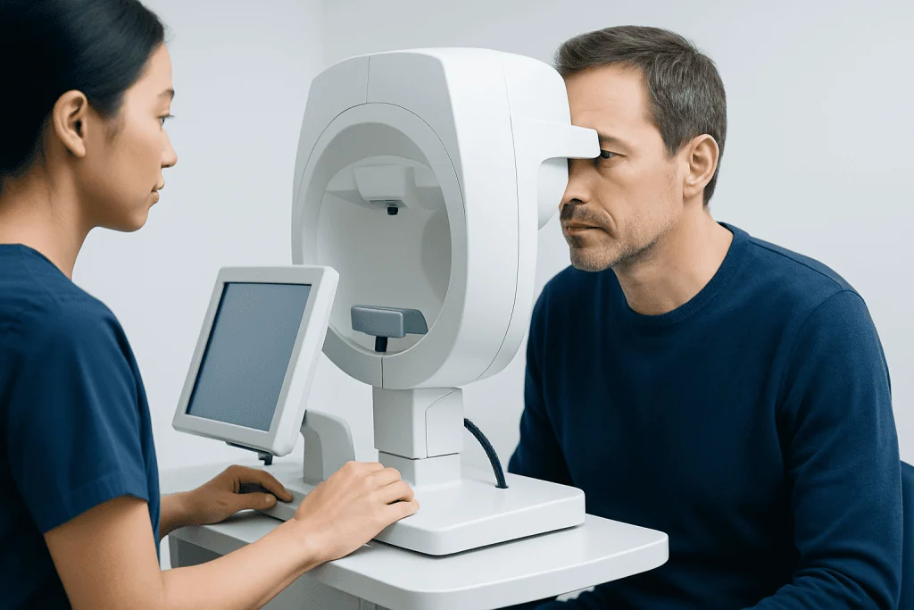
The main symptom of hemianopia is missing vision on one side. Depending on the exact pattern, a person may not notice objects, faces, or movement on the affected side. This can interfere with reading, watching screens, crossing streets, eating from one side of a plate, and finding items in a room.
Some people experience additional symptoms related to the underlying cause rather than hemianopia itself. These may include headache, weakness, numbness, difficulty speaking, confusion, dizziness, or imbalance. When such symptoms appear suddenly, urgent medical care is especially important.
Hemianopia can affect independence and confidence. Reading may become slow because the eyes repeatedly lose their place on the line, and navigating unfamiliar spaces may feel tiring or unsafe. People may also stop driving or need specialist assessment before driving is considered safe again.
- Missing objects or people on the left or right side
- Bumping into furniture, walls, or doorways
- Difficulty reading lines of text smoothly
- Trouble finding items in crowded spaces
- Feeling disoriented in busy environments
Causes and risk factors

Hemianopia is most often caused by damage along the visual pathways of the brain. A stroke is a leading cause, especially when the back part of the brain that processes vision is affected. Other possible causes include traumatic brain injury, brain tumors, bleeding in or around the brain, infections, inflammation, and complications after brain surgery.
In some cases, hemianopia appears with neurological conditions such as migraine aura or seizures, although these episodes may be temporary. Less commonly, demyelinating disease, reduced blood flow to the brain, or certain rare disorders can affect the optic tract, optic radiations, or occipital lobe and produce a similar pattern of visual loss.
Risk factors depend on the underlying condition. For example, high blood pressure, diabetes, smoking, high cholesterol, and heart disease can raise the risk of stroke. Falls, road traffic injuries, and sports accidents increase the chance of head trauma. The important point is that hemianopia is a symptom or sign of a problem elsewhere, so the cause always needs medical assessment rather than assumption.
Types of hemianopia and why the pattern matters
Doctors use the visual field pattern to help determine where the problem may be located. Homonymous hemianopia means the same side of the visual field is lost in both eyes, such as the right half or the left half. This usually points to a lesion behind the optic chiasm, in areas such as the optic tract, optic radiations, or occipital lobe.
Heteronymous hemianopia is less common and refers to opposite halves being affected in each eye. Bitemporal hemianopia, for example, can occur when the central crossing point of the optic nerves is compressed, such as by a pituitary region mass. A person may also have quadrantanopia, where only one quarter of the visual field is missing rather than an entire half.
These distinctions matter because they guide further testing and treatment. The pattern can help doctors decide whether the problem is more likely to arise from vascular disease, trauma, tumor, or another neurological process. In some cases, related evaluation may involve brain MRI to look closely at the visual pathways and surrounding structures.
How hemianopia is diagnosed
Diagnosis starts with a careful medical history and examination. The clinician asks when the visual change began, whether it came on suddenly or gradually, and whether there are associated symptoms such as weakness, headache, speech changes, or recent injury. A complete eye examination is usually combined with a neurological assessment because the issue may come from the brain rather than the eye itself.
Formal visual field testing is central to diagnosis. This may be done with computerized perimetry, which maps areas that the person can and cannot see. Testing helps confirm whether the defect is a true hemianopia, identifies whether it is complete or partial, and shows whether the pattern is stable or changing over time.
Brain imaging is often needed to find the cause. Depending on the situation, doctors may recommend urgent CT in emergencies or more detailed MRI scanning to assess stroke, bleeding, tumor, inflammation, or other structural changes. Some patients may also need blood tests, vascular imaging, heart evaluation, or specialist review in neuro-ophthalmology when the diagnosis is complex.
Treatment options and rehabilitation
There is no single treatment that fits all cases of hemianopia because management depends on the underlying cause. If the visual field loss is related to a recent stroke, treatment focuses on urgent stroke care and prevention of further events. If it is caused by a tumor, inflammation, or pressure on the visual pathways, treatment may involve medication, surgery, or other targeted therapies based on specialist assessment.
Some people improve over time, especially in the first weeks to months after a brain injury or stroke, but recovery varies. Even when the field loss does not fully return, rehabilitation can make a meaningful difference. Visual scanning therapy teaches a person to move their eyes and head more effectively toward the affected side, improving navigation, reading, and awareness of hazards.
Additional support may include occupational therapy, reading strategies, prism lenses for selected patients, and rehabilitation after neurological injury. When hemianopia occurs as part of a broader condition affecting movement, speech, or cognition, coordinated care can be especially helpful, including stroke rehabilitation where appropriate. Near the end of the care pathway, some patients seek multidisciplinary assessment; Acibadem International’s specialists in JCI-accredited hospitals diagnose and treat hemianopia and its underlying causes for international patients.
Prevention, self-care, and when to seek medical care
Because hemianopia is usually a result of another medical problem, prevention focuses on reducing the risk of those underlying conditions. Managing blood pressure, cholesterol, and diabetes, not smoking, staying physically active, and following medical advice for heart and vascular health may help lower the risk of stroke-related visual field loss. Head injury prevention, such as seatbelt use and fall prevention measures, also matters.
At home, practical strategies can improve safety and confidence. Good lighting, reducing clutter, placing important objects in predictable locations, and learning systematic scanning habits can make daily tasks easier. Family members may help by approaching from the person’s seeing side at first while rehabilitation skills are being learned.
Medical care should be sought urgently if hemianopia starts suddenly, especially if it comes with weakness, numbness, severe headache, trouble speaking, confusion, or loss of balance. Prompt assessment is also important when visual loss worsens, follows a head injury, or is associated with symptoms that suggest a neurological condition such as a brain tumor or another acute brain disorder. Even gradual symptoms deserve timely evaluation, since treatment is most effective when the cause is identified early.
Frequently asked questions
Is hemianopia the same as blindness in one eye?
No. Hemianopia is loss of half of the visual field, often affecting the same side in both eyes. It is different from complete vision loss in one eye, which usually points to a problem in the eye or optic nerve before the visual pathways join deeper in the brain.
Can hemianopia improve?
Sometimes it can, especially if the cause is treated quickly and the brain recovers in the weeks or months afterward. The degree of improvement varies, and some people continue to have persistent field loss. Rehabilitation can still help improve daily function even when vision does not fully return.
What is the most common cause of hemianopia?
Stroke is one of the most common causes, particularly when it affects the visual processing areas of the brain. However, head injury, brain tumors, bleeding, inflammation, seizures, and migraine-related neurological events can also cause it.
How do doctors test for hemianopia?
Doctors usually combine a full eye examination with visual field testing, which maps what a person can and cannot see. They often also order brain imaging such as CT or MRI to identify the reason for the visual field loss.
Can someone drive with hemianopia?
Driving rules vary by country and by the severity of the visual field defect. Many people with hemianopia are not automatically safe to drive and need formal visual assessment and medical advice. It is important not to continue driving until a qualified clinician or licensing authority has reviewed the condition.
When is hemianopia an emergency?
Sudden hemianopia is an emergency, especially if it begins with weakness, speech difficulty, severe headache, confusion, or imbalance. These symptoms can suggest stroke or another urgent neurological problem. Immediate medical evaluation gives the best chance of identifying and treating the cause quickly.
This article is for general information only and is not a substitute for professional medical advice. Please consult a qualified doctor about your individual situation.
Explore treatments in Turkey — costs, top hospitals & a free quote
JCI-accredited · board-certified surgeons · reply within 24h
Add us as a Preferred Source to see more of our trusted health content across Google Search, AI Overviews and Discover.
Check your numbers in seconds
BMI, calories, due date, blood pressure and 30+ more clinical calculators — free, instant, doctor-reviewed ranges.

More from the Health Library

Liver Abscess: An Evidence-Based Guide for Patients

Cgrp Inhibitors: An Evidence-Based Guide for Patients

Healthy BMI: An Evidence-Based Guide for Patients

Im Needle Size: A Complete Medical Overview
Ubrelvy Cost: A Complete Medical Overview