Heterotopic Ossification: An Evidence-Based Guide for Patients

Heterotopic ossification means bone forms in soft tissue outside the normal skeleton. It most often appears after joint surgery, fractures, spinal cord injury, traumatic brain injury, or severe burns.
Key Takeaways
- Heterotopic ossification means bone forms in soft tissue outside the normal skeleton.
- It most often appears after joint surgery, fractures, spinal cord injury, traumatic brain injury, or severe burns.
- Common signs include pain, swelling, warmth, and reduced range of motion around a joint.
- Diagnosis usually involves a physical exam and imaging such as X-rays, ultrasound, CT, or bone scans.
- Treatment may include monitoring, physical rehabilitation, medicines, and in selected cases surgery.
Heterotopic ossification is the formation of bone in soft tissues such as muscles and connective tissue where bone does not normally grow. It often develops after trauma, surgery, burns, or neurologic injury, and early evaluation can help reduce pain, stiffness, and loss of movement.
Overview
Heterotopic ossification is abnormal bone formation in soft tissues, usually around joints, muscles, tendons, or other connective tissue. In simple terms, the body begins making bone in a place where bone should not be. This can lead to pain, stiffness, and difficulty moving the nearby joint, especially if the extra bone bridges normal tissues.
The condition is most often seen after a major trigger, such as orthopedic surgery, a fracture, spinal cord injury, traumatic brain injury, or severe burns. It can also occur after repeated local trauma. Not every person with these risk factors develops heterotopic ossification, but when it happens, symptoms often begin gradually over days to weeks.
Heterotopic ossification is not the same as calcium deposits, although the two may be confused. Calcium deposition and true bone formation are different processes. In heterotopic ossification, the tissue develops organized bone, sometimes with marrow-like structure, rather than just scattered mineral buildup.
The outlook depends on where the abnormal bone forms, how much motion it limits, and whether the underlying cause can be managed. Many people improve with a careful plan that combines symptom control, rehabilitation, and follow-up imaging. When symptoms are significant, specialists may also evaluate whether orthopedic rehabilitation or more advanced orthopedic care is appropriate.
How It Develops in the Body
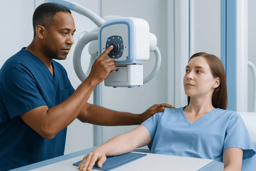
Heterotopic ossification appears to result from an abnormal healing response. After tissue injury or inflammation, the body releases signaling molecules that normally help repair damage. In some situations, these signals encourage certain immature cells in soft tissues to change into bone-forming cells.
This process usually happens in stages. At first, the area may be inflamed, swollen, warm, and painful. Later, immature bone begins to organize within the soft tissue. As the bone matures over time, the affected area may feel firm, and nearby movement can become more restricted.
The condition can be classified in different ways. One broad form is acquired heterotopic ossification, which follows trauma, surgery, burns, or neurologic injury. Much more rarely, there are inherited disorders that cause widespread extra bone formation. Those rare genetic conditions are distinct from the more common forms seen after injury or surgery.
Understanding this step-by-step development matters because treatment choices often depend on timing. Measures used early to control inflammation and protect mobility may differ from treatments considered later, when mature bone has already formed and function is clearly limited.
Symptoms and Common Locations

Symptoms of heterotopic ossification usually develop near a joint. People may notice increasing pain, warmth, tenderness, swelling, and stiffness in an area that had been recovering from surgery or injury. A previously improving joint may suddenly become harder to move, or exercises may become more uncomfortable than expected.
The hips are a common site, especially after hip surgery or neurologic injury. The elbows, shoulders, and knees can also be affected. In some people, the signs are subtle at first and are mistaken for routine postoperative discomfort, muscle strain, infection, or normal scar-related tightness.
Symptoms can include:
- Pain around a joint or injured area
- Reduced range of motion
- Swelling and local warmth
- A hard or firm feeling in the soft tissue
- Difficulty with walking, dressing, or daily tasks if a major joint is involved
Not all cases are severe. Some areas of extra bone are found incidentally on imaging and cause few symptoms. Others can interfere significantly with mobility, posture, hygiene, or rehabilitation progress, especially after spinal cord injury or brain injury.
Causes and Risk Factors
The most common causes of heterotopic ossification are tissue trauma and inflammation. It may develop after joint replacement, fracture repair, dislocation, muscle injury, or extensive soft-tissue damage. It is also well recognized after spinal cord injury, traumatic brain injury, and serious burns.
Joint surgery is a major setting in which this condition is discussed. For example, some people develop extra bone around the hip after orthopedic procedures. This does not mean surgery was performed incorrectly; rather, it reflects an individual healing response influenced by tissue injury, local inflammation, and personal risk factors.
Risk factors may include:
- Previous heterotopic ossification
- Major trauma or repeated injury to the same area
- Hip surgery or other orthopedic procedures
- Spinal cord injury or traumatic brain injury
- Severe burns
- Prolonged immobility
- Certain rare inherited bone-forming disorders
Some medical conditions may need to be considered when symptoms overlap with other causes of joint pain and stiffness, such as arthritis or problems related to recovery after hip replacement. A doctor will look at the full clinical picture rather than relying on a single symptom.
Diagnosis
Diagnosis starts with a medical history and physical examination. The doctor will ask about recent surgery, injuries, neurologic events, burns, and the timing of symptoms. They will also assess pain, swelling, warmth, muscle tone, and how much the nearby joint can move.
Imaging is often needed because early heterotopic ossification may not be visible on a standard X-ray right away. Depending on the stage and clinical question, a doctor may use ultrasound, X-rays, CT, MRI, or a bone scan. Repeat imaging can be helpful if symptoms strongly suggest the diagnosis but initial tests are not yet conclusive.
Blood tests are not enough to confirm heterotopic ossification on their own, but they may support the overall assessment or help exclude other concerns such as infection. In the early inflammatory phase, the main challenge is often distinguishing this condition from postoperative infection, deep vein thrombosis, hematoma, or routine healing discomfort.
When the diagnosis is uncertain or symptoms are affecting recovery, referral to specialists may be useful. This can involve orthopedic surgeons, rehabilitation physicians, neurologists, or radiologists. If advanced imaging or specialist evaluation is needed, some patients may undergo MRI as part of a broader diagnostic workup.
Treatment Options
Treatment depends on the cause, severity, location, and stage of the condition. In mild cases, doctors may recommend observation, symptom relief, and regular follow-up while monitoring whether the extra bone is progressing or stabilizing. The goals are to reduce pain, preserve function, and prevent further limitation of movement.
Rehabilitation is often a central part of care. Guided physical therapy may help maintain joint range of motion and support safe recovery after injury or surgery. Exercises should be individualized, because overly aggressive stretching can worsen pain and may not be helpful. A rehabilitation team can also advise on positioning, mobility aids, and daily activity adjustments.
Medicines may be used in selected cases to reduce inflammation or lower the risk of heterotopic ossification after certain procedures. The decision to use them depends on the person’s medical history, surgery type, and potential side effects. In specific high-risk situations, radiation therapy may be considered as a preventive measure under specialist guidance.
Surgery to remove mature heterotopic bone may be considered if symptoms are substantial and the extra bone clearly limits function, causes persistent pain, or interferes with hygiene or mobility. Timing is important and varies by case, since doctors generally want to balance symptom burden against the risk of recurrence and surgical complications. If surgery is being discussed, evaluation by orthopedic specialists can help determine the best approach.
Prevention, Self-care, and Recovery
Prevention is most relevant for people known to be at higher risk, such as those with previous heterotopic ossification or those undergoing certain orthopedic surgeries. Doctors may recommend preventive measures based on the procedure, the body area involved, and the person’s overall health. These measures are tailored individually rather than used routinely for everyone.
Self-care during recovery focuses on following the rehabilitation plan, protecting the affected area, and reporting new symptoms early. People should take prescribed medicines only as directed and attend follow-up appointments so the care team can watch for changes in movement or pain. Consistent, gentle movement under professional guidance is often more helpful than pushing through severe discomfort.
Practical steps that may support recovery include:
- Keeping follow-up visits and imaging appointments
- Doing recommended therapy exercises regularly
- Avoiding unsupervised forceful stretching
- Monitoring for increasing stiffness, swelling, or warmth
- Asking for help if daily activities become harder
For international patients who need coordinated evaluation, Acibadem International’s multidisciplinary specialists and JCI-accredited hospitals diagnose and treat conditions such as heterotopic ossification with orthopedic, rehabilitation, and imaging support. The emphasis is on individualized assessment and evidence-based care.
When to Seek Medical Care
Medical review is appropriate if pain, swelling, warmth, or stiffness around a joint seems to be getting worse instead of better during recovery from surgery or injury. A person should also seek care if range of motion suddenly decreases, walking becomes more difficult, or rehabilitation progress stalls without a clear reason.
Urgent medical attention may be needed if symptoms are accompanied by fever, severe redness, drainage from a surgical wound, chest symptoms, or calf swelling, because these signs can suggest problems other than heterotopic ossification, such as infection or a blood clot. It is important not to assume that all postoperative pain or stiffness is part of normal healing.
People with spinal cord injury, traumatic brain injury, or severe burns should mention any new loss of movement around the hips, knees, shoulders, or elbows to their care team promptly. Early assessment can help doctors distinguish heterotopic ossification from other causes and plan the next steps safely.
If symptoms are persistent or a diagnosis remains unclear, consultation with orthopedics, rehabilitation medicine, or related specialists may be recommended. Timely evaluation often helps preserve function and improves planning for treatment and recovery.
Frequently asked questions
Is heterotopic ossification serious?
It can be mild or more function-limiting depending on where it forms and how much movement it affects. Some people have little discomfort, while others develop significant stiffness or pain around a joint. A doctor's evaluation helps determine how important it is in a specific case.
Can heterotopic ossification go away on its own?
The extra bone itself does not usually simply disappear once it has matured. However, symptoms may improve over time with rehabilitation, pain management, and reduced inflammation. Some cases remain stable and do not require surgery.
How long after surgery or injury does heterotopic ossification appear?
It often begins within weeks after a triggering event, though the exact timing varies. Early symptoms may include warmth, swelling, and decreasing range of motion before clear bone is visible on X-ray. Follow-up imaging may be needed if symptoms strongly suggest the condition.
What is the difference between heterotopic ossification and calcification?
Calcification means calcium salts collect in tissue, but that does not always mean true bone has formed. Heterotopic ossification involves the organized formation of bone in soft tissue. Doctors use imaging and clinical assessment to tell the difference.
Does everyone with hip or joint surgery get heterotopic ossification?
No. It is a recognized complication after some surgeries and injuries, but many people never develop it. Risk depends on factors such as the procedure type, prior history, local tissue trauma, and individual healing response.
When is surgery needed for heterotopic ossification?
Surgery may be considered when mature extra bone causes major pain, blocks movement, or interferes with daily function, mobility, or hygiene. The decision is individualized and usually made after imaging, physical assessment, and discussion of recurrence risk. Conservative measures are often tried first when appropriate.
This article is for general information only and is not a substitute for professional medical advice. Please consult a qualified doctor about your individual situation.
Explore treatments in Turkey — costs, top hospitals & a free quote
JCI-accredited · board-certified surgeons · reply within 24h
Add us as a Preferred Source to see more of our trusted health content across Google Search, AI Overviews and Discover.

More from the Health Library

Omphalocele — Explained by Medical Evidence, Not Myths

Nutmeg Benefits, Uses, and Safe Amounts

High Sugar Fruits to Avoid: What Patients Need to Know

Sophia Burns Md: What Patients Need to Know

Cytotec: An Evidence-Based Guide for Patients