Holosystolic Murmur: What Patients Need to Know

A holosystolic murmur is heard continuously from the first heart sound to the second heart sound. Common causes include mitral regurgitation, tricuspid regurgitation, and ventricular septal defect.
Key Takeaways
- A holosystolic murmur is heard continuously from the first heart sound to the second heart sound.
- Common causes include mitral regurgitation, tricuspid regurgitation, and ventricular septal defect.
- Some people have no symptoms, while others may experience breathlessness, tiredness, palpitations, or swelling.
- An echocardiogram is usually the main test used to identify the cause and assess its severity.
- Treatment depends on the underlying heart condition and may range from monitoring and medicines to catheter-based or surgical procedures.
A holosystolic murmur, also called a pansystolic murmur, is an extra heart sound that continues through the whole pumping phase of a heartbeat. It is not a diagnosis itself, but it can point to conditions such as a leaking heart valve or an opening between the lower heart chambers, so assessment by a qualified clinician is important.
Overview: What Is a Holosystolic Murmur?
A holosystolic murmur is a whooshing or blowing sound that a clinician hears through a stethoscope during the entire systolic phase of the heartbeat. Systole is the phase in which the heart’s lower chambers, called ventricles, contract to pump blood to the lungs and the rest of the body. Because the sound starts with the first heart sound and continues until the second, it is also called a pansystolic murmur.
A murmur is a physical examination finding rather than a disease. It reflects turbulent blood flow in or around the heart. A holosystolic pattern can occur when blood flows backward through a valve that should be closed or passes through an abnormal opening between heart chambers. The importance of a murmur depends on its cause, intensity, location, associated symptoms, and imaging findings.
Some causes are mild and can be monitored for years without affecting daily life. Others may gradually strain the heart or lead to complications if untreated. For this reason, a newly detected holosystolic murmur should be evaluated, especially when it is accompanied by shortness of breath, chest discomfort, fainting, swelling, or reduced exercise tolerance.
How the Sound Relates to Heart Function
The heart has four valves that keep blood moving in one direction. During ventricular contraction, the mitral and tricuspid valves normally close to prevent blood from flowing backward into the upper chambers. If either valve does not close fully, some blood can leak backward throughout systole, creating a holosystolic murmur.
The location where the murmur is loudest helps clinicians narrow down the possible cause. A murmur from mitral valve leakage is often heard best near the apex of the heart, toward the lower left side of the chest, and may radiate toward the left armpit. A murmur due to tricuspid valve leakage is commonly strongest near the lower left edge of the breastbone and may become louder during a deep breath in.
A holosystolic murmur can also occur with a ventricular septal defect, an opening in the wall between the two lower heart chambers. In this situation, blood may move from the left ventricle to the right ventricle during contraction. Listening findings provide useful clues, but they cannot confirm the diagnosis on their own; heart imaging is usually needed.
Common Causes and Risk Factors

Mitral regurgitation is one of the most common causes of a holosystolic murmur. It occurs when the mitral valve allows blood to leak from the left ventricle back into the left atrium. The valve may leak because of age-related changes, mitral valve prolapse, prior rheumatic fever, infection of the heart lining or valves, enlargement of the heart, or damage to heart muscle after a heart attack.
Tricuspid regurgitation is another cause. It may result from enlargement of the right side of the heart, often related to conditions that increase pressure in the lung circulation. It can also occur due to valve infection, congenital valve differences, certain implanted cardiac device leads, or less commonly, direct injury to the valve.
Ventricular septal defects are often congenital, meaning they are present from birth, although they can rarely develop after a heart attack, trauma, or certain medical conditions. Smaller defects may produce a prominent murmur while causing few symptoms; larger defects may affect blood flow to the lungs and place extra work on the heart.
Risk factors vary with the cause. They may include a history of valve disease, rheumatic fever, infective endocarditis, congenital heart disease, high blood pressure, coronary artery disease, heart failure, or conditions affecting the lungs and pulmonary blood vessels. Having a risk factor does not mean a person will develop a significant murmur, but it can guide appropriate assessment.
Possible Symptoms and What They Mean
A person with a holosystolic murmur may feel completely well. Mild valve leakage or a small congenital defect can sometimes be discovered during a routine examination. In these cases, follow-up recommendations depend on echocardiogram results and the person’s overall health rather than on the murmur sound alone.
When the underlying condition is more significant, symptoms can develop gradually. These may include shortness of breath during activity or when lying flat, reduced stamina, unusual tiredness, awareness of a fast or irregular heartbeat, dizziness, or a persistent cough. Some people may notice ankle or leg swelling, abdominal fullness, or weight gain from fluid retention, particularly with right-sided heart problems.
Symptoms do not always match the loudness of a murmur. A loud murmur does not automatically mean severe disease, and a softer murmur can still require attention. Changes in symptoms, daily activity tolerance, heart rhythm, and heart structure are all considered when deciding whether treatment is needed.
How a Holosystolic Murmur Is Diagnosed
Evaluation usually begins with a detailed medical history and physical examination. A clinician may ask about breathlessness, chest pain, palpitations, fainting, past infections, childhood heart conditions, family history, and medicines. They will listen to the heart in several positions and may check for signs of fluid retention, irregular rhythm, or changes in blood pressure.
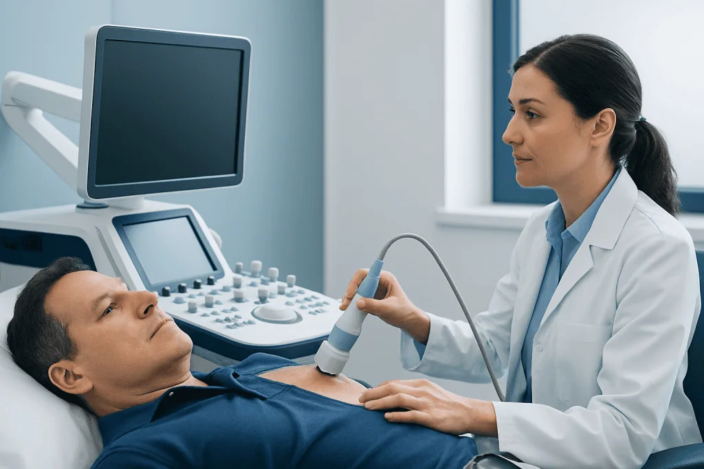
An echocardiogram is the key test for most people with a suspected significant murmur. This painless ultrasound examination shows the heart chambers, valves, blood flow direction, and pumping function. It can identify valve narrowing or leakage, measure the size of a septal defect, and help determine whether the heart is under strain.
Other tests may include an electrocardiogram to assess heart rhythm, a chest X-ray to look at heart and lung changes, blood tests, or exercise testing when appropriate. Some patients need transesophageal echocardiography, which provides more detailed valve images using an ultrasound probe passed into the esophagus under medical supervision. Cardiac MRI, CT, or cardiac catheterization may be used in selected cases when further detail is required for treatment planning.
Treatment Options and Ongoing Care
There is no single treatment for a holosystolic murmur because treatment is directed at its cause. If imaging shows mild valve leakage, normal heart function, and no symptoms, regular clinical reviews and repeat echocardiograms may be all that is needed. Monitoring helps detect changes before they cause substantial effects on the heart.
Medicines may be recommended to manage related conditions such as high blood pressure, heart failure symptoms, fluid retention, or an abnormal heart rhythm. These medicines can improve symptoms and reduce strain on the heart, but they do not always correct a structurally damaged valve. People should take prescribed medicines as directed and should not stop heart medicines without discussing this with their clinician.
When valve leakage is severe, symptoms are persistent, or the heart begins to enlarge or weaken, repair or replacement of the affected valve may be considered. Depending on the valve problem and individual anatomy, options may include surgery or a catheter-based procedure. Some ventricular septal defects can be monitored, while others may need closure using a catheter device or surgery.
Care is often coordinated by a cardiologist, with input from cardiac imaging specialists, interventional cardiologists, cardiac surgeons, and other professionals when needed. Acibadem International’s multidisciplinary specialists and JCI-accredited hospitals evaluate and treat heart valve disorders and congenital heart conditions for international patients.
Living Well and When to Seek Medical Care
Healthy daily habits support heart health regardless of the cause of a murmur. These include not smoking, staying physically active within the limits advised by a clinician, eating a balanced diet, maintaining a healthy weight where possible, managing blood pressure and diabetes, and attending scheduled follow-up visits. People with known valve disease should ask their cardiologist what kinds and levels of exercise are suitable for them.
Good oral hygiene and routine dental care are also important, as they help reduce the risk of bacteria entering the bloodstream. Preventive antibiotics before dental procedures are not routinely needed for most people with heart murmurs. However, certain people at high risk for infective endocarditis may require them, so individual advice should come from a cardiologist or dentist.
Medical care should be arranged promptly for a newly identified murmur, worsening breathlessness, declining exercise capacity, new ankle swelling, frequent palpitations, or unexplained fatigue. Urgent medical assessment is appropriate for severe or sudden shortness of breath, chest pain, fainting, blue or gray lips, new confusion, or symptoms that occur suddenly and feel severe. These symptoms may have several causes, but they should not be ignored.
Frequently asked questions
Is a holosystolic murmur dangerous?
A holosystolic murmur is not automatically dangerous, but it can be a sign of a structural heart condition that needs evaluation. Its significance depends on the underlying cause, the severity of blood flow changes, heart function, and whether symptoms are present. An echocardiogram usually helps clarify the situation.
What is the difference between holosystolic and systolic murmur?
A systolic murmur is any murmur heard while the ventricles are contracting. A holosystolic murmur is a specific type of systolic murmur that continues evenly from the first to the second heart sound. It is often associated with mitral regurgitation, tricuspid regurgitation, or ventricular septal defect.
Can a holosystolic murmur go away?
Whether it goes away depends on the cause. A murmur caused by a temporary change in blood flow or a treatable underlying problem may lessen, while one caused by a structural valve issue or congenital defect may persist. Even when the sound remains, the condition may be stable and manageable with appropriate follow-up.
What test confirms the cause of a holosystolic murmur?
An echocardiogram is usually the main test used to confirm the cause. It uses sound waves to show how the valves and heart chambers are working and to measure blood flow. Additional tests may be needed if the echocardiogram suggests a complex valve problem or if intervention is being considered.
Can exercise make a holosystolic murmur worse?
Exercise does not necessarily worsen the underlying condition, and many people with mild valve disease can remain active. However, the safe level of activity depends on the cause and severity of the murmur, symptoms, heart rhythm, and heart function. A cardiologist can provide individualized guidance, particularly for strenuous exercise.
Does a holosystolic murmur always require surgery?
No. Many people only need observation and periodic echocardiograms, especially when valve leakage is mild and the heart is functioning normally. Surgery or catheter-based treatment is generally considered when a defect or valve problem is severe, symptoms are troublesome, or tests show that the heart is being affected.
References
- American Heart Association
- American College of Cardiology
- European Society of Cardiology
- National Heart, Lung, and Blood Institute
- Merck Manual Professional Edition
This article is for general information only and is not a substitute for professional medical advice. Please consult a qualified doctor about your individual situation.
Explore treatments in Turkey — costs, top hospitals & a free quote
JCI-accredited · board-certified surgeons · reply within 24h
Add us as a Preferred Source to see more of our trusted health content across Google Search, AI Overviews and Discover.
Check your numbers in seconds
BMI, calories, due date, blood pressure and 30+ more clinical calculators — free, instant, doctor-reviewed ranges.

More from the Health Library

Paleo Meal Delivery: A Complete Medical Overview

Celebrex and Alcohol: What Patients Need to Know

Why Your Cervix May Hurt After Intercourse

Poblano Peppers: A Complete Medical Overview

Hair Plugs: A Complete Medical Overview