How Does MRI Work: Preparation, Procedure and Results
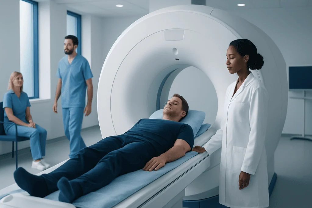
MRI creates detailed internal images using magnetic fields and radio waves rather than X-rays. Patients must tell the imaging team about implants, metal fragments, pregnancy, kidney problems, allergies and claustrophobia before the scan.
Key Takeaways
- MRI creates detailed internal images using magnetic fields and radio waves rather than X-rays.
- Patients must tell the imaging team about implants, metal fragments, pregnancy, kidney problems, allergies and claustrophobia before the scan.
- Most MRI scans take about 20 to 60 minutes, depending on the body area and whether contrast is needed.
- Remaining still is important because movement can blur the images; normal swallowing is safe.
- A radiologist interprets the images and sends a report to the referring clinician, who explains what the findings mean in context.
MRI, or magnetic resonance imaging, uses a powerful magnet, radio waves and a computer to make detailed images of organs, soft tissues, bones and blood vessels. The scan does not use ionizing radiation, and preparation is usually simple, although metal implants, certain devices and contrast dye need careful review beforehand.
How does MRI work?
MRI stands for magnetic resonance imaging. It works by placing the body in a strong magnetic field that temporarily aligns tiny particles called hydrogen protons, which are abundant in water and fat. The MRI scanner sends carefully controlled radiofrequency pulses, changing that alignment. As the protons return to their usual state, they release signals that the scanner detects.
A computer processes these signals into detailed cross-sectional images. Because different tissues release signals in different ways, MRI can distinguish soft tissues very clearly. It is often useful for evaluating the brain, spine, joints, muscles, abdomen, pelvis, heart and blood vessels.
Unlike X-rays and CT scans, MRI does not use ionizing radiation. However, its strong magnet means that safety screening is essential. The examination is painless, but the scanner makes loud tapping or knocking sounds while images are being acquired.
Who may need an MRI and how is safety assessed?
A clinician may request MRI when detailed information is needed after symptoms, examination findings or other tests. For example, it may help assess persistent joint pain, nerve-related symptoms, headaches with concerning features, possible spinal conditions, abdominal or pelvic concerns, or changes involving blood vessels and organs. MRI is one part of clinical assessment; its results are interpreted alongside medical history, examination and other tests.
Before scheduling or arriving for the scan, patients should tell the team about any pacemaker, defibrillator, neurostimulator, cochlear implant, aneurysm clip, insulin pump, implanted medication pump, artificial joint, vascular stent, metal plate, surgical clip, bullet or shrapnel injury. Many modern implants are MRI-compatible under specific conditions, but this must be checked carefully.
Pregnancy, breastfeeding, reduced kidney function, prior reaction to MRI contrast and severe anxiety in enclosed spaces should also be discussed. MRI contrast, usually gadolinium-based, is not needed for every scan. When it is useful, the radiology team considers individual safety factors and explains the process.
What should you not do before your MRI?

Patients should not bring metal objects into the MRI scan room. This includes jewelry, watches, hairpins, hearing aids, removable dental appliances, coins, keys, credit cards, phones and clothing with metal zips, snaps or underwires. Makeup, nail products, tattoos and transdermal patches can occasionally contain metallic components, so it is sensible to ask the imaging center for its specific instructions.
Most routine MRI scans do not require fasting. However, patients should follow any food and drink instructions provided for abdominal, pelvic, cardiac or contrast-enhanced imaging. They should not stop prescribed medicines unless a clinician specifically advises it. It is helpful to bring details of implants, previous imaging and relevant medical records when available.
Before the appointment, patients should inform the center if they may be pregnant, have kidney disease, have ever had a contrast reaction, or have difficulty lying flat. People with claustrophobia can ask in advance about practical support, including relaxation strategies, a support person where permitted, or clinician-prescribed medicine when appropriate. If sedating medicine is used, the patient will generally need someone else to take them home.
MRI procedure: what happens step by step?
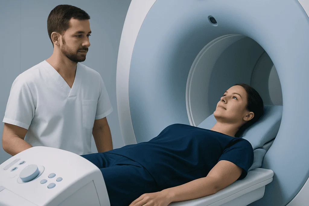
On arrival, the patient completes a detailed safety questionnaire and may change into a gown. A radiographer reviews the answers, checks for metal and explains the scan. If contrast is planned, an intravenous cannula may be placed in the arm or hand. The patient is then positioned on a padded table, often with a receiver coil placed around or near the body part being imaged.
The table moves slowly into the scanner. The radiographer operates the equipment from a nearby control room but can see, hear and speak with the patient throughout the examination. An alarm call button is usually provided. Earplugs or headphones protect hearing from the repetitive loud sounds produced during scanning.
The key task is to remain as still as possible during each image sequence. The radiographer may give simple instructions, such as holding the breath briefly for some chest or abdominal images. If contrast is used, it may be injected partway through the scan; some people notice a cool sensation at the injection site, but significant reactions are uncommon and staff are prepared to respond.
What if I have to pee during an MRI?
Patients should use the bathroom before the scan whenever possible, particularly if they expect a longer examination. If the need to urinate becomes uncomfortable during MRI, they should use the call button or tell the radiographer. The scan can often be paused safely so that the patient can take a bathroom break.
It is best not to try to endure severe discomfort, as this can make it harder to remain still and may affect image quality. For certain pelvic examinations, the imaging center may give specific instructions about bladder fullness. In that situation, patients should follow the instructions provided and ask the team if they are uncertain.
How long does an MRI actually take?
Most MRI examinations take around 20 to 60 minutes. A focused scan of one area may be shorter, while studies involving several body regions, specialized sequences, contrast dye or cardiac imaging can take longer. The appointment itself may last longer than the scan because it includes registration, safety screening, changing clothes and positioning.
The duration depends on the area being examined, the number of image sequences required and whether images need to be repeated because of movement. The radiographer can usually provide a realistic estimate before the scan starts. Patients should allow enough time and avoid planning an urgent commitment immediately afterward, especially if sedation may be used.
Can I swallow saliva during MRI?
Yes. Swallowing saliva during MRI is safe and normal. For many scans, especially of areas away from the head and neck, swallowing has little effect on image quality. The patient should stay as relaxed and still as possible without causing discomfort.
During detailed imaging of the brain, face, neck or throat, repeated swallowing can sometimes create motion on certain images. The radiographer may ask the patient to keep still or avoid swallowing briefly during a particular sequence if possible. Patients should never feel that they must suppress swallowing to the point of discomfort; they can communicate with the team at any time.
Recovery, benefits, risks and when to seek medical care
After a standard MRI without sedation, most people can return to normal activities immediately. If contrast was used, the cannula is removed and the patient may be observed briefly when clinically appropriate. People who received sedating medication should follow their clinician’s instructions, avoid driving, alcohol and important decisions for the advised period, and arrange assistance getting home.
The principal benefit of MRI is its ability to provide high-detail images, particularly of soft tissues, without ionizing radiation. Limitations include the need to lie still in a confined, noisy environment and the fact that not every implant or device can be scanned safely. Rare risks include contrast reactions and, in people with severe kidney impairment, specific concerns related to gadolinium contrast; this is why screening is important.
Patients should seek urgent medical care for symptoms such as sudden weakness or numbness on one side, new trouble speaking, severe sudden headache, chest pain, severe breathing difficulty, or loss of bladder or bowel control with back pain. These symptoms may need prompt assessment and should not wait for a routine MRI appointment. For non-urgent concerns, a clinician can decide whether MRI or another test is appropriate.
A radiologist reviews the images and prepares a report for the referring clinician. Results may be available within days, though timing varies by facility and clinical urgency. Acibadem International’s multidisciplinary specialists and JCI-accredited hospitals can coordinate diagnostic imaging and follow-up care for international patients.
Frequently asked questions
Is an MRI painful?
MRI is usually painless. The main challenges are lying still, hearing the loud scanning sounds and being in a confined space for some examinations. Padding, ear protection and communication with the radiographer can make the experience more comfortable.
Do I need contrast dye for an MRI?
Not every MRI requires contrast. A clinician and radiologist decide whether contrast may provide useful additional information based on the reason for the scan. Patients should report kidney problems, pregnancy and any previous contrast reaction before the examination.
Can someone with a pacemaker have an MRI?
Some people with certain modern pacemakers and implanted devices can undergo MRI under carefully controlled conditions. The device model, leads and clinical circumstances must be reviewed in advance. Patients should never assume an implant is safe for MRI without confirmation from the imaging and device teams.
What should I wear to an MRI appointment?
Wear comfortable clothing without metal, such as metal-free zips, buttons, hooks or underwires. Many centers ask patients to change into a gown to ensure safety and avoid image artifacts. The imaging center can provide specific clothing guidance before the appointment.
Can I eat and drink before an MRI?
For many MRI scans, normal eating and drinking are allowed. Some scans, especially certain abdominal or pelvic studies, may require fasting or other preparation. Patients should follow the instructions from their imaging center and continue medicines unless told otherwise by their clinician.
When will I get my MRI results?
A radiologist interprets the images and sends a report to the clinician who ordered the scan. Timing varies according to the facility and urgency of the findings, but results are often discussed within several days. The referring clinician explains whether any finding is relevant to the patient's symptoms and whether further assessment is needed.
References
- Radiological Society of North America
- American College of Radiology
- U.S. Food and Drug Administration
- National Institute of Biomedical Imaging and Bioengineering
This article is for general information only and is not a substitute for professional medical advice. Please consult a qualified doctor about your individual situation.
Explore treatments in Turkey — costs, top hospitals & a free quote
JCI-accredited · board-certified surgeons · reply within 24h
Add us as a Preferred Source to see more of our trusted health content across Google Search, AI Overviews and Discover.