How Neuroimmune Diseases Are Diagnosed: MRI, Spinal Tap, Blood Tests, and Antibodies

Diagnosing neuroimmune diseases usually requires a combination of history, examination, MRI, blood tests, and sometimes a spinal tap. Antibody tests can be very helpful, but they must be interpreted together with symptoms and imaging findings.
Key Takeaways
- Diagnosing neuroimmune diseases usually requires a combination of history, examination, MRI, blood tests, and sometimes a spinal tap.
- Antibody tests can be very helpful, but they must be interpreted together with symptoms and imaging findings.
- Doctors also rule out infections, vitamin deficiencies, stroke, and other neurological conditions that can look similar.
- Early and accurate diagnosis can help guide treatment and reduce the risk of ongoing inflammation or nerve damage.
- A normal test does not always exclude disease, so follow-up and repeat testing may sometimes be needed.
Neuroimmune disease diagnosis combines a careful neurological exam with tests such as MRI, spinal fluid analysis, blood work, and antibody studies. Because symptoms can overlap with many other conditions, diagnosis is usually based on patterns from several tests rather than one result alone.
Overview: why diagnosis takes more than one test
Neuroimmune diseases are conditions in which the immune system mistakenly affects the brain, spinal cord, optic nerves, nerves, or muscles. Examples include multiple sclerosis, autoimmune encephalitis, neuromyelitis optica spectrum disorder, and other inflammatory disorders of the nervous system. These conditions can cause symptoms such as weakness, numbness, vision changes, seizures, confusion, balance problems, or changes in bladder control.
Diagnosis can be complex because many symptoms overlap with infection, migraine, stroke, metabolic problems, vitamin deficiencies, and other neurological diseases. For that reason, doctors do not usually rely on a single scan or blood test. Instead, they look for a pattern that matches inflammation or immune-related injury in the nervous system.
A full diagnostic workup often includes a medical history, neurological examination, MRI, blood tests, and sometimes a spinal tap, also called lumbar puncture. In some cases, additional tests such as EEG, nerve studies, or visual testing are needed. The main goal is to identify whether inflammation is present, where it is located, what may be causing it, and which treatment is likely to help.
Symptoms and clues doctors look for
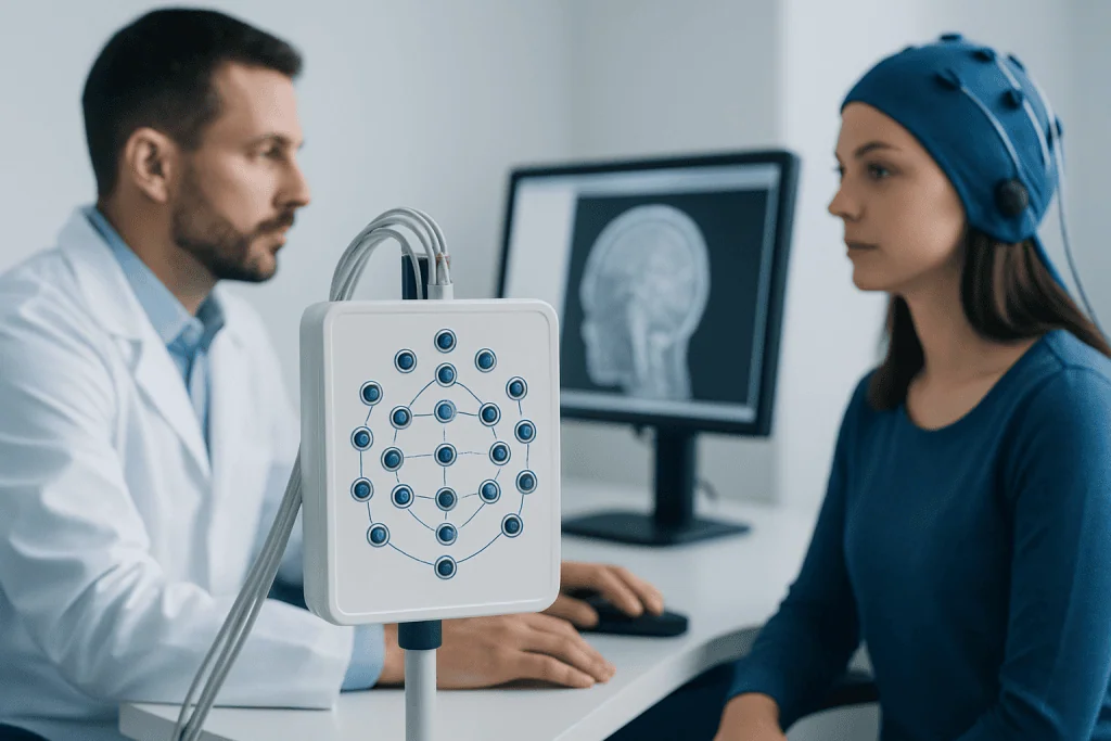
The first step in neuroimmune disease diagnosis is understanding the symptom pattern. Doctors ask when symptoms began, whether they came on suddenly or gradually, how long they lasted, and whether they affected one area or several parts of the nervous system. They also ask about recent infections, vaccinations, autoimmune diseases, cancer history, medication use, and family history.
Some symptom patterns raise suspicion for a neuroimmune condition. These include optic neuritis with painful vision loss, repeated episodes of numbness or weakness, inflammation of the spinal cord, unexplained seizures, rapid memory or behavior changes, and new balance or coordination problems. Symptoms that occur in attacks and then partly improve can suggest relapsing inflammation, while steady worsening may point to ongoing disease activity or another cause.
A neurological examination helps localize the problem. During the exam, the doctor checks strength, reflexes, sensation, coordination, eye movements, speech, and walking. This can show whether the issue is more likely to involve the brain, spinal cord, peripheral nerves, neuromuscular junction, or muscles, which then guides the next tests.
- Vision loss or eye pain may suggest optic nerve inflammation.
- Weakness and sensory change in both legs may suggest spinal cord involvement.
- Confusion, personality change, or seizures may suggest brain inflammation.
- Tingling, reduced reflexes, or burning pain may point toward peripheral nerve disease.
MRI: seeing inflammation in the brain and spinal cord

MRI is one of the most important tools in neuroimmune disease diagnosis because it can show inflammation, demyelination, swelling, or scarring in the central nervous system. MRI uses magnets rather than radiation and can image the brain, optic pathways, and spinal cord in detail. Contrast dye is sometimes used to show areas of active inflammation.
Doctors study where lesions are located, how many there are, and whether they look old or new. Certain patterns can support specific diagnoses. For example, lesions typical of multiple sclerosis often appear in characteristic areas of the brain and spinal cord, while long segments of spinal cord inflammation can raise concern for neuromyelitis optica or related disorders. In suspected autoimmune encephalitis, MRI may show inflammation in brain regions linked with memory or behavior, although MRI can also be normal early on.
MRI is valuable, but it does not answer every question by itself. Some findings are nonspecific and can be seen with aging, migraine, vascular changes, or past infections. A normal MRI also does not always rule out neuroimmune disease, especially in the early stages or in disorders that affect structures not easily seen on standard imaging. That is why MRI results are interpreted together with the examination and laboratory tests.
Spinal tap: what cerebrospinal fluid can show
A spinal tap, or lumbar puncture, collects a small sample of cerebrospinal fluid, the clear fluid surrounding the brain and spinal cord. This test can provide direct clues about inflammation inside the central nervous system. It is often used when doctors need to distinguish immune-mediated disease from infection, bleeding, or other causes of neurological symptoms.
The fluid is tested for cell count, protein, glucose, immune markers, and sometimes oligoclonal bands. Oligoclonal bands can support inflammatory activity in disorders such as multiple sclerosis, although they are not specific to one diagnosis. Cerebrospinal fluid may also be examined for infections, cancer cells, or specific antibodies depending on the clinical concern.
Many people worry about discomfort or complications, but the procedure is generally brief and commonly performed. The care team explains how to prepare and what to expect afterward. A spinal tap is not needed in every patient, but when used appropriately it can meaningfully narrow the diagnosis and help decide whether prompt treatment is necessary.
Blood tests and antibody studies
Blood tests help doctors look for signs of immune activity and exclude other illnesses that can mimic a neuroimmune disorder. Routine tests may include blood counts, electrolytes, liver and kidney function, thyroid testing, inflammatory markers, vitamin levels, and screening for infections. Doctors may also check for systemic autoimmune conditions such as lupus or Sjogren syndrome when symptoms suggest a broader immune disorder.
Antibody testing has become especially important in neuroimmunology. Some antibodies are linked with specific conditions, such as aquaporin-4 antibodies in neuromyelitis optica spectrum disorder or MOG antibodies in myelin oligodendrocyte glycoprotein-associated disease. In suspected autoimmune encephalitis, doctors may order panels that look for antibodies associated with inflammation affecting memory, behavior, movement, or seizures.
Even so, antibody tests are not perfect. A positive result does not always mean that the antibody is causing the symptoms, and a negative result does not fully exclude disease. Results are most useful when they match the patient’s symptoms, MRI findings, spinal fluid results, and examination. In selected cases, doctors may repeat tests or compare blood and spinal fluid antibody findings for a clearer answer.
Other tests that may be needed
Some patients need more than MRI, blood work, and spinal fluid analysis. If a person has seizures, confusion, or episodes of unresponsiveness, an electroencephalogram may help detect abnormal brain activity. If there is concern about optic nerve involvement, visual evoked potentials or a detailed eye examination can provide added information. Nerve conduction studies and electromyography can help when symptoms point to peripheral nerve or muscle involvement rather than the brain or spinal cord.
Doctors may also order CT scans, chest imaging, or cancer screening in situations where an immune neurological disorder could be triggered by an underlying tumor. This is particularly relevant in some forms of autoimmune encephalitis or paraneoplastic syndromes. The choice of tests depends on age, symptoms, risk factors, and how rapidly the condition is evolving.
Because several inflammatory disorders can resemble one another, specialists often think in terms of ruling conditions in and out. For example, a person with weakness may need testing to distinguish central nervous system inflammation from myasthenia gravis, neuropathy, or muscle disease. This stepwise approach helps prevent misdiagnosis and supports more targeted treatment planning.
How doctors combine results to reach a diagnosis
The final diagnosis usually comes from putting all the pieces together rather than focusing on one abnormal result. Doctors consider the timing of symptoms, neurological exam findings, MRI pattern, spinal fluid markers, and antibody results. They also assess whether the presentation fits established diagnostic criteria for a specific disease.
Sometimes the answer is clear early on. In other cases, the initial evaluation suggests inflammation but not yet a precise diagnosis. A doctor may describe the condition as probable or suspected neuroimmune disease while arranging follow-up imaging, repeat antibody tests, or monitoring over time. This can be frustrating for patients, but careful observation is often the safest way to avoid the wrong label or unnecessary treatment.
Once the diagnosis is established, the next step is choosing treatment based on the specific disorder, severity, and pace of symptoms. Depending on the situation, this may include plasmapheresis, intravenous immunoglobulin, corticosteroids, or longer-term immune therapies. For international patients who need advanced assessment, Acibadem International’s multidisciplinary specialists in JCI-accredited hospitals evaluate neuroimmune diseases using coordinated imaging, laboratory, and neurological testing.
When to seek medical attention and why follow-up matters
Prompt medical evaluation is important when neurological symptoms are new, worsening, or unexplained. Urgent assessment is especially important for sudden vision loss, rapidly increasing weakness, trouble walking, severe confusion, seizures, loss of bladder or bowel control, or symptoms affecting breathing or swallowing. These problems may have several causes, but they should not be ignored.
Even after the first tests are completed, follow-up remains a key part of neuroimmune disease diagnosis. Some conditions declare themselves more clearly over time, and repeat MRI or repeat examination may show a pattern that was not visible at the beginning. Monitoring also helps doctors see whether treatment is working or whether another explanation has become more likely.
Patients can help by keeping a record of symptoms, bringing prior scan reports, and sharing a full list of medicines and supplements. Questions about test results are always appropriate, especially when terms such as lesions, antibodies, or inflammation are unfamiliar. Clear communication with a neurologist or neuroimmunology specialist can make the diagnostic process easier to understand and less stressful.
Frequently asked questions
Can one blood test diagnose a neuroimmune disease?
Usually not. Blood tests and antibody studies can provide strong clues, but doctors generally need to interpret them alongside symptoms, MRI findings, and sometimes spinal fluid results. A single result rarely tells the whole story.
Why is an MRI so important in neuroimmune disease diagnosis?
MRI can show areas of inflammation or damage in the brain and spinal cord that may match a patient’s symptoms. It also helps doctors tell immune-related disease apart from some other neurological conditions. Still, MRI is only one part of the evaluation.
Is a spinal tap always necessary?
No. Some people can be diagnosed without a spinal tap, while others benefit from it because it can detect inflammation, immune markers, or infection-related clues in cerebrospinal fluid. The decision depends on the suspected condition and how clear the diagnosis is after other tests.
Can antibody tests be negative even if the disease is present?
Yes. A negative antibody test does not completely rule out a neuroimmune disorder. Some diseases do not have a known antibody marker, and sometimes antibodies are not detectable early or with certain testing methods.
How long does it take to reach a diagnosis?
The timeline varies. Some diagnoses are made quickly when symptoms and test results fit a clear pattern, while others require follow-up visits, repeat imaging, or additional laboratory studies. Careful evaluation can take time, but it helps improve accuracy.
What conditions can look like a neuroimmune disease?
Several disorders can cause similar symptoms, including infections, stroke, migraine, vitamin deficiencies, thyroid disease, metabolic problems, and other neurological conditions. That is why doctors often order tests to rule out common mimics before confirming a neuroimmune diagnosis.
References
- National Institute of Neurological Disorders and Stroke
- National Multiple Sclerosis Society
- American Academy of Neurology
- Mayo Clinic
- Cleveland Clinic
This article is for general information only and is not a substitute for professional medical advice. Please consult a qualified doctor about your individual situation.
Joint and spine care in Turkey — expert assessment & treatment
JCI-accredited · board-certified surgeons · reply within 24h
Add us as a Preferred Source to see more of our trusted health content across Google Search, AI Overviews and Discover.