How to Read Biopsy Report: Procedure, Recovery and Results

The final diagnosis section is the most important part of a biopsy or pathology report. A positive result means the test found the condition being assessed for; it does not always mean cancer.
Key Takeaways
- The final diagnosis section is the most important part of a biopsy or pathology report.
- A positive result means the test found the condition being assessed for; it does not always mean cancer.
- A negative result can be reassuring but may not rule out disease if the sample was limited or did not represent the area of concern.
- Reports may include tissue type, microscopic findings, margins, grade, biomarkers and recommendations for additional tests.
- Biopsy recovery depends on the location and technique, but worsening pain, bleeding, fever or signs of infection should be assessed promptly.
To read a biopsy report, start with the final diagnosis, which states whether the sampled tissue is normal, inflamed, benign, precancerous or cancerous. Terms such as “positive” and “negative” must always be interpreted in context by the doctor who requested the biopsy, alongside symptoms, imaging and medical history.
How do I read my biopsy report?
Reading a biopsy report begins with the final diagnosis, usually located near the top or end of the document. This section summarizes what a pathologist found after examining cells or tissue under a microscope. It may describe normal tissue, infection, inflammation, a benign growth, changes that may raise future risk, or cancer.
A biopsy report is not usually designed to be interpreted alone. The pathologist evaluates the sample, while the clinician who ordered the biopsy considers why it was taken, where it came from, scan or laboratory findings, symptoms and medical history. A word that sounds concerning can have a limited meaning in context, and a result that seems normal may still need follow-up if the sample does not fully explain an abnormal finding.
Most reports contain several technical sections. The specimen or clinical history identifies the body site and reason for testing. The gross description records what the tissue looked like before it was processed, while the microscopic description provides detailed observations. The final diagnosis is generally the clearest place to start.
- Benign: non-cancerous.
- Malignant: cancerous.
- Atypia or dysplasia: cells look unusual; significance varies by tissue type and degree.
- Inflammation: may occur with infection, injury, immune conditions or other causes.
- Insufficient or nondiagnostic: there was not enough suitable tissue for a definite conclusion.
How a biopsy works and who may need one
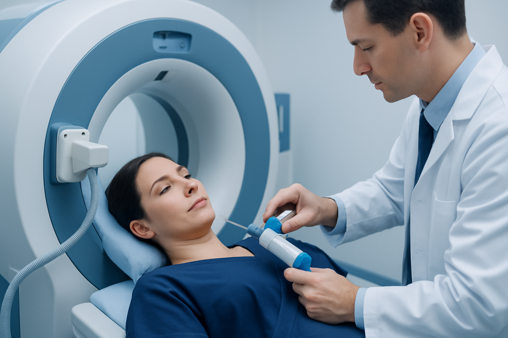
A biopsy is a procedure in which a clinician removes a small sample of cells, fluid or tissue for laboratory examination. It helps clarify an abnormality found during an examination, screening test, blood test or imaging study. Biopsies are used in many specialties, including dermatology, breast care, gastroenterology, gynecology, urology, lung medicine and oncology.
Not every lump, skin change or imaging finding requires a biopsy. A clinician may recommend one when examination and imaging cannot provide a reliable diagnosis, when infection or inflammatory disease needs confirmation, or when it is important to determine whether a suspicious area is benign or malignant. The type of biopsy depends on the location, size and appearance of the finding.
Common approaches include a needle biopsy, in which a hollow needle removes cells or tissue; a punch, shave or excisional biopsy for a skin lesion; an endoscopic biopsy collected during a procedure such as colonoscopy; and a surgical biopsy when a larger or deeper sample is needed. Imaging guidance with ultrasound, CT or MRI may help the clinician place a needle accurately. The goal is to obtain an adequate, representative sample while minimizing unnecessary tissue removal.
What happens during the biopsy procedure

Before a biopsy, the healthcare team reviews medications, allergies, bleeding risks, relevant medical conditions and pregnancy status where applicable. Some medicines, including blood-thinning medicines, may need special planning; patients should not stop prescribed medication unless the clinician who manages it provides instructions. Depending on the procedure, fasting or arranging transport home may be advised.
Many minor biopsies are performed with local anesthetic, which numbs the area while the person remains awake. Needle biopsies and endoscopic biopsies may involve local anesthetic, sedation or anesthesia according to the site and technique. The clinician cleans the skin or prepares the relevant area, collects the sample, controls bleeding and applies a dressing or closes the incision if needed.
The sample is sent to a pathology laboratory. It is preserved, prepared into thin sections and examined under a microscope. The pathologist may order special stains, immunohistochemistry, molecular testing or other studies if these are needed to identify the condition more precisely or guide treatment. These additional analyses can add time but may provide clinically important detail.
For people undergoing assessment of a possible malignancy, cancer treatment planning is based on the confirmed diagnosis and the full clinical picture rather than on one isolated report phrase. The treating team can explain which findings affect next steps and which details are simply part of standard laboratory documentation.
How to see biopsy results and understand timing
Biopsy results are commonly shared through a follow-up appointment, telephone call or a secure patient portal, depending on local practice and the urgency of the finding. Some results are available within several days, but more complex specimens can take longer when special testing or expert review is required. A longer wait does not, by itself, indicate a serious result.
When viewing a report online, patients may see technical language before their clinician has had an opportunity to explain it. It can be helpful to write down the exact diagnosis, the specimen location and any unfamiliar terms, then discuss them at the planned review. The healthcare team can also confirm whether all expected samples have resulted, especially when tissue was collected from more than one location.
A useful discussion with the clinician may include: what was found, whether the sample adequately represents the area of concern, whether further testing is needed, and what follow-up is recommended. If cancer is diagnosed, the doctor may discuss staging investigations, referral to relevant specialists and individualized treatment options. A pathology result is an important step, but it is not always the final step in diagnosis.
How do I understand my pathology report results?
Pathology reports vary by organ and condition, but several terms occur frequently. The diagnosis identifies the condition found in the sample. If an abnormal growth is present, the report may describe its type and subtype. In cancer reports, the grade describes how closely cells resemble normal cells and can provide information about behavior; it is different from the stage, which describes the extent of disease in the body.
If a lesion was removed surgically, the report may comment on margins. Margins are the edges of the removed tissue. “Negative” or “clear” margins generally mean no abnormal or cancer cells were seen at the examined edge, while “positive” margins mean abnormal cells extend to an edge of the specimen. The practical importance varies and should be discussed with the surgeon or specialist.
Some pathology reports include biomarker, receptor, genetic or molecular findings. These tests can help classify a disease and, for certain cancers, may help identify treatments that are more likely to be useful. They do not always indicate inherited risk; some genetic changes occur only within the sampled cells. A clinician or genetic counselor can explain whether family assessment is relevant.
Pathologists sometimes use wording such as “consistent with,” “suggestive of,” or “cannot exclude.” These phrases communicate the level of certainty based on the tissue available. Further sampling, imaging, laboratory testing or clinical follow-up may be needed when findings are uncertain or do not match the wider clinical picture.
How to check if a biopsy report is positive or negative?
“Positive” and “negative” are shorthand terms, not universal labels of good or bad news. A positive biopsy result means the test detected the specific finding being investigated. For example, a biopsy may be positive for cancer cells, an infection, an inflammatory condition or another abnormality. The final diagnosis states exactly what was identified.
A negative biopsy result means the tested sample did not show the targeted abnormality. For example, no malignancy may be seen in the tissue examined. This is often reassuring, but it should be interpreted with the original concern in mind. If a scan, examination or symptoms remain suspicious, the clinician may recommend repeat sampling, specialist review, additional imaging or monitoring.
It is also possible for a report to be inconclusive. “Nondiagnostic,” “inadequate,” “limited sample,” or “insufficient tissue” can mean the laboratory could not make a definite assessment. This may happen if too few cells were collected, the sample was damaged, or the area of concern was difficult to reach. It does not confirm or exclude the suspected condition.
Patients should avoid deciding what a result means based only on one word. Asking the clinician, “What did this result show, and what does it mean for me?” is appropriate and can help turn a technical report into a clear care plan.
Recovery, benefits and possible risks
Recovery after a biopsy depends on the technique and body area. After a small skin or needle biopsy, mild soreness, bruising or a small amount of bleeding may occur for a few days. Keeping the area clean, following dressing instructions and avoiding strenuous activity when advised can support healing. If stitches are used, the team explains when and how they will be removed or when they will dissolve.
Endoscopic, deep-organ and surgical biopsies may require a longer observation period and more specific aftercare. Sedation can temporarily affect coordination and judgment, so patients may need someone to take them home and should follow instructions about driving, work and alcohol. The care team provides procedure-specific guidance before discharge.
The principal benefit of a biopsy is diagnostic clarity. It can distinguish between conditions that look similar on imaging or examination and may guide monitoring, medication, surgery or other treatment. Risks are generally uncommon but can include bleeding, infection, pain, scarring, injury to nearby structures and reactions to anesthesia or sedation. The exact risk profile depends on the biopsy site and method.
Medical teams aim to balance the value of obtaining a diagnosis with the risks of the procedure. Patients can ask why a particular technique is recommended, whether alternatives are appropriate, and what the expected recovery plan will involve.
When to seek medical care
After a biopsy, prompt medical advice is important for heavy or persistent bleeding, increasing swelling, severe or worsening pain, fever, chills, spreading redness, pus-like drainage, shortness of breath, chest pain or fainting. People should also contact their care team if a dressing becomes soaked with blood or if they are unsure how to care for the biopsy site.
It is also important to arrange follow-up if results have not been received within the timeframe given by the healthcare team, or if symptoms persist or worsen despite a negative or inconclusive result. Do not delay discussing a report because it contains unfamiliar or concerning terms; clarification is part of safe care.
Acibadem International’s multidisciplinary specialists and JCI-accredited hospitals support international patients who need diagnostic assessment, pathology review and coordinated treatment planning. A qualified clinician can explain biopsy findings in relation to the individual’s health, tests and personal priorities.
Frequently asked questions
What is the most important part of a biopsy report?
The final diagnosis is usually the most important section because it summarizes what the pathologist identified in the sample. However, the specimen site, microscopic findings, margins and any additional tests may also matter. The clinician who ordered the biopsy can explain which details affect care.
Does a positive biopsy always mean cancer?
No. A positive result means the biopsy found the specific condition being tested for. It may identify cancer, but it can also show infection, inflammation, benign disease or another abnormality. The report’s final diagnosis provides the necessary context.
Can a biopsy be negative even if cancer is present?
A negative biopsy means no cancer was seen in the tissue sampled, but no test is perfect. Occasionally, a sample may be too small or may not capture the relevant area. If clinical examination or imaging remains concerning, the doctor may recommend further assessment.
What does “benign” mean on a pathology report?
Benign means non-cancerous. Some benign conditions need no treatment, while others may require monitoring or removal because of symptoms, location, growth or uncertainty. The recommended approach depends on the specific diagnosis.
What does “inconclusive” or “nondiagnostic” mean in a biopsy report?
These terms mean the sample did not provide enough reliable information for a definite diagnosis. This can occur when there is too little tissue, the cells are not well preserved, or the targeted area was difficult to sample. The clinician may suggest repeating the biopsy or using another test.
How long does it take to get biopsy results?
Timing varies by biopsy type, laboratory workflow and whether special tests are needed. Straightforward results may be ready in several days, while more complex testing can take longer. The healthcare team can provide the most accurate expected timeframe for the individual procedure.
References
- National Cancer Institute
- American Cancer Society
- College of American Pathologists
- Mayo Clinic
- National Health Service
This article is for general information only and is not a substitute for professional medical advice. Please consult a qualified doctor about your individual situation.
Oncology care in Turkey — second opinion and treatment plan
JCI-accredited · board-certified surgeons · reply within 24h
Add us as a Preferred Source to see more of our trusted health content across Google Search, AI Overviews and Discover.