How to Repair White Matter in the Brain: Procedure, Recovery and Results
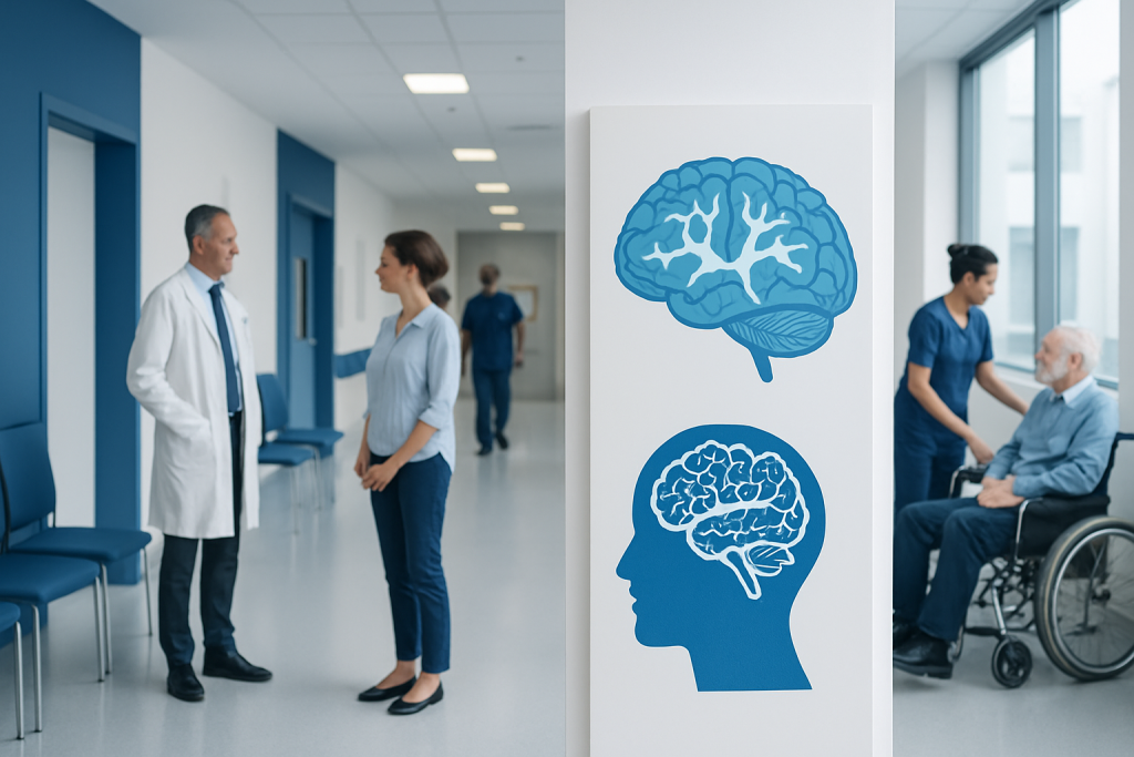
White matter is the brain’s communication network, made largely of nerve fibers and their protective myelin coating. There is no proven universal procedure that regrows white matter, but targeted treatment and rehabilitation can improve function for many people.
Key Takeaways
- White matter is the brain’s communication network, made largely of nerve fibers and their protective myelin coating.
- There is no proven universal procedure that regrows white matter, but targeted treatment and rehabilitation can improve function for many people.
- The outlook depends on the cause, location and extent of changes, as well as a person’s age, symptoms and overall vascular health.
- Controlling blood pressure, diabetes, cholesterol, smoking and sleep problems can help protect white matter health.
- New or sudden neurological symptoms require urgent medical assessment, especially possible stroke symptoms.
White matter cannot usually be repaired through one standard procedure. Recovery focuses on identifying the cause of damage, treating reversible contributors, preventing further injury and using rehabilitation to help the brain adapt and regain function.
Overview: How to Repair White Matter in the Brain
How to repair white matter in the brain depends on why it has been damaged. There is no single procedure that reliably restores white matter in every situation; care usually combines treatment of the underlying cause, rehabilitation and measures that protect the brain from further injury.
White matter consists mainly of bundles of nerve fibers, called axons, covered by myelin. These pathways connect different brain regions and help signals travel efficiently. Changes in white matter may be seen on magnetic resonance imaging (MRI) after small-vessel disease, stroke, inflammation, multiple sclerosis, head injury, migraine or other conditions.
An MRI report mentioning “white matter lesions,” “white matter hyperintensities” or “chronic microvascular changes” does not by itself establish a diagnosis or predict a person’s future. A neurologist interprets the scan alongside symptoms, medical history, examination findings and, when needed, further tests.
Can You Recover From White Matter Damage?
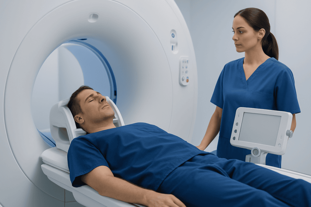
Recovery from white matter damage is possible in some circumstances, but it varies considerably. The brain can adapt by strengthening remaining connections and using alternative pathways, a process known as neuroplasticity. Improvement may occur after stroke, traumatic brain injury or an inflammatory episode, particularly when rehabilitation begins early and is tailored to the person’s needs.
Some causes are partly reversible. For example, correcting vitamin deficiencies, treating infections, managing an autoimmune condition or addressing toxic exposures may prevent additional injury and sometimes improve symptoms. In contrast, white matter changes related to long-standing small-vessel disease or established scarring may not disappear on imaging, although symptoms and daily function can still improve.
Recovery goals are individual. They may include walking more safely, improving hand use, reducing fatigue, supporting memory and attention, managing speech or swallowing difficulties, and maintaining independence. Physical therapy, occupational therapy, speech and language therapy, neuropsychological support and supervised exercise can each have an important role.
Finding the Cause and Deciding on Treatment
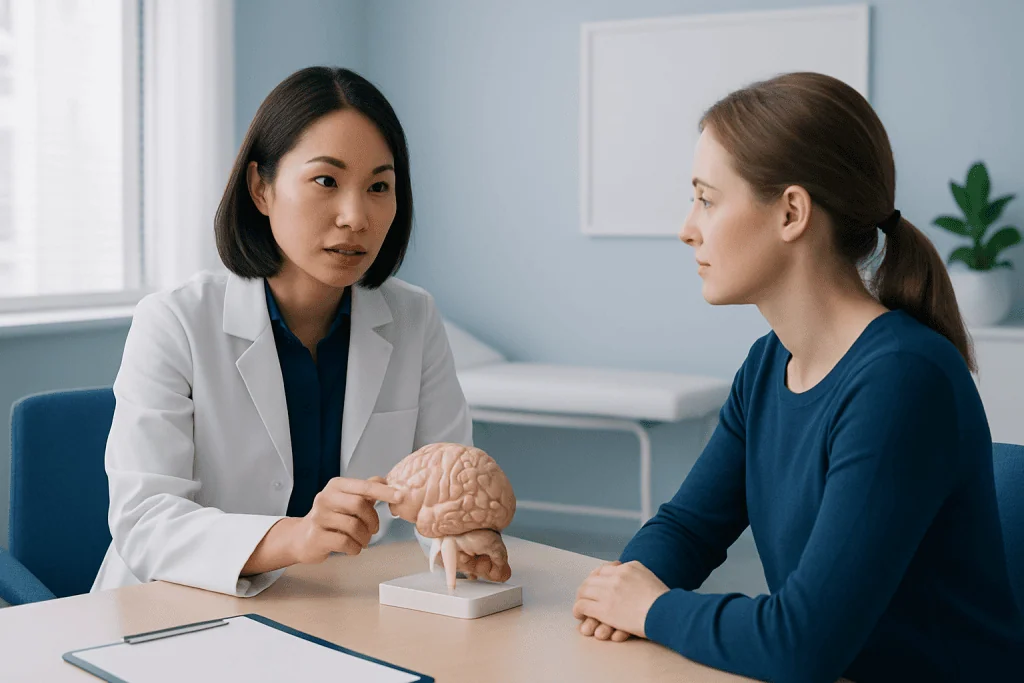
White matter changes are not a condition with one operation or one medication. The first step is a careful evaluation to determine whether the finding is linked to vascular risk factors, a previous stroke, demyelinating disease, migraine, trauma, infection, inherited disease or another explanation. The pattern and location of MRI changes can provide useful clues, but diagnosis should not rest on imaging alone.
Assessment may include a neurological examination, MRI with appropriate sequences, blood tests and review of medications and lifestyle factors. Depending on the clinical picture, clinicians may recommend cognitive testing, spinal fluid analysis, heart rhythm assessment, ultrasound of neck arteries or other investigations. This helps distinguish common age-related vascular changes from conditions that need specific treatment.
People may be candidates for targeted treatment when an active, treatable cause is identified. For example, stroke prevention may involve controlling vascular risks, while inflammatory demyelinating disorders require specialist-directed treatment. If symptoms are related to impaired function, a rehabilitation assessment can identify practical goals and the most appropriate therapies.
- Vascular causes: control blood pressure, diabetes, cholesterol and other risk factors.
- Inflammatory causes: use disease-specific treatment under neurological supervision.
- Stroke or injury: combine medical treatment with structured rehabilitation.
- Metabolic or nutritional causes: correct the underlying abnormality safely and appropriately.
How Treatment and Rehabilitation Work
For most people, “repair” is a treatment plan rather than a surgical procedure. The plan works by stopping or slowing the process causing injury, reducing risks of future damage and helping the nervous system regain skills. Surgery or catheter-based procedures are considered only for selected underlying conditions, such as certain vascular abnormalities, hydrocephalus or brain lesions; they are not routine treatments for nonspecific white matter changes.
A typical pathway begins with confirming the likely cause and discussing priorities, such as balance, mobility, concentration, vision, mood or fatigue. The medical team then treats contributing conditions and creates a rehabilitation program. Therapy may include repetitive movement practice, balance and strength training, activities for hand function, cognitive strategies, communication therapy and education for family members or caregivers.
Recovery plans should be reviewed over time. Progress can be gradual, and treatment may be adjusted as symptoms, activity tolerance and imaging findings change. For people recovering after a neurological event, stroke rehabilitation can coordinate physical, occupational and speech therapies with medical follow-up.
Acibadem International’s multidisciplinary specialists and JCI-accredited hospitals can assess neurological symptoms and coordinate diagnosis, rehabilitation and treatment planning for international patients.
Recovery Timeline, Benefits and Possible Risks
The recovery timeline depends on the cause and severity of injury. Following an acute event such as stroke or head trauma, the most noticeable gains often occur in the first weeks to months, but meaningful progress may continue for much longer with regular practice and appropriate support. Chronic white matter changes may be managed over years, with the focus on preserving function and reducing future risk.
Potential benefits of treatment include better mobility, balance, thinking skills, mood, communication, fatigue management and ability to complete everyday tasks. Stabilizing blood pressure and other vascular factors may also help limit progression of small-vessel injury. A repeat MRI may be useful in some cases, but how a person feels and functions is at least as important as scan appearances.
Risks depend on the treatment being considered. Medicines may have side effects or interactions, and intensive therapy can cause temporary fatigue, soreness or frustration. Procedures directed at an underlying brain or blood-vessel problem have their own risks and should be discussed with the responsible specialist. Unproven “brain repair” supplements, stem-cell clinics and intensive programs outside standard medical care may be costly or unsafe and should be approached cautiously.
What Rebuilds White Matter in the Brain?
There is currently no established medication, supplement or procedure that can reliably rebuild white matter for everyone. Myelin repair is an active area of research, particularly in demyelinating diseases, but experimental approaches should not be confused with proven routine care.
The best-supported approach is to address the underlying cause and create conditions that support brain health and neuroplasticity. This can include treating high blood pressure, diabetes, sleep apnea, high cholesterol, depression, vitamin deficiency or an inflammatory neurological disease when present. A clinician can advise whether medications are appropriate for the individual condition.
Rehabilitation helps the brain make effective use of intact pathways. Skill-focused practice, gradual aerobic activity where medically suitable, good sleep and management of pain, anxiety or depression can all support participation in recovery. No food, vitamin or over-the-counter product should replace an evaluation for new neurological symptoms.
What Is the Prognosis for White Matter Lesions on the Brain?
The prognosis for white matter lesions on the brain is highly individual. Small, incidental changes are common with aging and may never cause noticeable symptoms. However, a greater burden of white matter disease, especially when accompanied by vascular risk factors, can be associated with slower walking, balance problems, cognitive changes, depression and a higher risk of stroke.
Prognosis is influenced by the lesion pattern, whether changes are stable or progressing, their cause, and the person’s symptoms and health conditions. Lesions caused by a single past event may remain stable, while active inflammation or uncontrolled vascular disease may require closer monitoring. The treating clinician may recommend follow-up MRI when it would change management.
Importantly, an MRI finding is not a forecast of inevitable decline. Risk-factor management, activity, rehabilitation and regular medical care can make a meaningful difference. A neurologist can explain what the scan means in the context of the individual rather than relying on a report phrase alone.
How to Increase White Matter in Brain Naturally and When to Seek Medical Care
People often ask how to increase white matter in brain naturally. While lifestyle measures cannot guarantee that white matter volume will increase or erase existing lesions, they can support overall brain and blood-vessel health. Helpful habits include regular physical activity suited to ability, a balanced eating pattern rich in vegetables, fruit, whole grains, legumes and healthy fats, adequate sleep, social and mental activity, and avoiding tobacco and recreational drugs.
It is also important to manage blood pressure, blood sugar and cholesterol with a healthcare professional. Limiting alcohol, treating sleep apnea if present and following advice for migraine or other medical conditions may reduce potentially modifiable risks. Before taking supplements for “myelin repair,” people should speak with a clinician, particularly if they use prescription medicines or have kidney, liver or neurological conditions.
Medical care should be sought urgently for sudden weakness or numbness on one side, facial drooping, speech difficulty, sudden severe headache, new vision loss, confusion, loss of balance or a seizure. These symptoms may indicate a stroke or another emergency. Non-urgent medical review is also appropriate for new or worsening memory problems, persistent walking changes, tremor, frequent falls, unexplained weakness, or an MRI report that has not been discussed with a clinician.
Frequently asked questions
Can white matter lesions be reversed?
Some white matter changes related to treatable causes may improve or stabilize when the cause is addressed. Long-standing vascular changes or scarring may remain visible on MRI, but symptoms can still improve through rehabilitation and risk-factor management. A neurologist can explain what the lesion pattern means for an individual.
Is there a procedure to repair white matter in the brain?
There is no standard procedure that directly repairs all forms of white matter damage. Treatment is directed at the underlying cause, such as stroke, inflammation, nutritional deficiency or vascular disease, and often includes rehabilitation. Procedures may be relevant only when a specific structural or vascular problem is found.
What causes white matter damage?
Common causes include small-vessel disease associated with aging and vascular risk factors, stroke, head injury, inflammatory disorders, migraine and some infections or metabolic conditions. The cause cannot be determined from an MRI phrase alone. Medical history, symptoms and neurological assessment are essential.
Can exercise help white matter health?
Regular physical activity supports cardiovascular health and may support brain function and neuroplasticity. The safest exercise type and intensity depend on a person’s symptoms, balance, heart health and underlying diagnosis. A rehabilitation professional can help create an appropriate plan after stroke or injury.
Should I take supplements to rebuild myelin?
No supplement has been proven to rebuild myelin or white matter reliably in all people. Supplements can also interact with medicines or be inappropriate for certain medical conditions. A clinician may test for and treat a confirmed nutritional deficiency when relevant.
When are white matter lesions an emergency?
White matter lesions themselves are usually an imaging finding rather than an emergency. However, sudden symptoms such as weakness, facial drooping, trouble speaking, new severe headache, confusion, vision loss or severe imbalance need emergency assessment because they may signal a stroke or another urgent neurological condition.
References
- American Heart Association
- National Institute of Neurological Disorders and Stroke
- National Institute on Aging
- World Health Organization
- American Academy of Neurology
This article is for general information only and is not a substitute for professional medical advice. Please consult a qualified doctor about your individual situation.
Explore treatments in Turkey — costs, top hospitals & a free quote
JCI-accredited · board-certified surgeons · reply within 24h
Add us as a Preferred Source to see more of our trusted health content across Google Search, AI Overviews and Discover.
Check your numbers in seconds
BMI, calories, due date, blood pressure and 30+ more clinical calculators — free, instant, doctor-reviewed ranges.