Hydrops Ultrasound: Preparation, Procedure and Results

Hydrops fetalis describes abnormal fluid accumulation in at least two fetal compartments. Ultrasound can show skin swelling, fluid around the heart or lungs, abdominal fluid and placental changes.
Key Takeaways
- Hydrops fetalis describes abnormal fluid accumulation in at least two fetal compartments.
- Ultrasound can show skin swelling, fluid around the heart or lungs, abdominal fluid and placental changes.
- The examination is noninvasive, usually needs little preparation and does not expose the fetus to ionizing radiation.
- Hydrops can develop over a short period or more gradually, so follow-up scans may be important.
- Outlook depends mainly on the underlying cause, gestational age and whether effective treatment is available.
A hydrops ultrasound is a detailed pregnancy scan used to look for abnormal fluid buildup in two or more areas of a fetus’s body. It is safe, does not use radiation, and helps the care team identify possible causes, assess severity and plan timely follow-up.
Hydrops Ultrasound: What It Is and Why It Is Used
A hydrops ultrasound is a detailed prenatal ultrasound performed when a clinician needs to check for fetal hydrops, also called hydrops fetalis. The scan looks for unusual fluid accumulation in two or more fetal areas, such as beneath the skin, in the chest, around the heart or within the abdomen. It can also assess the placenta, amniotic fluid and blood flow in the umbilical cord and fetal vessels.
Hydrops is a finding rather than a single disease. It may result from immune causes, such as red blood cell incompatibility, or from nonimmune causes including fetal anemia, heart rhythm problems, structural heart conditions, infections, chromosomal conditions, lung or chest abnormalities, and genetic or metabolic disorders. A detailed ultrasound is usually one part of a broader assessment of hydrops fetalis.
The examination helps the fetal medicine team understand how much fluid is present, look for clues to the cause and decide whether further blood tests, fetal echocardiography, genetic testing or closer monitoring may be appropriate. An ultrasound result alone may not identify the exact cause, but it provides essential information for the next steps.
How Hydrops Ultrasound Works and Who May Need It
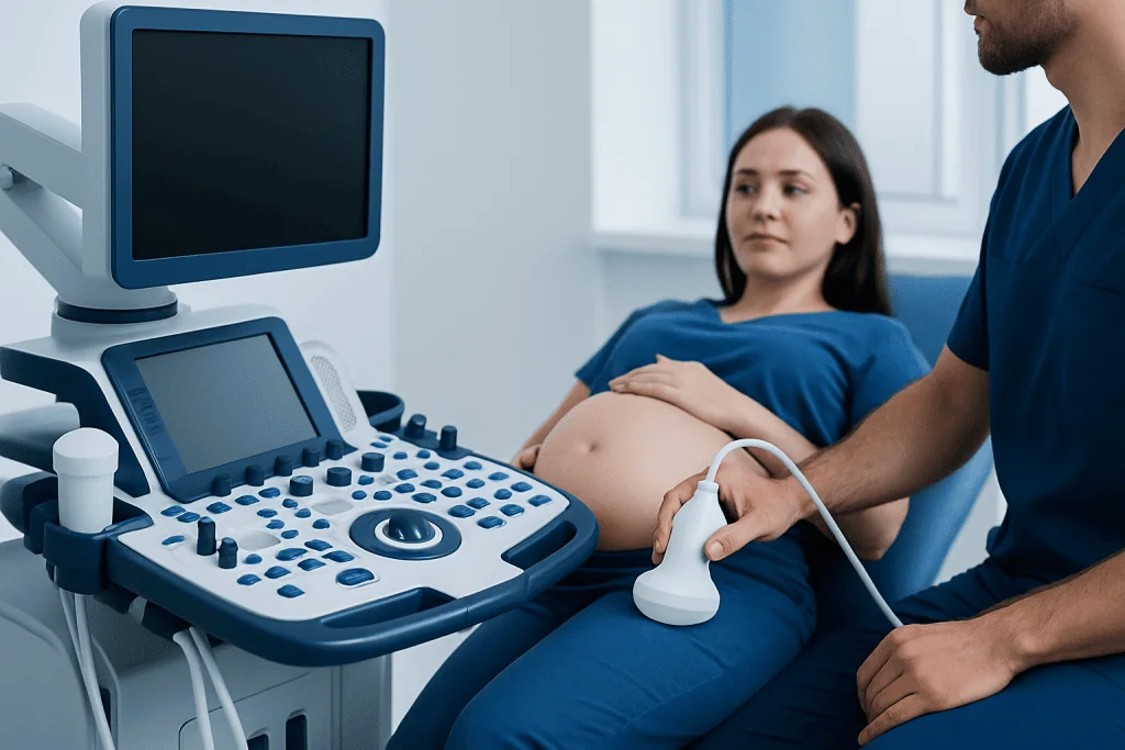
Ultrasound uses high-frequency sound waves to create real-time images. A trained sonographer or fetal medicine specialist moves a small handheld probe over the abdomen. The probe sends sound waves into the body and receives returning echoes, which are converted into images on a monitor. No radiation is used.
A hydrops ultrasound may be recommended when a routine pregnancy scan shows fluid around the fetus, the uterus measures larger than expected, fetal movements have changed, or there is a known risk of fetal anemia or a heart condition. It may also be advised after certain maternal infections, in pregnancies with red blood cell antibodies, or when an earlier examination suggests a fetal structural or genetic concern.
In many cases, the assessment includes Doppler ultrasound. Doppler measures blood flow and can provide indirect evidence of fetal anemia, heart strain or circulatory changes. A detailed heart assessment may be arranged through fetal echocardiography when a cardiac cause is possible.
There is generally no special candidacy requirement: the scan is tailored to the pregnancy and the clinical question. A referral may come from an obstetrician, maternal-fetal medicine specialist or pediatric cardiology team.
Preparation and Step-by-Step Procedure

Most people do not need special preparation for a hydrops ultrasound. The imaging center may ask the patient to arrive with a comfortably full bladder, particularly early in pregnancy, although this is not always necessary. Wearing a two-piece outfit can make abdominal scanning more convenient. Patients should bring previous ultrasound reports, blood test results and relevant pregnancy records when available.
At the appointment, the clinician will review the pregnancy history and reason for the scan. The patient lies on an examination bed, usually on the back with positioning adjusted for comfort. Gel is placed on the abdomen to help transmit sound waves, and the transducer is moved over the skin to obtain images from different angles.
The examination commonly includes measurements of fetal growth, an assessment of fetal anatomy, the heart rate and rhythm, fluid around the fetus, placental thickness, amniotic fluid and Doppler blood-flow studies. The clinician may spend additional time examining the chest, abdomen, skin and heart. Depending on fetal position and the complexity of the assessment, the visit may take longer than a standard routine scan.
Images are reviewed by an appropriately trained clinician, often during the visit or soon afterward. If findings suggest hydrops, the team may discuss further testing, referral to a fetal medicine center and an individualized monitoring plan. In selected cases, testing or treatment may involve fetal medicine care.
What Are the Ultrasound Findings of Fetal Hydrops?
The defining ultrasound finding is fluid in at least two fetal compartments. These compartments include the skin, the space around the lungs, the sac around the heart and the abdominal cavity. Fluid in only one area may have a different explanation and is assessed in its own clinical context.
- Skin edema: diffuse thickening or swelling under the skin, often visible around the scalp, body wall or limbs.
- Pleural effusion: fluid surrounding one or both lungs within the chest.
- Pericardial effusion: fluid around the heart.
- Ascites: free fluid within the fetal abdomen.
- Placental edema: an unusually thick placenta, which may occur with more severe fluid imbalance.
- Polyhydramnios: increased amniotic fluid, which can sometimes accompany hydrops.
The scan may also reveal findings that point toward an underlying cause, such as an abnormal heart rhythm, structural heart difference, chest mass, enlarged organs, signs of anemia or features associated with a genetic condition. These clues guide additional investigations, but they do not always provide a definite diagnosis by themselves.
What Does Hydrops Look Like on an Ultrasound?
On ultrasound, fluid usually appears as dark or black areas because fluid allows sound waves to pass through with relatively few echoes. For example, ascites may appear as a dark rim or collection around the abdominal organs, while pleural effusion appears as dark fluid around the lungs. Pericardial effusion appears as a dark space surrounding the heart.
Skin edema may look like a thickened, darker layer beneath the outer contour of the fetal skin. When several signs are present together, the image pattern raises concern for hydrops. The clinician also examines whether swelling is generalized, whether the placenta is thickened and whether amniotic fluid is increased.
Images can be emotionally difficult to discuss, especially when findings are unexpected. The ultrasound team should explain what has been seen in clear language, distinguish confirmed findings from possibilities and describe which further tests may help clarify the situation. A detailed consultation with maternal-fetal medicine and other relevant specialists can support informed decision-making.
How Quickly Can Fetal Hydrops Develop?
Fetal hydrops can develop at different speeds. In some situations, such as severe fetal anemia or certain rapid heart rhythm disturbances, fluid may accumulate over days to a few weeks. In other cases, it develops more gradually as an underlying condition affects the fetal circulation or protein balance over time.
Because the pace is unpredictable and depends on the cause, clinicians may recommend repeat ultrasounds at intervals based on the individual situation. Follow-up scans can assess whether fluid is increasing, stable or improving, as well as track fetal growth, heart function, blood flow and amniotic fluid.
Rapid changes are one reason why suspected hydrops is assessed promptly by an experienced fetal medicine team. Early identification of a treatable cause may alter management, but the appropriate plan depends on gestational age, scan findings and the results of further evaluation.
Benefits, Risks, Results and Recovery Timeline
The main benefit of a hydrops ultrasound is that it provides detailed, real-time information without an incision, anesthesia or radiation exposure. It can help identify fluid collections, evaluate fetal wellbeing and support decisions about referral, testing and possible treatment. Serial scanning also allows the team to monitor changes over time.
Diagnostic ultrasound is widely used in pregnancy when medically indicated. It is considered noninvasive and has no known direct recovery period. The patient can usually return to normal daily activities immediately after the appointment. Mild pressure from the probe or the need to lie still may be uncomfortable for some people, but the examination itself should not be painful.
Results may be available the same day, although a full specialist interpretation and care plan can take additional time. If hydrops is suspected or confirmed, clinicians may recommend maternal blood tests, infection screening, antibody testing, fetal echocardiography, genetic counseling or diagnostic procedures such as amniocentesis when appropriate. In selected cases, treatment may be directed at the underlying cause, including monitoring or intervention for fetal anemia, arrhythmia or fluid in the chest.
Recovery after any additional test or fetal procedure depends on that specific procedure. The care team will explain expected symptoms, activity guidance and warning signs before a procedure is undertaken.
Can a Fetus Survive With Hydrops?
Some fetuses with hydrops survive, particularly when the cause can be identified and treated or when fluid improves with close monitoring. However, hydrops can be a serious finding, and outcomes vary widely. Prognosis depends on the cause, the degree of fluid buildup, the presence of structural or genetic conditions, the fetal response to treatment and how far along the pregnancy is at diagnosis.
For example, outcomes may be more favorable in some treatable causes, including certain cases of fetal anemia or abnormal fetal heart rhythm. Other causes can be more complex and may carry a higher risk of pregnancy loss, premature birth or newborn illness. The care team will avoid making predictions from one scan alone whenever possible and will use the complete evaluation to discuss the individual outlook.
Care may involve maternal-fetal medicine, obstetrics, pediatric cardiology, neonatology, genetics, hematology and other specialists. Acibadem International’s multidisciplinary specialists and JCI-accredited hospitals support diagnosis and treatment planning for international patients with complex fetal conditions.
When to Seek Medical Care
Patients should contact their obstetrician or maternity unit promptly if they have been told that an ultrasound may show fetal fluid buildup or if they are asked to attend an urgent fetal medicine assessment. Keeping scheduled ultrasound appointments is especially important when there is a known risk of fetal anemia, maternal red blood cell antibodies, infection or a fetal heart concern.
Urgent maternity advice is appropriate for vaginal bleeding, leaking fluid, painful regular contractions, severe abdominal pain, severe headache with vision changes, sudden swelling with feeling unwell, or a noticeable reduction in fetal movements after movements have become established. These symptoms do not necessarily mean hydrops, but they require timely assessment in pregnancy.
People who receive an uncertain or unexpected scan result may also benefit from asking for a clear explanation of the findings, the possible causes, recommended next tests and when results will be available. A qualified obstetric and fetal medicine team can provide individualized guidance and emotional support.
Frequently asked questions
Is a hydrops ultrasound safe during pregnancy?
A hydrops ultrasound uses sound waves rather than ionizing radiation and is noninvasive. It is commonly used when there is a medical reason to evaluate the fetus in detail. The scan should be performed and interpreted by appropriately trained professionals.
How long does a hydrops ultrasound take?
The time varies according to fetal position, pregnancy stage and how much detail is needed. A targeted scan for suspected hydrops can take longer than a routine prenatal ultrasound because the clinician may examine multiple organs and use Doppler blood-flow measurements. The imaging center can provide a more specific estimate before the appointment.
Does fetal hydrops always mean there is a genetic condition?
No. Genetic and chromosomal conditions are among the possible causes, but they are not the only causes. Fetal anemia, heart rhythm problems, structural conditions, infections and other medical conditions may also lead to hydrops. Further testing may be offered to help identify the cause.
Can hydrops resolve before birth?
In some cases, fluid can lessen or resolve, particularly if the underlying cause is treatable or temporary. In other cases, hydrops may persist or worsen. Repeat ultrasound and specialist follow-up are important for tracking changes and planning care.
Will a hydrops ultrasound show the cause of the fluid buildup?
The scan can sometimes identify clues to the cause, such as a heart rhythm issue, structural difference, signs of anemia or fluid in the chest. However, ultrasound may not provide a complete diagnosis on its own. Blood tests, fetal echocardiography, genetic counseling or diagnostic testing may be recommended.
What happens after hydrops is found on ultrasound?
The next step is usually referral or review by a maternal-fetal medicine specialist. The team may arrange additional imaging, laboratory tests and consultations with other specialists, depending on the findings. Management is individualized and may range from close monitoring to treatment directed at an identified cause.
References
- American College of Obstetricians and Gynecologists
- Society for Maternal-Fetal Medicine
- International Society of Ultrasound in Obstetrics and Gynecology
- National Institutes of Health
- Merck Manual Professional Edition
This article is for general information only and is not a substitute for professional medical advice. Please consult a qualified doctor about your individual situation.
Ultrasound in Turkey — costs, top hospitals & a free quote
JCI-accredited · board-certified surgeons · reply within 24h
Add us as a Preferred Source to see more of our trusted health content across Google Search, AI Overviews and Discover.
Check your numbers in seconds
BMI, calories, due date, blood pressure and 30+ more clinical calculators — free, instant, doctor-reviewed ranges.

Check-up & Preventive Medicine
Comprehensive health screenings designed for early detection and prevention.
1 specialists in this unitMore from the Health Library



Related Specialists

Dt. Beril Karakaya Gökçimen
Oral & Dental Health
Prof. Dr. Burak Akan
Orthopedic Surgery & Traumatology
Assoc. Prof. Dr. İsmail Selçuk
Cardiovascular Surgery
Fzt. Ahsen Ercan
Physical Medicine & Rehabilitation