Hypertelorism: An Evidence-Based Guide for Patients

Hypertelorism refers to an increased distance between the eye sockets. It can occur on its own or as part of a genetic, craniofacial, or developmental condition.
Key Takeaways
- Hypertelorism refers to an increased distance between the eye sockets.
- It can occur on its own or as part of a genetic, craniofacial, or developmental condition.
- Diagnosis usually includes a physical examination, measurements, and sometimes imaging or genetic evaluation.
- Treatment depends on the cause, the severity, and whether there are functional or cosmetic concerns.
- Many people do not need urgent treatment, but a medical assessment is important when hypertelorism appears with other symptoms.
Hypertelorism means the eyes are set farther apart than expected based on standard facial measurements. It is a physical finding rather than a disease itself, and care focuses on identifying any underlying cause and deciding whether treatment is needed.
What hypertelorism means
Hypertelorism is the medical term for an increased distance between the eyes, more precisely between the bony eye sockets. In everyday language, people may describe this as wide-set eyes. On its own, this feature does not always mean there is a health problem, because facial appearance varies naturally from person to person and among families.
Doctors use the term because it can sometimes be a clue to how the bones of the face and skull developed before birth. In some people, hypertelorism is an isolated finding with no effect on vision, brain development, or general health. In others, it may occur together with other facial, eye, dental, or skull differences that deserve further assessment.
It is also helpful to distinguish true hypertelorism from telecanthus. Telecanthus means the inner corners of the eyelids are farther apart, while the bony eye sockets themselves are not unusually separated. This distinction matters because the causes, evaluation, and treatment planning may differ.
How hypertelorism may appear
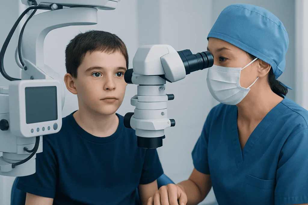
The most noticeable feature is a wider-than-expected space between the eyes. Some children or adults have no symptoms beyond appearance. Others may have associated findings depending on the underlying cause, such as a broad nasal bridge, differences in forehead shape, a cleft lip or palate, or eyelid changes.
Hypertelorism itself does not automatically cause vision problems, but related eye concerns can occur in some cases. These may include eye misalignment, refractive errors, double vision, blocked tear drainage, or difficulty with depth perception. For this reason, an eye examination is often part of the assessment, especially in children.
If hypertelorism is part of a broader craniofacial condition, there may also be symptoms outside the eye area. These can include developmental delay, hearing changes, dental crowding, breathing issues, or abnormal skull shape. The presence or absence of these associated findings helps doctors decide what tests are needed and whether specialist care is appropriate.
- Wider spacing between the eyes
- Broad nasal bridge or unusual forehead contour
- Eye alignment or vision concerns
- Other facial or skull differences present at birth
Why hypertelorism happens

Hypertelorism develops when the bones of the face and skull form differently during fetal development. This can happen for several reasons. In some people, it appears as a familial trait without any disease. In others, it is linked to a genetic syndrome, a craniofacial difference, or a structural issue affecting skull growth.
Conditions associated with hypertelorism can include craniosynostosis syndromes, frontonasal developmental differences, certain chromosomal conditions, and other rare congenital disorders. A doctor may also consider whether it occurs with cleft lip and palate or other craniofacial findings. The goal is not to assume the worst, but to understand whether the facial appearance is isolated or part of a recognizable pattern.
Risk factors are not always identifiable. Some cases arise from spontaneous genetic changes, while others may run in families. In pregnancy, most parents did nothing to cause the condition. Understanding this can be reassuring, as many families worry unnecessarily that they are responsible for a child’s appearance or diagnosis.
How doctors evaluate hypertelorism
Evaluation begins with a detailed medical history and physical examination. The doctor looks at facial proportions, skull shape, eyelids, nose, and jaw, and asks about family traits, pregnancy history, growth, and development. In children, the timing of milestones and the presence of other congenital findings can provide important clues.
Measurements are a key part of diagnosis. Specialists may measure the distance between the inner corners of the eyes, the pupils, and the outer eye corners, then compare these values with age- and sex-based norms. This helps distinguish true hypertelorism from telecanthus and from normal facial variation.
Imaging may be recommended if doctors suspect a structural skull or facial difference. Depending on the situation, this may include CT or MRI to assess the bones and surrounding structures. Eye specialists may evaluate vision and alignment, and genetics specialists may suggest testing if there are signs of a syndrome. When facial bone anatomy needs closer assessment, imaging may be coordinated with MRI or CT scanning as part of a broader workup.
Treatment options and when they are used
Treatment for hypertelorism depends on the cause, severity, age of the patient, and whether there are functional concerns. Not everyone needs treatment. If hypertelorism is mild, stable, and not linked to vision, breathing, or developmental problems, doctors may simply recommend observation and follow-up.
When treatment is needed, it is usually directed at associated problems rather than the spacing alone. This may include vision correction, treatment for strabismus, management of airway or dental issues, or support for developmental needs. In some cases, a multidisciplinary team that includes pediatrics, genetics, ophthalmology, maxillofacial surgery, neurosurgery, and plastic surgery is involved.
Surgery may be considered in selected cases, especially when hypertelorism is severe, affects function, or is part of a craniofacial abnormality. Surgical planning is highly individualized and may involve reconstructing facial bones to improve eye position and facial balance. If reconstructive care is appropriate, specialists may discuss options related to craniofacial surgery or plastic and reconstructive surgery. Surgery is usually planned carefully after imaging, growth assessment, and a detailed discussion of goals, benefits, and risks.
Living with hypertelorism and supportive care
For many patients and families, the most important part of care is understanding what hypertelorism does and does not mean. An isolated facial difference may not affect health, intelligence, or daily function. Clear communication from the care team can reduce anxiety and help families focus on the child’s overall growth, vision, comfort, and confidence.
Psychological and social support can also matter. Children and adults with visible facial differences may sometimes face questions or unwanted attention. Age-appropriate explanations, support from counselors when needed, and school-based understanding can help protect self-esteem. Families may find it useful to connect with craniofacial support groups or patient organizations.
Regular follow-up may be advised to monitor vision, skull growth, dental development, or any associated condition over time. If hypertelorism is part of a broader diagnosis, long-term care may involve several specialists working together. Acibadem International’s multidisciplinary specialists in JCI-accredited hospitals evaluate and treat craniofacial and related conditions for international patients when coordinated specialist care is needed.
When to seek medical care
A medical evaluation is a good idea whenever hypertelorism is noticed in a baby, child, or adult for the first time, especially if it appears alongside other facial differences. Prompt assessment is particularly important if there are concerns about vision, abnormal head shape, feeding problems, delayed development, or breathing difficulties.
Parents should seek medical advice if a child has wide-set eyes together with a rapidly changing skull shape, bulging eyes, crossed eyes, repeated eye infections, hearing concerns, or developmental delay. These signs do not always indicate a serious problem, but they do mean a more complete evaluation may be needed.
Adults should also speak with a doctor if facial changes are new, if there is double vision or reduced vision, or if a previously known craniofacial condition has not been assessed recently. Early review can help clarify the cause, arrange the right referrals, and provide reassurance when no treatment is needed.
Frequently asked questions
Is hypertelorism a disease?
No. Hypertelorism is a physical finding that describes increased distance between the eye sockets. It may be a normal facial variation or a sign associated with another condition, so the meaning depends on the full medical evaluation.
Can hypertelorism be normal in some families?
Yes. Some people have naturally wider facial spacing that runs in families and does not cause health problems. A doctor can help determine whether the appearance is within normal variation or suggests an underlying condition.
Does hypertelorism affect vision?
Not always. Many people with hypertelorism have normal vision, but some may have related eye issues such as strabismus, refractive errors, or tear drainage problems. That is why an eye examination is often recommended.
How is hypertelorism diagnosed?
Doctors diagnose hypertelorism through a physical examination and facial measurements compared with standard norms for age and sex. If needed, they may also order imaging studies or genetic testing to look for associated craniofacial or inherited conditions.
Can hypertelorism be treated without surgery?
Yes, in many cases no surgery is needed. Treatment may simply involve monitoring, vision care, or managing any related medical issues. Surgery is usually reserved for severe cases or for situations involving function or significant craniofacial differences.
At what age is surgery considered for hypertelorism?
There is no single age that fits everyone. Timing depends on the cause, the child’s growth, the severity of the finding, and whether there are functional concerns such as eye or skull-related problems. Decisions are usually made by a specialist team after careful planning.
References
- National Institutes of Health
- American Academy of Ophthalmology
- American Academy of Pediatrics
- MedlinePlus
- National Organization for Rare Disorders
This article is for general information only and is not a substitute for professional medical advice. Please consult a qualified doctor about your individual situation.
Explore treatments in Turkey — costs, top hospitals & a free quote
JCI-accredited · board-certified surgeons · reply within 24h
Add us as a Preferred Source to see more of our trusted health content across Google Search, AI Overviews and Discover.

More from the Health Library

Mini Heart Attack: An Evidence-Based Guide for Patients

Connective Tissue — Explained by Medical Evidence, Not Myths

Nose Hair: An Evidence-Based Guide for Patients

Penrose Drain — Explained by Medical Evidence, Not Myths

Parenteral: An Evidence-Based Guide for Patients