Image-Guided Tumor Ablation: How Heat or Cold Can Destroy Small Tumors
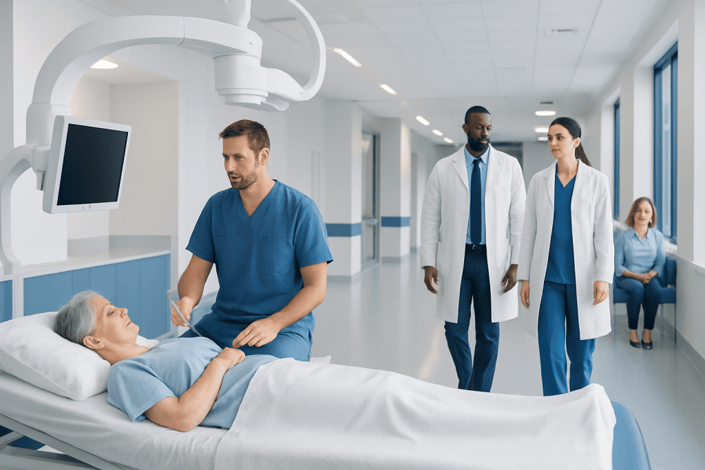
Image-guided tumor ablation can treat certain small tumors without open surgery. Different methods include radiofrequency ablation, microwave ablation, and cryoablation.
Key Takeaways
- Image-guided tumor ablation can treat certain small tumors without open surgery.
- Different methods include radiofrequency ablation, microwave ablation, and cryoablation.
- Imaging helps doctors place treatment needles accurately and monitor the procedure.
- Recovery is often shorter than with major surgery, but follow-up imaging is still essential.
- This treatment is not right for every tumor, so careful evaluation by specialists is important.
Image-guided tumor ablation is a minimally invasive treatment that uses heat or extreme cold to destroy small tumors. It is guided by imaging such as ultrasound, CT, or MRI, helping doctors target abnormal tissue while protecting nearby healthy structures.
Overview of image-guided tumor ablation
Image-guided tumor ablation is a procedure that destroys tumor tissue by applying extreme heat or cold through a thin needle-like probe. The treatment is called “image-guided” because doctors use imaging tools such as ultrasound, computed tomography (CT), or magnetic resonance imaging (MRI) to guide the probe into the exact location of the tumor. This approach allows treatment to be delivered with precision and without a large surgical incision.
Ablation is most often used for selected small tumors in organs such as the liver, kidneys, lungs, bones, and sometimes the thyroid or soft tissues. It may be recommended when a tumor is difficult to remove surgically, when a patient is not a good candidate for surgery, or when a less invasive option is preferred. In some situations, it is also used to control symptoms, such as pain caused by a tumor in bone.
Several forms of ablation exist. Radiofrequency ablation and microwave ablation destroy tissue with heat, while cryoablation destroys tissue by freezing it. Each technique has advantages depending on the tumor’s size, location, and blood supply. The goal is to destroy the targeted tissue while minimizing damage to nearby healthy structures.
Image-guided tumor ablation is not a single treatment for all cancers. Instead, it is one part of a broader cancer care plan that may also include surgery, medical oncology treatment, radiation therapy, or careful imaging follow-up. A multidisciplinary team helps decide whether ablation is appropriate for a person’s specific diagnosis.
How heat or cold destroys tumors
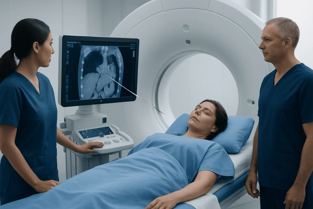
Heat-based ablation works by raising the temperature of tumor cells high enough to cause irreversible damage. In radiofrequency ablation, electrical energy generates heat around the tip of the probe. In microwave ablation, electromagnetic waves create heat more quickly and may treat a somewhat larger area. As the temperature rises, tumor proteins break down and the cancer cells lose the ability to survive.
Cold-based ablation, called cryoablation, works in the opposite way. The probe creates an “ice ball” around the tumor, freezing the abnormal tissue. Freezing and thawing damage cell membranes, reduce blood flow, and lead to cell death. One advantage of cryoablation is that the treated zone can often be seen clearly on imaging during the procedure, which can help in some anatomical locations.
The choice between heat and cold depends on several factors. Doctors consider the tumor’s size, shape, exact position, and whether it is close to nerves, major blood vessels, bile ducts, bowel, or other sensitive structures. Some methods may be preferred in one organ more than another, and sometimes more than one ablation session is needed.
Although ablation can be very effective for carefully selected small tumors, it does not replace all other cancer treatments. Some tumors are too large, too numerous, or too close to critical structures for ablation alone. In such cases, other therapies may be safer or more effective, or ablation may be combined with other treatments.
When tumor ablation may be used
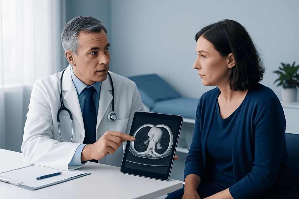
Doctors may consider image-guided tumor ablation for primary cancers that begin in an organ or for tumors that have spread from another part of the body. It is commonly discussed for selected liver tumors, kidney tumors, lung nodules, and painful bone lesions. In many cases, the best candidates are people with one or a few small tumors that can be reached safely with a probe.
Ablation can have different goals. In some people, it is intended to destroy all visible disease in a small area. In others, it is used to reduce tumor burden, delay progression, or relieve symptoms. For example, a painful lesion related to cancer may sometimes be treated with ablation to improve comfort and function.
This approach may be especially useful for patients who would face higher risks with major surgery because of age, other medical conditions, or reduced organ function. It may also be considered when a person has already had surgery in the same area, making another operation more complex. Because the procedure is minimally invasive, hospital stays are often shorter and recovery may be easier for some patients.
Even so, ablation is not suitable for every situation. Tumors near major structures may carry a higher risk of injury, and some cancers respond better to surgical removal or systemic treatment. Careful case selection is essential, often with input from interventional radiologists, oncologists, surgeons, and imaging specialists.
How the procedure is performed
Before treatment, the care team reviews imaging scans and medical history to plan the safest route to the tumor. Blood tests may be done to check kidney function, liver function, blood counts, and clotting. Patients are usually told when to stop eating and drinking before the procedure and whether any medications, especially blood thinners, need to be adjusted.
During the procedure, the patient may receive local anesthesia, sedation, or general anesthesia depending on the tumor site and the expected complexity. The doctor uses imaging guidance to insert a thin probe through the skin and into the tumor. Once the probe is in the correct position, heat or cold is applied for a carefully measured period to cover the target area.
In some cases, more than one probe is used, or the probe is repositioned to treat the entire tumor plus a small margin around it. Imaging may be repeated during or immediately after the procedure to assess the treatment zone and look for early complications. The patient is then observed for a period of time, and some people go home the same day while others stay overnight.
Because imaging is central to the procedure, image-guided tumor ablation is closely linked to advanced radiology services. When a tumor is in a challenging location, highly detailed planning and real-time guidance help improve accuracy. In international centers such as Acibadem International, multidisciplinary specialists in JCI-accredited hospitals evaluate and treat appropriate patients using these coordinated techniques.
Benefits, limitations, and possible risks
One of the main benefits of image-guided tumor ablation is that it is minimally invasive. Compared with open surgery, it may involve less pain, smaller scars, shorter hospital stays, and a faster return to daily activities. For some people, it can preserve more normal organ tissue, which is especially important in organs such as the liver or kidney.
Another advantage is precision. Imaging guidance allows doctors to target tumors that may be difficult to feel or see directly. In selected cases, ablation can be repeated if a new small lesion appears or if a treated area needs additional therapy. It may also be used alongside interventional radiology treatments and other cancer therapies as part of a tailored plan.
However, ablation has limitations. It may be less effective for larger tumors, for tumors with irregular borders, or for lesions close to large blood vessels where heat can dissipate. Not all treated tissue can be assessed immediately, so follow-up imaging is necessary to confirm whether the tumor has been fully destroyed. Some patients may still need surgery, systemic therapy, or radiation afterward.
Possible risks vary by the organ being treated and the technique used. They can include bleeding, infection, pain, injury to nearby organs, fluid or air leaks in lung procedures, or damage to bile ducts or nerves in certain locations. Serious complications are uncommon in experienced hands, but they are important to discuss in advance so that informed decisions can be made.
Recovery and follow-up after ablation
After image-guided tumor ablation, most patients are monitored for several hours or overnight. Mild pain, fatigue, bruising, or temporary discomfort around the treatment site can occur and usually improves over a short period. The care team provides instructions about wound care, activity, bathing, pain relief, eating, and when regular medications can be restarted.
Recovery depends partly on which organ was treated. Someone who has had ablation in the liver or kidney may feel tired for a few days, while lung procedures may require observation for breathing-related issues such as a small pneumothorax. Many people can return to light activity relatively soon, but strenuous exercise and heavy lifting may need to wait until the doctor confirms it is safe.
Follow-up is a key part of treatment. Imaging scans are usually scheduled after the procedure to see whether the ablation zone fully covers the tumor and whether any residual or recurrent disease is present. These checks may continue at intervals because ablation treats a specific target area, while the underlying cancer type may still require ongoing monitoring.
If the follow-up scan suggests incomplete treatment, the team may recommend another ablation session or a different therapy. For some patients, ablation fits into a long-term plan that includes surveillance for tumor-related conditions elsewhere in the body. Close communication with the treating team helps patients understand what the results mean and what the next steps should be.
Who should ask a doctor about this treatment
People who have been told they have a small tumor, a limited number of tumors, or a tumor in a location that may be accessible through the skin may wish to ask whether ablation is an option. It can be particularly relevant for those who want to understand alternatives to surgery or who have health conditions that make a major operation more challenging. A doctor can explain whether the size, location, and type of tumor make this approach realistic.
Questions to ask may include which ablation method is being considered, what imaging will be used, whether the goal is cure or control, and what follow-up is needed afterward. Patients can also ask how the procedure compares with surgery, radiation therapy, or systemic treatments for their situation. This discussion helps align treatment choices with both medical needs and personal preferences.
It is important to seek medical advice promptly if there are new symptoms such as unexplained weight loss, persistent pain, coughing up blood, jaundice, blood in the urine, or worsening fatigue, especially in someone with a known tumor. After ablation, urgent medical attention may be needed for fever, severe pain, heavy bleeding, shortness of breath, chest pain, confusion, or signs of infection at the puncture site.
Because treatment decisions can be complex, many patients benefit from review by a multidisciplinary team. Imaging specialists, oncologists, surgeons, and other doctors work together to weigh safety, effectiveness, and long-term follow-up. This team-based approach helps ensure that image-guided tumor ablation is used in the right patient, at the right time, and for the right reason.
Frequently asked questions
Is image-guided tumor ablation the same as surgery?
No. Image-guided tumor ablation is usually performed through the skin using one or more thin probes rather than through a large incision. It is considered minimally invasive, although it still requires careful planning and medical monitoring.
Which tumors can be treated with ablation?
Ablation is most often used for selected small tumors in organs such as the liver, kidneys, lungs, and bones. Whether it is suitable depends on the tumor’s size, number, location, and cancer type, as well as the patient’s overall health.
Does tumor ablation use heat or cold?
It can use either. Radiofrequency ablation and microwave ablation use heat, while cryoablation uses extreme cold. The doctor chooses the method based on the tumor and the surrounding anatomy.
Is image-guided tumor ablation painful?
The procedure is usually done with local anesthesia, sedation, or general anesthesia, so discomfort during treatment is controlled. Some soreness or fatigue afterward is common, but the care team provides guidance for relief and recovery.
Can a tumor come back after ablation?
Yes, it is possible for a treated tumor to be only partially destroyed or for cancer to recur later. That is why follow-up imaging is essential after ablation, even when the initial treatment appears successful.
How long is recovery after tumor ablation?
Recovery is often shorter than recovery after major surgery, but it depends on the organ treated and the person’s general health. Some patients go home the same day, while others stay overnight for observation.
References
- World Health Organization
- National Cancer Institute
- Radiological Society of North America
- Society of Interventional Radiology
- American Cancer Society
This article is for general information only and is not a substitute for professional medical advice. Please consult a qualified doctor about your individual situation.
Oncology care in Turkey — second opinion and treatment plan
JCI-accredited · board-certified surgeons · reply within 24h
Add us as a Preferred Source to see more of our trusted health content across Google Search, AI Overviews and Discover.