Kidney Stones: Flank Pain, Urine Tests, and Prevention Strategies

Kidney stones may cause severe one-sided flank pain that can move toward the lower abdomen or groin. Urine tests help detect blood, infection, crystals, and chemical patterns linked to stone formation.
Key Takeaways
- Kidney stones may cause severe one-sided flank pain that can move toward the lower abdomen or groin.
- Urine tests help detect blood, infection, crystals, and chemical patterns linked to stone formation.
- Imaging tests, especially ultrasound or CT when appropriate, help confirm stone size and location.
- Many small stones pass naturally, but some require medication, procedures, or urgent care.
- Prevention usually starts with drinking enough fluids and may include diet changes or prescribed medicines based on stone type.
Kidney stones are hard mineral deposits that can form in the urinary tract and may cause sudden flank pain, urinary symptoms, nausea, or blood in the urine. Diagnosis often combines urine tests, blood tests, and imaging, while prevention focuses on hydration, diet, and treating underlying risk factors.
Overview
Kidney stones are solid deposits made from minerals and salts that form when urine becomes concentrated or when certain substances collect in the urinary tract. They can develop in the kidney and may remain there without symptoms, or they can move into the ureter, the narrow tube that carries urine from the kidney to the bladder. When a stone blocks urine flow or irritates the urinary tract, it can cause pain and other symptoms.
Stones vary in size, shape, and composition. Some are as small as grains of sand and pass without medical procedures, while others are larger and may need treatment. The most common types include calcium oxalate stones, calcium phosphate stones, uric acid stones, struvite stones related to certain infections, and cystine stones linked to a rare inherited condition.
Although kidney stones can be painful, they are a treatable condition. A careful evaluation can identify the size and location of a stone, check for complications such as infection or blockage, and guide prevention strategies. People who have had one stone may have a higher chance of forming another, so follow-up and prevention are important parts of care.
Symptoms and Flank Pain
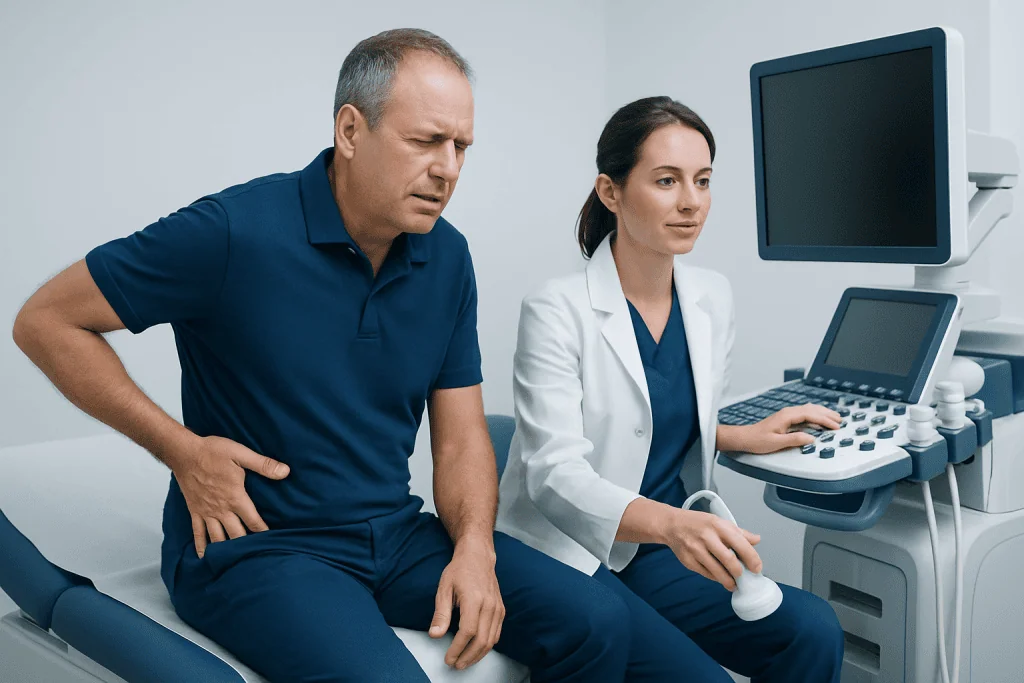
The classic symptom of a kidney stone is sudden, intense pain in the flank, which is the area on the side of the back below the ribs. This pain often comes in waves and may shift as the stone moves. It can travel toward the lower abdomen, groin, or, in men, the testicle. The pain may be difficult to relieve by changing position because it is caused by urinary tract irritation or blockage rather than muscle strain.
Other symptoms can include burning during urination, frequent or urgent urination, cloudy or foul-smelling urine, and visible blood in the urine. Sometimes blood is not visible but is found on a urine test. Nausea and vomiting may occur because the nerves serving the kidney and digestive tract are closely linked.
Not every kidney stone causes severe symptoms. A small stone inside the kidney may be discovered during imaging for another reason. However, symptoms are more likely when the stone enters the ureter, blocks urine flow, or is associated with infection. Fever, chills, weakness, or feeling very unwell may suggest infection and should be assessed promptly.
Causes and Risk Factors
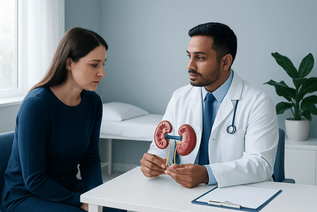
Kidney stones form when urine contains more stone-forming substances than it can dilute. Calcium, oxalate, uric acid, cystine, and phosphate can crystallize under certain conditions. Low urine volume is one of the most common contributors because concentrated urine allows minerals to come together more easily. This is why dehydration, hot climates, heavy sweating, or not drinking enough fluids can increase risk.
Diet and medical conditions can also influence stone formation. A high intake of salt can increase calcium in the urine, while high intake of animal protein may raise uric acid and lower citrate, a natural substance that helps prevent stones. Some people are advised to moderate high-oxalate foods, depending on their stone type and urine chemistry, but this should be individualized rather than overly restrictive.
Risk factors include a personal or family history of stones, recurrent urinary tract infections, obesity, gout, certain bowel diseases or surgeries, and some medications or supplements. Conditions that affect calcium or uric acid metabolism may also contribute. Because the causes differ from person to person, a prevention plan works best when it is based on test results and medical history.
- Low fluid intake or frequent dehydration
- High dietary sodium
- Previous kidney stones
- Family history of stones
- Recurrent urinary tract infections
- Metabolic or digestive conditions that change urine chemistry
Diagnosis: Urine Tests, Blood Tests, and Imaging
Diagnosis begins with a medical history, physical examination, and review of symptoms such as flank pain, urinary changes, nausea, or fever. A doctor may ask about previous stones, diet, fluid intake, medications, supplements, infections, and family history. This information helps determine which tests are most appropriate and whether urgent evaluation is needed.
Urine tests are central to kidney stone assessment. A urinalysis can check for blood, white blood cells, bacteria, crystals, urine pH, and other signs that help distinguish stone disease from infection or other urinary problems. If infection is suspected, a urine culture may be ordered. In people with recurrent stones or higher-risk features, a 24-hour urine collection may measure urine volume, calcium, oxalate, citrate, uric acid, sodium, and other factors that guide prevention.
Blood tests may evaluate kidney function, electrolytes, calcium, uric acid, and markers of infection when relevant. Imaging helps confirm whether a stone is present, where it is located, and whether it is causing blockage. Ultrasound is commonly used because it avoids radiation and can detect swelling of the kidney, while non-contrast CT is highly sensitive and may be used when diagnosis is uncertain or detailed planning is needed. The choice depends on the patient, symptoms, pregnancy status, previous imaging, and clinical urgency.
Treatment Options
Treatment depends on stone size, location, symptoms, kidney function, and whether infection or significant obstruction is present. Many small stones can pass naturally with time, adequate fluids, and pain control under medical guidance. A doctor may recommend medicines to manage pain, nausea, or urinary tract spasm, and in selected cases may use medication that helps relax the ureter to support stone passage.
If a stone is too large to pass, causes ongoing severe symptoms, blocks urine flow, or is associated with infection, a procedure may be needed. Common options include shock wave lithotripsy, which uses energy waves to break stones into smaller pieces, ureteroscopy, which uses a thin scope passed through the urinary tract to remove or fragment the stone, and percutaneous nephrolithotomy, which is usually reserved for larger or more complex kidney stones. The best approach is individualized.
Stone analysis is helpful when a stone is passed or removed. Knowing whether it is made of calcium oxalate, uric acid, struvite, cystine, or another material can guide long-term prevention. After treatment, follow-up imaging or urine testing may be advised to confirm clearance, check kidney drainage, and reduce the risk of future stones.
Prevention and Self-Care Strategies
Prevention often begins with increasing fluid intake so that urine stays pale and well diluted throughout the day. The exact amount needed varies with body size, activity level, climate, and medical conditions, so people with heart, kidney, or liver disease should ask their doctor how much fluid is safe. Water is usually the best choice, while frequent sugary drinks may not be ideal for overall health.
Diet changes can reduce risk for many people. A common recommendation is to limit excess salt because sodium can increase urinary calcium. Maintaining normal dietary calcium from foods, rather than severely restricting calcium, may help reduce oxalate absorption in the gut for many calcium oxalate stone formers. People may also be advised to moderate large amounts of animal protein, avoid excessive vitamin C supplements, and adjust high-oxalate foods if testing shows oxalate is a problem.
Prevention should be personalized. For example, uric acid stones may be related to acidic urine and high uric acid levels, while struvite stones are linked to certain infections and require infection-focused management. Some patients benefit from prescription medicines that change urine chemistry or reduce stone-forming substances. These decisions should be made with a qualified clinician after reviewing urine, blood, imaging, and stone analysis results.
- Drink fluids regularly rather than only when thirsty.
- Reduce excess salt in processed and restaurant foods.
- Keep a balanced intake of calcium-rich foods unless told otherwise.
- Discuss supplements with a doctor, especially calcium or vitamin C.
- Attend follow-up testing after a first stone or any recurrent stones.
When to See a Doctor
Medical evaluation is recommended for new flank pain, pain with blood in the urine, painful urination, repeated vomiting, or symptoms that do not improve. Prompt assessment is especially important if pain is accompanied by fever, chills, confusion, weakness, or difficulty passing urine. These features can indicate infection, significant blockage, or another condition that needs timely care.
People with one kidney, known kidney disease, pregnancy, recurrent urinary tract infections, or a history of complicated stones should seek care early when symptoms appear. Children with suspected stones should also be evaluated by a clinician, as their causes and prevention needs may differ from adults. Anyone who is passing a stone should follow medical advice about pain relief, hydration, and whether a strainer should be used to collect the stone for analysis.
International patients who need evaluation for kidney stones may benefit from coordinated care involving urology, nephrology, emergency medicine, radiology, and laboratory services. Acibadem International provides diagnosis and treatment for kidney stones within multidisciplinary teams and JCI-accredited hospitals, supporting patients through assessment, treatment planning, and follow-up.
Frequently asked questions
What does kidney stone pain usually feel like?
Kidney stone pain often starts suddenly in the flank, on one side of the back below the ribs. It may come in waves and move toward the lower abdomen or groin as the stone travels. Some people also have nausea, vomiting, blood in the urine, or urinary urgency.
Can a urine test diagnose kidney stones?
A urine test can strongly support the diagnosis by showing blood, crystals, infection signs, urine pH, and other findings. However, imaging is often needed to confirm the stone, identify its size and location, and check for blockage. In recurrent stones, a 24-hour urine test can guide prevention.
Do all kidney stones need surgery?
No. Many small stones pass on their own with appropriate pain control, fluids, and monitoring. Procedures are considered when a stone is large, unlikely to pass, causes ongoing obstruction or severe symptoms, or is linked to infection or kidney function concerns.
How can someone reduce the chance of another kidney stone?
The most common first step is drinking enough fluids to keep urine diluted. Many people also benefit from reducing excess salt, keeping a balanced calcium intake, and adjusting animal protein or oxalate intake based on test results. A doctor may recommend specific medication if urine studies show a clear metabolic risk.
Is blood in the urine always caused by a kidney stone?
No. Blood in the urine can occur with kidney stones, urinary tract infection, kidney disease, prostate conditions, trauma, or other urinary tract problems. Even if a stone is suspected, medical evaluation is important to confirm the cause and decide whether further testing is needed.
When is a kidney stone an emergency?
Urgent care is needed if flank pain occurs with fever, chills, repeated vomiting, confusion, severe weakness, inability to pass urine, or known kidney disease. A blocked urinary tract with infection requires prompt treatment. People with a single kidney or pregnancy should also seek medical attention quickly if stone symptoms develop.
References
- National Institute of Diabetes and Digestive and Kidney Diseases
- European Association of Urology
- American Urological Association
- Mayo Clinic
- Cleveland Clinic
This article is for general information only and is not a substitute for professional medical advice. Please consult a qualified doctor about your individual situation.
Persistent digestive symptoms? Get evaluated in Turkey
JCI-accredited · board-certified surgeons · reply within 24h
Add us as a Preferred Source to see more of our trusted health content across Google Search, AI Overviews and Discover.