Legg Calve Perthes Disease Xray: Preparation, Procedure and Results
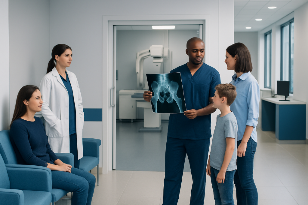
X-rays are a quick, painless way to examine the hip joint in suspected Perthes disease. Early X-rays can appear normal, so repeat X-rays or MRI may be needed when symptoms continue.
Key Takeaways
- X-rays are a quick, painless way to examine the hip joint in suspected Perthes disease.
- Early X-rays can appear normal, so repeat X-rays or MRI may be needed when symptoms continue.
- Typical changes include increased density, flattening, fragmentation, and later healing of the femoral head.
- Imaging results are interpreted alongside the child’s symptoms, physical examination, age, and hip range of motion.
- Treatment and recovery vary; regular follow-up imaging helps the care team protect hip shape and function.
A Legg Calve Perthes disease Xray is usually the first imaging test used when a child has a limp, hip pain, or reduced hip movement. It shows the shape and density of the femoral head and helps clinicians diagnose the condition, assess its stage, and follow hip development during treatment.
Legg Calve Perthes Disease Xray: Overview
Legg-Calvé-Perthes disease is a childhood hip condition in which the blood supply to the rounded top of the thigh bone, called the femoral head, is temporarily disrupted. The bone can weaken and change shape while the body gradually restores blood flow and rebuilds it. A Legg Calve Perthes disease Xray is a central part of evaluating this process.
The examination uses a small amount of ionizing radiation to create images of the pelvis and hips. It is fast, noninvasive, and does not require needles, contrast dye, or anesthesia in most children. Clinicians use X-rays to look for features of Perthes disease, estimate the extent of femoral-head involvement, and compare changes at follow-up visits.
Because the earliest stage may not yet cause visible bone changes, a normal first X-ray does not always rule out Perthes disease. If the child’s symptoms and examination remain suggestive, an orthopedic clinician may recommend repeat X-rays after an interval or magnetic resonance imaging (MRI), which can detect changes in bone marrow and blood supply earlier.
How the X-ray Works and Who May Need It
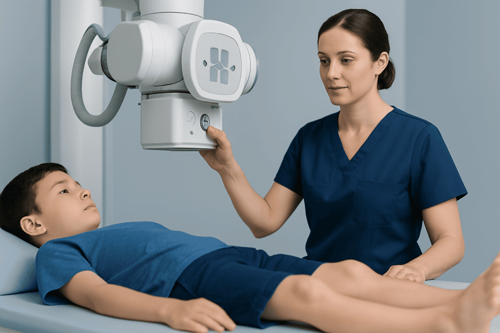
An X-ray beam passes through the body and is captured by a detector. Dense structures such as bone absorb more radiation and appear lighter on the image, while soft tissues appear in shades of gray. For a suspected hip disorder, images commonly include a front-facing view of the pelvis and additional views that show each hip from a different angle.
A child may be considered for hip X-rays when there is an unexplained limp, activity-related pain in the hip, groin, thigh, or knee, limited hip motion, or a leg that seems to turn outward. Symptoms can be subtle, and some children have little pain despite a noticeable limp. The test may also be used after diagnosis to monitor healing and guide decisions about activity, physiotherapy, bracing, or surgery.
Clinical assessment remains important. A clinician will ask about symptom onset, recent injuries or illness, activity limitations, and general health. They will also assess walking pattern, leg length, and hip movement. This combination of history, examination, and imaging helps distinguish Perthes disease from other causes of hip pain or limping in children.
Preparing for and Having a Legg Calve Perthes Disease Xray
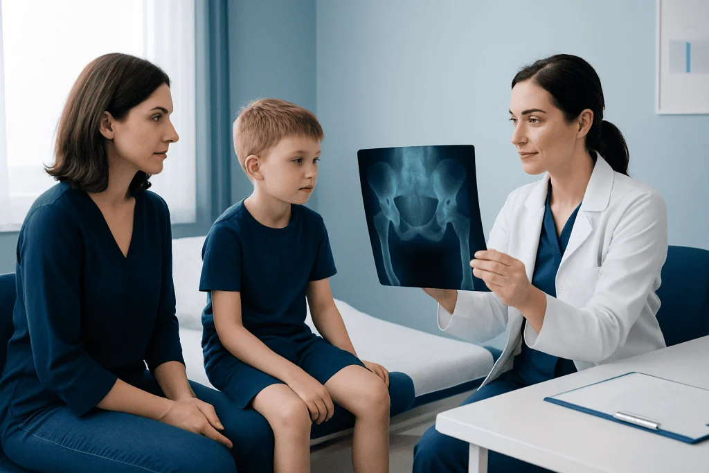
There is usually very little preparation. The child can generally eat, drink, and take usual medicines unless the healthcare team has given different instructions. A parent or caregiver should tell the imaging staff about any possibility of pregnancy in an adolescent patient and about any prior imaging that may be useful for comparison.
Before the images are taken, the child may be asked to change into a gown and remove metal objects around the waist or pelvis, such as belts, jewelry, or clothing with metal fasteners. A radiographer positions the child on an X-ray table or standing platform, depending on the views needed. Parents can often remain nearby to help the child feel comfortable, following the imaging department’s safety guidance.
The child will need to stay still for a few seconds while each image is made. Positioning can be uncomfortable if hip movement is painful, but the procedure itself should not hurt. The imaging portion commonly takes only a few minutes, although the overall visit may take longer for registration, positioning, and review of image quality.
X-rays use the lowest radiation exposure reasonably needed to obtain useful images, especially in children. The expected benefit of accurately evaluating a potentially important hip condition generally outweighs the small radiation risk. Families can ask the radiology team any questions they have about safety and the planned views.
What Are the Radiographic Features of Perthes Disease?
The radiographic features of Perthes disease evolve over time. In the very early phase, an X-ray may be normal or show subtle widening of the hip joint space. As the condition progresses, the femoral head may look denser or whiter than the other side because weakened bone is being resorbed and reshaped.
Later, the femoral head can develop areas of fragmentation, where the bone appears irregular or divided into denser and less dense portions. Flattening, widening, loss of the normally round contour, and changes in the growth region beneath the femoral head may also be seen. The hip socket may change as it responds to the altered shape of the femoral head.
During reossification and healing, new bone forms and the femoral head gradually remodels. Follow-up images help the orthopedic team assess whether the femoral head remains well contained within the hip socket and whether its shape is likely to support good long-term hip function. Clinicians may use recognized classification systems, but these are only one part of individualized decision-making.
MRI may be helpful when plain films are normal but clinical concern remains, or when more detailed assessment is needed. Ultrasound can sometimes identify fluid in the hip joint, but it does not provide the same information about femoral-head bone structure as X-ray or MRI.
What Are the Clinical Findings of Legg-Calvé-Perthes Disease?
The most common clinical finding is a limp that develops gradually. It may become more obvious after physical activity and can be painless or accompanied by discomfort in the hip, groin, thigh, or knee. Knee pain in a child can sometimes originate from the hip, so a hip assessment is important even when the knee seems to be the main area of concern.
On examination, clinicians often find reduced hip movement, especially internal rotation and abduction, which is moving the leg away from the body. The child may hold the leg in a slightly outward-turned position or have muscle tightening around the hip. Some children show a difference in leg length as the hip changes or as the pelvis tilts during walking.
General illness symptoms such as high fever, marked fatigue, or a very unwell appearance are not typical of Perthes disease and need prompt medical assessment for other possible conditions. Blood tests are not used to diagnose Perthes disease directly, but they may be considered when the care team needs to exclude infection, inflammatory disease, or another explanation for the child’s symptoms.
Results, Treatment Decisions and Recovery Timeline
A radiologist interprets the images, and the treating clinician explains what the results mean in the context of the child’s examination and symptoms. If Perthes disease is diagnosed or strongly suspected, referral to a pediatric orthopedic specialist is common. The goals are to preserve hip movement, keep the femoral head as well positioned in the socket as possible, manage symptoms, and monitor growth.
Management may include activity adjustment, pain relief recommended by a clinician, physiotherapy to maintain range of motion, and periodic clinical and imaging reviews. Some children may need bracing, casting, traction in selected circumstances, or surgery when age, hip movement, or the extent of femoral-head involvement suggests that additional containment is needed. Treatment planning is individualized and may involve orthopedic surgery when non-surgical measures are not sufficient.
How long does it take to recover from Legg-Perthes disease? The active disease and remodeling process commonly take several years, often around two to four years, although the course can be shorter or longer. A child’s age at onset, hip shape, range of motion, and degree of femoral-head involvement all influence recovery. Even after the bone has healed, periodic follow-up may be advised to assess hip function and development.
The benefits of regular imaging include earlier recognition of changes that could alter the treatment plan and a clearer picture of healing over time. The main limitation is that X-ray cannot show every early change and involves low-dose radiation. MRI may add detail when needed, while avoiding ionizing radiation, though it takes longer and some younger children may need support to remain still.
What Are the Nursing Interventions for Legg-Calvé-Perthes Disease?
Nursing care focuses on comfort, mobility, education, and helping the child and family follow the orthopedic care plan. Nurses may assess pain, walking ability, sleep, medication effects, skin condition if a brace or cast is used, and the child’s ability to manage daily activities. They can also reinforce safe use of crutches, walkers, wheelchairs, or other prescribed mobility supports.
Education is an important intervention. Families may need practical guidance on activity restrictions, school participation, physiotherapy appointments, positioning, and when to contact the clinical team. Encouraging age-appropriate involvement can help a child understand the plan and express concerns about pain, mobility, sports, or social activities.
When braces, casts, or surgery are part of care, nurses monitor for pressure areas, swelling, numbness, unusual pain, fever, wound concerns, or changes in circulation. They also support rehabilitation and coordinate with orthopedic clinicians, physiotherapists, and school services where appropriate. Care plans should be adapted to the child’s needs rather than based on a single standard pathway.
Acibadem International’s multidisciplinary specialists and JCI-accredited hospitals support diagnosis, imaging, orthopedic treatment, and rehabilitation planning for international patients with childhood hip conditions.
When to Seek Medical Care
A child with a new limp that lasts more than a few days, recurring hip or knee pain, reduced hip movement, or difficulty participating in usual activities should be assessed by a qualified healthcare professional. Earlier evaluation can help identify the cause and determine whether imaging is appropriate.
Urgent medical assessment is needed if a child cannot bear weight, has severe or rapidly worsening pain, fever, looks unwell, has a red or swollen joint, or develops symptoms after a significant injury. These signs can occur with conditions other than Perthes disease that require prompt care.
Families should keep scheduled orthopedic and imaging follow-up appointments if Perthes disease is diagnosed. Contact the treating team sooner if pain changes significantly, a mobility device or brace is causing problems, or the child has new symptoms. Ongoing communication helps the care team adjust support as the child grows and heals.
Frequently asked questions
Can an X-ray diagnose Legg-Calvé-Perthes disease?
An X-ray is commonly the first imaging test used to assess suspected Legg-Calvé-Perthes disease. It can show characteristic bone changes and help monitor progression, but early X-rays may be normal. If symptoms persist despite normal films, the clinician may recommend repeat X-rays or an MRI.
Does a Legg Calve Perthes disease Xray hurt?
The X-ray itself is painless and does not involve injections. Some positions may be briefly uncomfortable if the child has hip pain or restricted movement. The radiographer can help position the child as gently and safely as possible.
How should a child prepare for a hip X-ray?
Most children do not need special preparation and can eat and drink normally. They may need to remove clothing or metal items around the pelvis and wear a gown. Bringing a familiar toy or having a caregiver nearby may help a young child remain calm and still.
What happens if the first X-ray is normal?
A normal early X-ray does not always exclude Perthes disease because visible bone changes can take time to develop. The clinician will consider the child’s symptoms and physical examination. Repeat imaging or MRI may be recommended if concern remains.
How often are follow-up X-rays needed in Perthes disease?
The schedule varies according to the child’s age, disease stage, symptoms, and treatment plan. Follow-up images may be taken at intervals to assess femoral-head shape, containment in the hip socket, and healing. The orthopedic specialist will recommend an individualized imaging schedule.
Will a child with Perthes disease need surgery?
Not every child needs surgery. Many are managed with observation, activity modification, physiotherapy, and regular follow-up, while others may benefit from procedures that improve hip containment. The decision depends on factors such as age, hip movement, and the pattern and severity of femoral-head involvement.
References
- American Academy of Orthopaedic Surgeons
- OrthoInfo
- National Institute of Arthritis and Musculoskeletal and Skin Diseases
- RadiologyInfo.org
- Royal College of Radiologists
This article is for general information only and is not a substitute for professional medical advice. Please consult a qualified doctor about your individual situation.
Joint and spine care in Turkey — expert assessment & treatment
JCI-accredited · board-certified surgeons · reply within 24h
Add us as a Preferred Source to see more of our trusted health content across Google Search, AI Overviews and Discover.