Lid Laceration Repair: Procedure, Recovery and Results

Any eyelid cut should be assessed promptly because apparently small wounds can involve important structures. Repair may be completed with local anesthetic, sedation or general anesthesia depending on age, injury complexity and comfort needs.
Key Takeaways
- Any eyelid cut should be assessed promptly because apparently small wounds can involve important structures.
- Repair may be completed with local anesthetic, sedation or general anesthesia depending on age, injury complexity and comfort needs.
- Accurate alignment of the eyelid edge is important for blinking, eye protection and cosmetic healing.
- Most uncomplicated wounds improve substantially over several weeks, while scar maturation can take months.
- Urgent care is needed for vision changes, a deep wound, an eyelid-edge cut, uncontrolled bleeding or possible foreign material in the eye.
Lid laceration repair is a procedure used to carefully close a cut in the eyelid and restore its protective position against the eye. The approach depends on the depth and location of the injury, especially whether the eyelid edge, tear drainage system, eye muscles or eyeball may be involved.
Overview: What Is Lid Laceration Repair?
Lid laceration repair is the careful cleaning and closure of a tear or cut in the upper or lower eyelid. Its aims are to protect the surface of the eye, restore normal blinking and eyelid position, reduce the chance of infection, and support the best possible scar outcome. In many cases, the wound can be repaired with fine sutures after a detailed eye examination.
Eyelid injuries may occur after falls, sports injuries, traffic accidents, work-related trauma, animal scratches or contact with sharp objects. Although some superficial cuts can be treated simply, eyelid tissue is delicate and contains structures that are essential for eyelid movement and tear drainage. Cuts involving the lid margin, the inner corner of the eye, deeper tissues or the tear duct usually need assessment by an ophthalmologist, oculoplastic surgeon or an appropriately trained specialist.
The timing and method of repair are individualized. A clinician first makes sure the eye itself is safe, checks vision and eye movement, and looks for retained material or injury around the orbit. Treatment is not only about closing the skin; it is about restoring the eyelid as a functioning protective structure.
When to Seek Medical Care
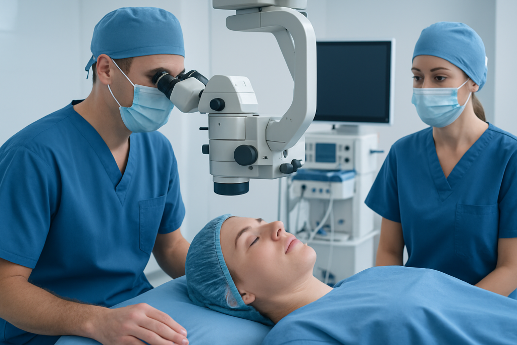
Medical assessment should be sought promptly for any eyelid laceration, particularly in children or after a high-impact injury. It is important not to rub the eye, pull on tissue, attempt to remove embedded objects, or use adhesive strips or glue near the eye unless a clinician has advised it. If an object is stuck in the eye or eyelid, it should be left in place and urgent care should be arranged.
Emergency evaluation is especially important if there is blurred, reduced or double vision; severe eye pain; inability to open or close the eye; bleeding that does not stop with gentle pressure to the surrounding area; a cut through the eyelid edge; visible fat or deeper tissue; loss of sensation; or a suspected penetrating injury. A wound near the inner corner of the eye can affect the tear drainage channels and should be assessed urgently.
Before review, a clean dressing can be placed lightly over the closed eye if tolerated. Food and drink may need to be avoided if a procedure under sedation or general anesthesia is possible; local emergency guidance should be followed. Tetanus vaccination status may also be reviewed, particularly after contaminated injuries.
How the Procedure Works and Who May Need It

Lid laceration repair may be appropriate when an eyelid cut is gaping, deep, contaminated, poorly aligned, or located where healing without careful closure could affect eyelid function. Simple superficial lacerations may sometimes be closed by an emergency clinician. More complex wounds generally require specialist repair, especially those involving the eyelid margin, the muscle that lifts the upper lid, the canaliculi that drain tears, tissue loss, bite wounds, or possible injury to the globe.
Assessment usually includes checking visual acuity, pupils, eye movement, corneal integrity and the position of the eyelid. The clinician may turn the eyelid gently to inspect its inner surface. Imaging may be considered when there is concern about an orbital fracture, a foreign body or deeper facial injury. If the eye is injured, protecting vision takes priority over eyelid closure.
Local anesthetic is often suitable for cooperative adults with straightforward injuries. Children, very anxious patients and people with extensive or complex wounds may need sedation or general anesthesia. The team also considers allergies, current medicines, bleeding risk, medical conditions and the mechanism of injury when planning care.
Step-by-Step: What Happens During Lid Laceration Repair
The area is first examined and gently irrigated to remove blood, dirt and small particles. Numbing medicine is applied or injected when appropriate. The skin around the wound is cleaned carefully while protecting the eye. If there is concern that the eyeball has been penetrated, pressure on the eye is avoided and urgent ophthalmic management is arranged.
The surgeon then identifies each injured layer and aligns it precisely. For an eyelid-edge wound, the lash line and lid margin require meticulous positioning so that the lid sits smoothly against the eye after healing. Deeper injuries may need repair of muscle, supportive tissue or the tear drainage channels before the skin is closed. Very fine absorbable or removable sutures are commonly used, and some tear-duct repairs require a temporary silicone stent.
After closure, an antibiotic ointment or protective dressing may be used. The patient receives instructions about wound cleaning, eye protection, medicines, follow-up and suture removal if non-absorbable stitches were placed. Clinical documentation records the wound location, dimensions, depth, involved structures, contamination, examination findings, anesthesia, repair layers and closure material. Terms such as lid laceration repair CPT or lid laceration repair CPT code relate to billing classification and are selected by the treating service according to the documented complexity; they do not describe whether an injury is medically serious.
Recovery Timeline, Benefits and Possible Risks
Swelling, bruising, tenderness, mild tearing and temporary blurred vision from ointment are common during the first days after repair. Cold compresses may help reduce swelling when recommended by the treating team, but direct pressure on the eye should be avoided. Patients are usually advised to keep the wound clean, use prescribed ointment as directed, avoid rubbing the eye, and limit activities that could cause another injury while the wound is healing.
In an uncomplicated injury, the skin typically seals within about one to two weeks. Bruising and visible swelling often improve noticeably over the first two to three weeks, while the scar may remain pink, firm or slightly uneven for several months as it matures. Follow-up is important because sutures may need removal and the clinician needs to confirm that the lid closes well, turns neither inward nor outward, and does not irritate the eye.
Benefits of repair include restoration of eyelid contour, better protection of the cornea, reduced wound separation and improved appearance as healing progresses. Possible complications include infection, bleeding, scarring, eyelid notching, asymmetry, persistent tearing, dry eye symptoms, ingrown lashes, reduced lid movement or the need for later revision. These risks are higher with severe trauma, delayed treatment, tissue loss, contamination or associated eye injury, but timely specialist assessment helps identify and address them.
How Long Does It Take for Eyelid Laceration to Heal?
Most uncomplicated eyelid lacerations heal enough for the skin to close within one to two weeks, although the exact timing depends on the cut’s depth, location, cleanliness and repair method. A laceration involving the eyelid margin, deeper muscle layers or the tear drainage system may require longer follow-up and can take more time to settle fully.
Early healing does not mean the final result is already visible. Redness, firmness and mild unevenness of the scar often soften gradually over several months. Protecting the area from further injury, attending follow-up visits and following the clinician’s aftercare instructions can support recovery.
Contact the care team sooner if redness spreads, pain worsens rather than improves, pus-like drainage develops, fever occurs, the wound opens, vision changes, or the eyelid no longer closes normally. These symptoms do not always indicate a serious problem, but they should be checked promptly.
How Painful Is Eyelid Surgery Recovery?
Recovery after eyelid laceration repair is usually described as soreness, tightness or irritation rather than severe pain. Local anesthetic prevents pain during many repairs, and discomfort afterward is commonly managed with the pain-relief plan recommended by the clinician. Some people experience stinging from the wound or temporary blurred vision after ointment is applied.
Severe eye pain, increasing pain, pain with vision loss, marked light sensitivity or nausea after an eye injury is not expected and needs urgent assessment. These symptoms may reflect a problem involving the eye rather than normal wound healing.
Patients should not use medicines, eye drops or herbal products near the eye unless their clinician has confirmed they are appropriate. Certain medicines can increase bleeding risk or may not be suitable for an individual medical history, so personalized advice is important.
How Long Does It Take to Look Normal After Upper Eyelid Surgery?
After upper eyelid laceration repair, many people look socially presentable once bruising and swelling have improved, often within two to three weeks. However, a wound caused by trauma can vary considerably from planned cosmetic eyelid surgery because there may be bruising, irregular tissue damage or associated facial injury.
The eyelid’s final contour and scar appearance can continue to improve for several months. A scar may initially look red or raised before gradually fading and flattening. If the injury involved the eyelid edge or deeper structures, the treating specialist may monitor healing over a longer period before deciding whether any additional treatment is needed.
Cosmetics, scar products and sun exposure should be discussed with the care team. Products should not be applied to an unhealed wound, and sun protection around a healed scar can help limit persistent darkening in some people.
Can Damaged Eyelids Heal?
Damaged eyelids can often heal well, especially when the eye and critical eyelid structures are assessed early and the wound is repaired accurately. The eyelids have a good blood supply, which supports healing, but their complex anatomy means even a small misalignment can affect blinking, comfort or tear flow.
Some injuries need more than simple stitches. Tissue loss, nerve or muscle damage, eyelid-edge disruption, tear-duct injury and scarring may require specialized reconstructive care. The outlook depends on the severity of the original trauma and whether the cornea, globe or surrounding facial bones were also affected.
For international patients needing coordinated assessment, Acibadem International’s multidisciplinary specialists and JCI-accredited hospitals diagnose and treat eyelid injuries and related eye conditions. Follow-up with a qualified eye-care professional remains important to monitor vision, lid position and scar healing over time.
Frequently asked questions
Do all eyelid cuts need stitches?
No. Very small, superficial cuts may not require sutures, but every eyelid cut should be assessed carefully because deeper injury can be difficult to recognize from the outside. A clinician can determine whether closure, specialist referral or observation is most appropriate.
Can an eyelid laceration affect vision?
The cut itself may not affect vision, but the same injury can damage the cornea, eyeball, eye muscles or surrounding orbit. Any blurred vision, double vision, reduced vision, severe pain or light sensitivity after an eyelid injury requires urgent evaluation.
When are stitches removed after eyelid laceration repair?
If removable skin sutures are used, they are often removed relatively early because eyelid skin heals quickly. The precise timing depends on the wound, suture type and clinician's assessment, so patients should follow their individual follow-up plan.
Will an eyelid laceration leave a scar?
Most lacerations leave some degree of scar, though eyelid scars often become less noticeable as they mature. Precise repair, good wound care and avoiding unnecessary tension or rubbing can support the best possible result.
What should be avoided after eyelid laceration repair?
Patients are commonly advised to avoid rubbing the eye, swimming until cleared, strenuous activity that increases swelling, and activities with a risk of another facial injury. They should also avoid applying makeup or non-prescribed products to an unhealed wound.
What information belongs in a laceration repair procedure template?
A clinical laceration repair procedure template or laceration repair documentation example generally records examination findings, consent, anesthesia, irrigation, wound location and measurements, tissue layers involved, closure technique, suture material, complications and aftercare. A laceration repair PDF may be used by clinicians for documentation, but patients should rely on their treating team's personalized discharge instructions.
References
- American Academy of Ophthalmology
- American Society of Ophthalmic Plastic and Reconstructive Surgery
- Merck Manual Professional Edition
- Mayo Clinic
This article is for general information only and is not a substitute for professional medical advice. Please consult a qualified doctor about your individual situation.
Explore treatments in Turkey — costs, top hospitals & a free quote
JCI-accredited · board-certified surgeons · reply within 24h
Add us as a Preferred Source to see more of our trusted health content across Google Search, AI Overviews and Discover.

More from the Health Library



Related Specialists

Prof. Dr. Taner Yavuz
Pediatric Cardiology
Dr. Destina Suay Sarı
Oral & Dental Health
Dt. Cüneyt Işıker
Oral & Dental Health
Assoc. Prof. Dr. Burak Özturan
Orthopedic Surgery & Traumatology