

Liver Hemangioma MRI: Preparation, Procedure and Results
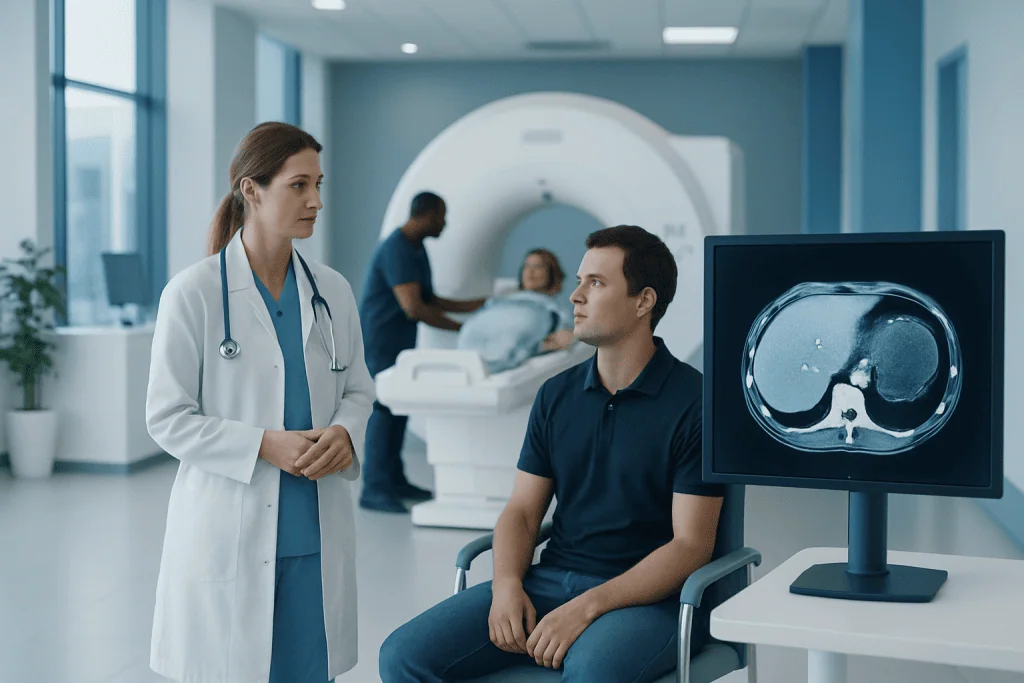
MRI is one of the most useful tests for confirming a suspected liver hemangioma without surgery or biopsy. A contrast-enhanced MRI can show the characteristic way a hemangioma fills with contrast over time.
Key Takeaways
- MRI is one of the most useful tests for confirming a suspected liver hemangioma without surgery or biopsy.
- A contrast-enhanced MRI can show the characteristic way a hemangioma fills with contrast over time.
- Most liver hemangiomas cause no symptoms, do not become cancer, and need observation rather than treatment.
- Preparation is usually simple, but people should tell the imaging team about kidney disease, pregnancy, implants, allergies, and anxiety in enclosed spaces.
- Liver surgery is uncommon and is generally considered only for selected large, symptomatic, or uncertain lesions.
Liver hemangioma MRI is a noninvasive imaging examination that can often identify a liver hemangioma by its characteristic appearance, especially when contrast material is used. Most liver hemangiomas are harmless and do not require treatment, but MRI helps clinicians distinguish them from other liver findings and decide whether follow-up is needed.
Overview: what a liver hemangioma MRI shows
A liver hemangioma MRI is a scan that uses a powerful magnet and radio waves to create detailed pictures of the liver. A hemangioma is a cluster of enlarged blood vessels within the liver. It is a benign, or non-cancerous, finding and is often discovered incidentally during imaging performed for another reason.
MRI is particularly helpful because it can assess the lesion’s fluid and blood-flow characteristics without using ionizing radiation. In many people, the image pattern is sufficiently typical to support a confident diagnosis and avoid invasive testing. A radiologist interprets the scan alongside the person’s symptoms, medical history, laboratory results, and any previous liver imaging.
Most hemangiomas do not affect liver function and do not need treatment. The aim of MRI is usually clarification rather than looking for an emergency problem: it helps determine whether the finding has the features expected of a hemangioma or whether additional assessment is appropriate.
How does MRI diagnose a liver hemangioma?
On MRI, a typical hemangioma usually appears darker than normal liver on some image sequences and very bright on fluid-sensitive sequences. After intravenous contrast is given, many hemangiomas show a distinctive pattern: contrast first appears at the outer edge in small nodules, then gradually moves inward on later images. This is often called peripheral nodular enhancement with progressive fill-in.
The radiologist also considers the lesion’s size, outline, location, and behavior across multiple image sequences. Comparing the MRI with an earlier ultrasound or CT scan can be useful, particularly if the finding is not entirely typical or if there is a history of liver disease or cancer.
Not every hemangioma has the classic appearance. Very small lesions, lesions with internal scarring or clotting, and hemangiomas in a liver affected by chronic disease may require more careful interpretation. In these situations, a specialist may recommend follow-up imaging or another test rather than drawing conclusions from a single image.
How accurate is MRI for liver hemangioma?
Contrast-enhanced MRI is highly accurate for liver hemangioma when the lesion has its classic imaging features. It provides excellent soft-tissue detail and captures how the lesion behaves at several points after contrast injection, which helps distinguish a typical hemangioma from many other focal liver lesions.
Accuracy depends on image quality, the MRI protocol, the size and appearance of the lesion, and the clinical setting. Interpretation may be more complex in people with cirrhosis, known cancer elsewhere in the body, multiple liver lesions, or an atypical imaging pattern. In these cases, radiologists may recommend comparison with earlier scans, short-term follow-up, contrast-enhanced ultrasound, CT, or consultation with a liver specialist.
Biopsy is not routinely used to diagnose a typical hemangioma because it can cause bleeding and is usually unnecessary when imaging is conclusive. Decisions about further testing should be individualized by a qualified clinician.
What is the MRI hemangioma protocol?
An MRI hemangioma protocol is a planned set of image sequences designed to characterize a liver lesion. The examination usually includes images obtained before contrast, fluid-sensitive sequences, in-phase and out-of-phase sequences that evaluate fat, diffusion-weighted imaging, and several contrast-enhanced phases.
If contrast is appropriate, images are commonly taken during the arterial phase shortly after injection, followed by portal venous, delayed, and sometimes later phases. These timed images allow the radiologist to observe the gradual enhancement pattern that can support the diagnosis of hemangioma. The exact protocol varies between imaging centers and may be adjusted for the person’s kidney function, clinical question, and prior scan findings.
Gadolinium-based contrast agents are widely used in MRI and are generally well tolerated. The imaging team will check for relevant kidney problems, previous contrast reactions, pregnancy, and other safety considerations. For some people, a non-contrast MRI can still provide useful information, although contrast may improve confidence in characterizing the lesion.
Preparation and step-by-step MRI procedure
Preparation is usually straightforward. The imaging center may ask the person not to eat for several hours before a liver MRI, particularly if contrast will be used; local instructions should always be followed. It is important to report pacemakers, implanted devices, metal fragments, cochlear implants, medication patches, kidney disease, pregnancy, breastfeeding, and any prior reaction to contrast material.
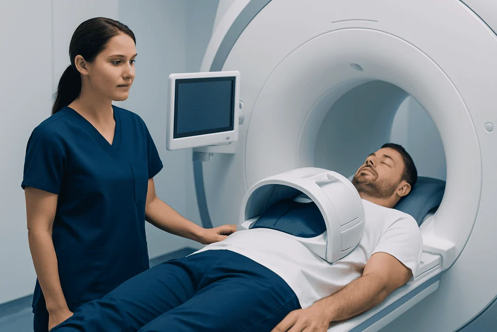
Before the scan, the person removes metal objects and changes into appropriate clothing. A small intravenous line may be placed in the arm for contrast. They lie on a moving table that slides into the MRI scanner, and the technologist provides ear protection because the machine makes loud tapping and knocking sounds.
During the scan, the person needs to remain still and may be asked to hold their breath briefly while pictures are taken. Contrast, if used, is injected through the IV during the examination. The scan commonly takes about 30 to 60 minutes, although timing varies with the protocol and whether additional images are needed.
MRI is painless, but some people find the enclosed space uncomfortable. The team can maintain communication throughout the examination and may discuss practical support or, when appropriate, medication for significant claustrophobia in advance. Anyone receiving a sedative will need arrangements for safe travel home.
Benefits, risks and understanding the results
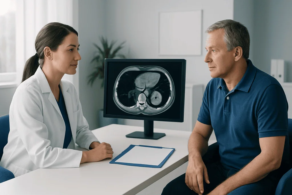
The main benefit of liver hemangioma MRI is detailed, radiation-free imaging that can often provide a clear diagnosis without an operation or tissue sample. It can also establish a useful baseline if a clinician recommends later comparison. Results are typically sent to the referring clinician, who explains what the finding means in the context of the individual’s health.
MRI has some limitations and risks. The strong magnetic field means that certain implanted or retained metal devices may make MRI unsuitable or require special precautions. Contrast reactions are uncommon, but they can occur. Gadolinium contrast is used cautiously in people with severe kidney impairment, and the radiology team assesses safety before the examination.
A report may describe a lesion as a typical hemangioma, probable hemangioma, or indeterminate lesion. A typical hemangioma often needs no further action. When results are uncertain, this does not automatically mean cancer; it means the images do not yet provide enough certainty, and the clinician may recommend targeted follow-up or specialist review.
How serious is a hemangioma on the liver?
For most people, a hemangioma on the liver is not serious. It is benign, does not usually turn into cancer, and commonly remains stable without causing symptoms. Many people learn about it only after an ultrasound, CT scan, or MRI performed for an unrelated concern.
Occasionally, a larger hemangioma may be associated with a feeling of fullness, upper abdominal discomfort, nausea, or pressure, although these symptoms can also have many other causes. A clinician should assess whether symptoms truly relate to the hemangioma before treatment is considered.
Complications such as rupture, significant bleeding, or effects on nearby structures are very unusual. Management is therefore usually conservative, with reassurance and selective imaging follow-up rather than routine intervention. Individual guidance may differ for a very large lesion, changing symptoms, uncertain diagnosis, or specific medical circumstances.
Treatment, recovery and when to seek medical care
Most liver hemangiomas do not require treatment. When treatment is considered, it is generally because a lesion is causing persistent, clearly related symptoms, has an uncertain diagnosis despite appropriate imaging, or is associated with rare complications. Options can include observation, selected image-guided approaches, or surgery, depending on the lesion and the person’s overall health.
How long does it take to recover from liver hemangioma surgery? Recovery varies according to the type and extent of surgery, whether it is performed through keyhole or open techniques, and the person’s general health. Hospital recovery may take several days, while return to usual energy levels and activities often takes weeks; open liver surgery generally requires a longer recovery than minimally invasive surgery. The surgical team provides individualized activity, wound-care, pain-management, and follow-up advice.
When to seek medical care: A person should contact a clinician for persistent or worsening upper abdominal pain, ongoing fullness, unexplained weight loss, jaundice, fever, vomiting, or a new concern after liver imaging. Sudden severe abdominal pain, fainting, marked weakness, or signs of significant illness warrant urgent medical assessment, although these symptoms are rarely caused by a hemangioma.
Acibadem International’s multidisciplinary specialists and JCI-accredited hospitals can assess liver lesions and coordinate imaging, gastroenterology, radiology, and surgical care for international patients when needed.
Frequently asked questions
Do I need contrast for a liver hemangioma MRI?
Contrast is often helpful because it shows the blood-flow pattern of a liver lesion over time. This pattern can make a hemangioma easier to identify with confidence. Whether contrast is appropriate depends on the clinical question and factors such as kidney health and prior reactions.
Do I need to fast before a liver MRI?
Many centers ask patients to avoid eating for several hours before a liver MRI, especially if contrast will be given. Instructions vary by facility and by the type of examination. The person should follow the directions provided by the imaging center.
Can a liver hemangioma become cancer?
A typical liver hemangioma is benign and is not considered a precancerous lesion. It does not usually transform into liver cancer. If imaging features are not typical, further evaluation may be advised to confirm the diagnosis.
Will a liver hemangioma need regular MRI scans?
A clearly typical, small, asymptomatic hemangioma often does not need repeated imaging. Follow-up may be considered if the lesion is large, has an atypical appearance, symptoms are present, or there is uncertainty on the initial study. The appropriate plan should be decided with the treating clinician.
Can I go home after a liver hemangioma MRI?
Yes. Most people can return home and resume usual activities immediately after an MRI. If sedation was used for anxiety or claustrophobia, the person will need someone else to drive them and should follow the sedation instructions.
What happens if MRI findings are inconclusive?
An inconclusive result means that the images do not show enough characteristic features to make a definite diagnosis. A clinician may recommend reviewing previous scans, repeating imaging after an interval, using another imaging method, or seeking input from liver and radiology specialists. Further testing is chosen to clarify the finding while avoiding unnecessary invasive procedures.
References
- American College of Radiology
- American College of Gastroenterology
- European Society of Gastrointestinal and Abdominal Radiology
- National Institute of Diabetes and Digestive and Kidney Diseases
This article is for general information only and is not a substitute for professional medical advice. Please consult a qualified doctor about your individual situation.
Explore treatments in Turkey — costs, top hospitals & a free quote
JCI-accredited · board-certified surgeons · reply within 24h
Add us as a Preferred Source to see more of our trusted health content across Google Search, AI Overviews and Discover.

More from the Health Library



Related Specialists

Prof. Dr. Zeynep Güven
Physical Medicine & Rehabilitation
Dr. Ahmet Bülent Demirbağ
Otorhinolaryngology
Prof. Dr. Aleks Değirmencioğlu
Cardiology
Prof. Dr. Halil Koyuncu
Physical Medicine & Rehabilitation