Liver Segments CT Scan: Preparation, Procedure and Results
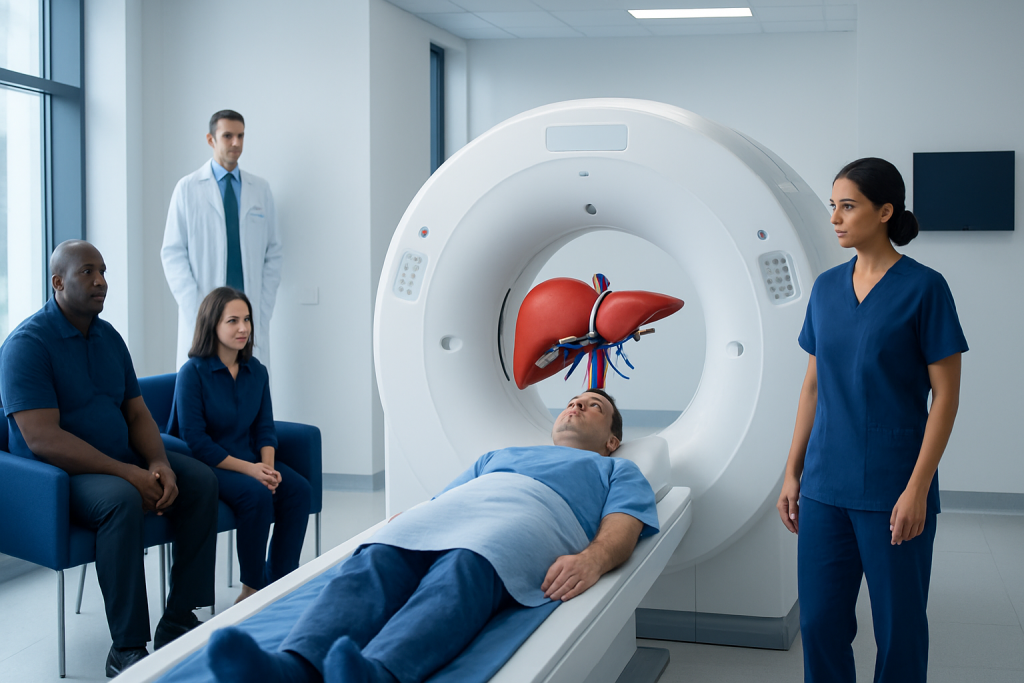
A liver segments CT scan creates cross-sectional images that map the liver into functionally important anatomical regions. Contrast material is often used to show liver arteries, veins, bile ducts and areas that differ from normal liver tissue.
Key Takeaways
- A liver segments CT scan creates cross-sectional images that map the liver into functionally important anatomical regions.
- Contrast material is often used to show liver arteries, veins, bile ducts and areas that differ from normal liver tissue.
- Preparation may include fasting for several hours and discussing allergies, kidney disease, pregnancy and medicines with the imaging team.
- The scan itself is usually quick, while the full appointment may take longer for preparation and contrast administration.
- A radiologist interprets the images in the context of symptoms, blood tests and other investigations; a scan result alone may not establish a diagnosis.
A liver segments CT scan is a detailed computed tomography examination that shows the liver in anatomical sections based on its blood vessels. It can help clinicians assess liver findings, evaluate injuries or disease, and plan surgery or other treatments with greater precision.
Overview: What Is a Liver Segments CT Scan?
A liver segments CT scan is a computed tomography examination designed to show the liver in detailed cross-sections and identify its functional segments. These segments are defined mainly by the branches of the portal vein, hepatic veins and bile ducts rather than by the outer shape of the liver. This detailed map helps clinicians describe where an abnormality is located and understand its relationship to important blood vessels.
CT uses rotating X-ray equipment and computer processing to produce images from many angles. In many liver examinations, an iodine-based contrast material is injected into a vein. Images are then obtained at carefully timed phases as contrast travels through the blood vessels and liver tissue. This can make certain lesions, inflammation, vascular changes or injuries easier to recognize.
The test may be requested after an ultrasound, abnormal liver blood tests, abdominal symptoms, trauma, or a previously identified liver finding. It may also support planning for liver surgery, ablation, embolization or other treatment. A liver segments CT scan is an imaging assessment, not a treatment, and its findings are considered alongside the person’s medical history and other results.
How the Scan Works and What It Can Show
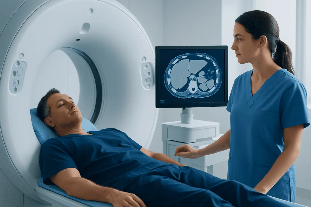
The liver receives blood from both the hepatic artery and portal vein, and it drains through hepatic veins. A multiphase CT study can capture these pathways at different times after contrast injection. Common image phases include an arterial phase, a portal venous phase and, when needed, a delayed phase. Each phase provides different information about blood flow and the appearance of liver tissue.
By displaying the vessels and their branches, CT software and radiologists can identify the liver segment containing a lesion. This is important because two abnormalities of a similar size may require different approaches depending on whether they are close to major vessels, bile ducts or the liver capsule. Three-dimensional reconstructions may be used when detailed surgical or interventional planning is needed.
CT may help assess focal liver lesions, fatty change, cirrhosis-related structural changes, infection, abscesses, cysts, bile duct dilatation and traumatic injury. It can also evaluate nearby organs and lymph nodes. Some findings need further characterization with magnetic resonance imaging, ultrasound, blood tests or, less commonly, a biopsy.
For people being assessed for a known liver condition, a segmental CT map can help a multidisciplinary team determine whether a local treatment is technically suitable. The appropriate next step depends on the imaging appearance, liver function, overall health and the suspected cause of the finding.
Who May Need It and How to Prepare
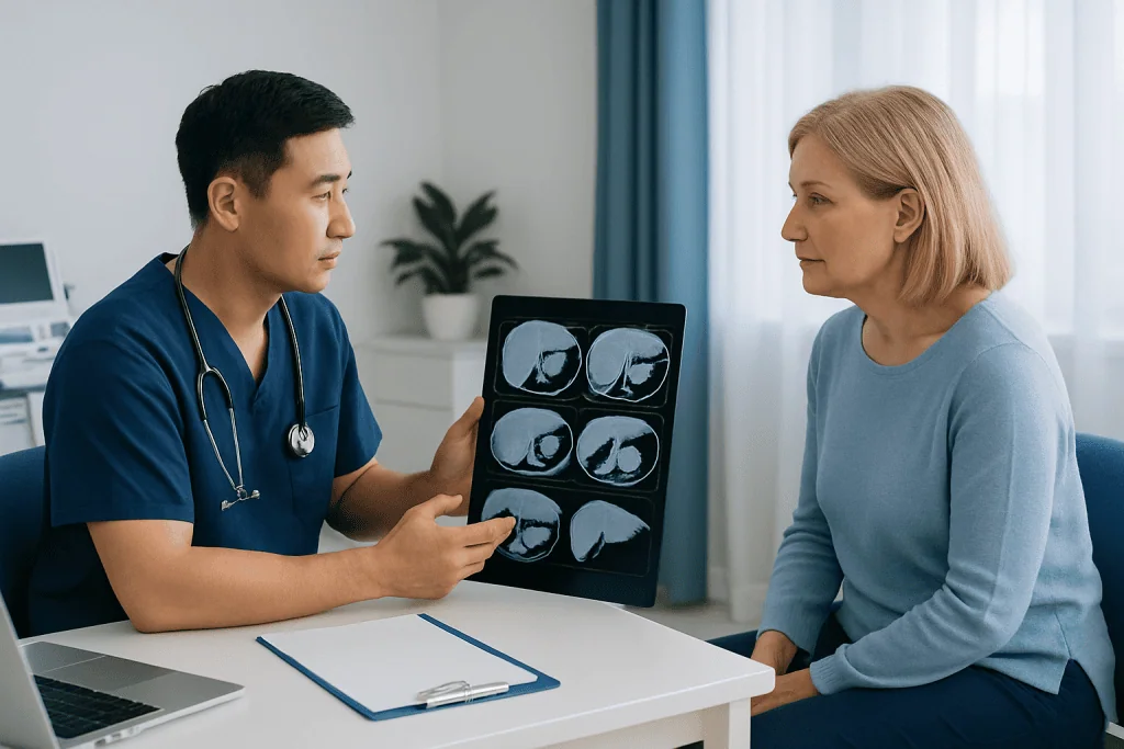
A clinician may recommend this examination for persistent upper abdominal discomfort, unexplained abnormal liver tests, a lesion seen on another scan, suspected liver infection, cancer staging, follow-up after treatment, or assessment after injury. It is also commonly used to define anatomy before complex procedures. Not everyone with a liver concern needs CT; ultrasound or MRI may sometimes be a better initial or follow-up test.
Preparation instructions vary by facility and by whether contrast will be used. Many people are asked not to eat for a few hours before a contrast-enhanced scan, although clear fluids may be allowed. The imaging department should provide the specific instructions to follow. Comfortable clothing without metal around the abdomen is helpful; some people change into a gown.
Before the appointment, the patient should tell the care team about prior reactions to contrast material, asthma or severe allergies, kidney disease, thyroid conditions, diabetes, pregnancy or possible pregnancy, breastfeeding, and all medicines or supplements. Kidney function testing may be needed before iodinated contrast, particularly for people with known kidney problems or other risk factors.
People should not stop prescribed medicines unless their doctor specifically advises it. If anxiety, difficulty lying flat, limited mobility or language needs could affect the appointment, informing the department beforehand can help the team arrange appropriate support.
Step by Step: What Happens During the Procedure
On arrival, staff confirm identity, review medical information and explain the examination. If contrast is planned, a small intravenous cannula is placed, usually in the arm or hand. The patient lies on a narrow table, most often on their back with the arms raised above the head when possible. The table moves slowly through the circular CT scanner.
The radiographer operates the scanner from an adjacent control area but can see, hear and speak with the patient throughout the examination. Clear instructions are given for brief breath-holds. Remaining still and following these instructions helps reduce motion blur and improves the accuracy of the images.
When contrast is injected, a temporary warm sensation throughout the body, a metallic taste or a feeling of needing to urinate can occur. These effects usually pass within seconds. The actual image acquisition often takes only a few minutes, although positioning, contrast preparation and multiple image phases can make the overall scan appointment longer.
After the images are checked for quality, the intravenous line is removed unless it is needed for other care. A radiologist, a doctor trained to interpret medical imaging, reviews the study and prepares a report for the referring clinician. Urgent findings are communicated promptly according to local clinical procedures.
Benefits, Limitations and Possible Risks
The key benefit of a liver segments CT scan is its speed and anatomical detail. It can show the location of a liver finding relative to vessels and other structures, which is valuable in urgent assessments and treatment planning. CT is widely available and can assess the liver and surrounding abdomen in one examination.
CT involves ionizing radiation. Imaging teams use protocols intended to keep radiation exposure as low as reasonably achievable while obtaining clinically useful images. The expected benefit of the information gained is weighed against this exposure. For some patients, MRI or ultrasound may be considered to avoid radiation or answer a more specific clinical question.
Iodinated contrast is generally well tolerated, but it can cause nausea, itching or hives in some people. Serious allergic-like reactions are uncommon, and imaging teams are trained to recognize and treat them. Contrast can also require special consideration in people with significantly reduced kidney function, so relevant medical history and recent tests should be shared before the scan.
A normal or unclear CT does not always rule out every liver condition. Very small findings, changes in liver tissue and lesions with similar imaging features may require comparison with older scans, repeat imaging or a different test. The treating clinician can explain what the result means for the individual situation.
Recovery Timeline and Understanding Results
Most people can return to normal daily activities immediately after a liver CT scan. If contrast was used and there are no fluid restrictions for another health reason, drinking water during the day may be encouraged. Mild warmth or a metallic taste during injection should resolve quickly. The puncture site may be briefly tender or bruised.
The report commonly describes liver size and contour, fat content, focal lesions if present, the relevant segment or segments, blood vessels, bile ducts and nearby abdominal structures. If a lesion is found, the report may describe its size, borders, density and behavior across contrast phases. These details help the referring clinician decide whether follow-up is required.
Results are often available after the radiologist has reviewed the images, but timing varies with the clinical setting. The clinician who ordered the scan should discuss the report in context rather than relying only on individual terms in the written result. A phrase such as “indeterminate” does not necessarily mean cancer; it often means additional imaging or interval follow-up would clarify the finding.
Depending on the result, next steps may include liver MRI, ultrasound surveillance, laboratory testing, referral to gastroenterology, hepatology, surgery or oncology, or no further testing. For complex cases, specialists may review the scans together to develop an individualized plan.
When to Seek Medical Care
People should contact a doctor if they have persistent or worsening upper abdominal pain, unexplained jaundice, dark urine, pale stools, ongoing vomiting, unintentional weight loss, fever with abdominal pain, or new abdominal swelling. These symptoms can have many causes, but timely medical assessment is important.
After a contrast-enhanced CT scan, urgent medical care is appropriate for trouble breathing, swelling of the face or throat, widespread hives, severe dizziness or fainting. These may indicate a significant allergic-like reaction and need prompt attention. Delayed rashes or other concerns should also be reported to a clinician.
Acibadem International’s multidisciplinary specialists and JCI-accredited hospitals can assess liver imaging findings and coordinate further evaluation or treatment for international patients. The most appropriate specialist and investigation depend on the scan result, symptoms, liver function and medical history.
Frequently asked questions
What does a liver segments CT scan look for?
A liver segments CT scan maps the liver’s anatomy and blood vessels while assessing liver tissue for abnormalities. It may be used to evaluate a lesion, injury, inflammation, infection, blood vessel changes or the extent of a known condition. It is also helpful when planning a procedure that needs precise knowledge of the lesion’s location.
Do I need contrast for a liver CT scan?
Many liver CT examinations use intravenous iodinated contrast because it improves visibility of blood vessels and helps characterize many liver findings. However, the need for contrast depends on the clinical question, kidney function, allergy history and other individual factors. The radiologist and referring clinician select the most appropriate protocol.
How long does a liver segments CT scan take?
The image acquisition itself is usually very quick, often only a few minutes. The full appointment can take longer because it may include registration, safety screening, placement of an intravenous line, contrast injection and checking the images. The imaging center can provide a more specific estimate before the appointment.
Can I eat before a liver CT scan?
Instructions differ between facilities and depend on whether contrast is planned. Patients are often asked to avoid food for several hours beforehand, while clear liquids may be permitted. They should follow the instructions provided by their imaging department and ask in advance if they are unsure.
Is a liver segments CT scan painful?
The scan is not usually painful. The person must lie still and may need to hold their breath briefly, and the intravenous cannula can cause a short pinch. Contrast injection may cause transient warmth or a metallic taste, which typically passes quickly.
How accurate is CT for liver lesions?
CT provides detailed, useful information about many liver lesions, particularly when multiphase contrast imaging is performed. Still, some findings cannot be fully identified on CT alone and may require MRI, ultrasound, follow-up imaging or other tests. Accuracy also depends on lesion size, liver background changes and the clinical context.
References
- American College of Radiology
- Radiological Society of North America
- National Institute of Diabetes and Digestive and Kidney Diseases
- European Society of Radiology
This article is for general information only and is not a substitute for professional medical advice. Please consult a qualified doctor about your individual situation.
Explore treatments in Turkey — costs, top hospitals & a free quote
JCI-accredited · board-certified surgeons · reply within 24h
Add us as a Preferred Source to see more of our trusted health content across Google Search, AI Overviews and Discover.