

MR-Guided Radiotherapy: Real-Time Imaging During Cancer Treatment
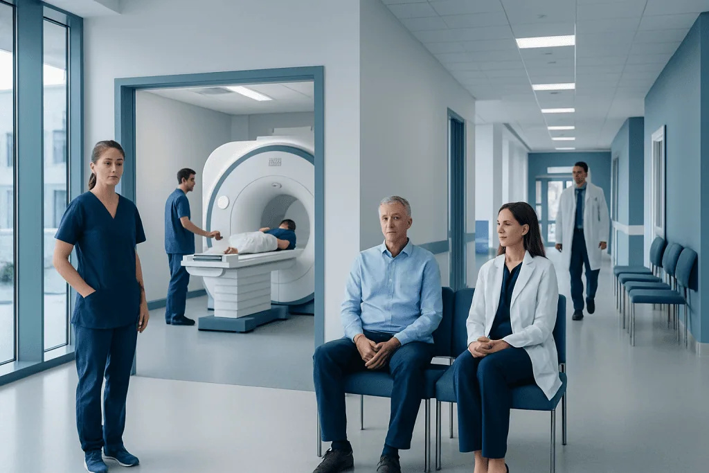
MR-guided radiotherapy uses MRI images before and sometimes during each treatment session to guide radiation more precisely. It is often considered for tumors in areas where organs move, such as the abdomen, pelvis, chest, and certain soft-tissue sites.
Key Takeaways
- MR-guided radiotherapy uses MRI images before and sometimes during each treatment session to guide radiation more precisely.
- It is often considered for tumors in areas where organs move, such as the abdomen, pelvis, chest, and certain soft-tissue sites.
- Real-time imaging may help doctors adapt treatment to daily anatomy changes, but it is not necessary or suitable for every cancer.
- Treatment is painless, but sessions may take longer than standard radiotherapy because imaging and planning checks are performed.
- A radiation oncology team decides whether MR-guided treatment is appropriate based on diagnosis, tumor location, treatment goals, and patient safety.
MR-guided radiotherapy combines radiation treatment with magnetic resonance imaging, allowing the care team to see the tumor and nearby organs during treatment. This technology can help personalize radiation delivery, especially for tumors that move or sit close to sensitive healthy tissues.
Overview
MR-guided radiotherapy, also called MRI-guided radiation therapy, is an advanced form of cancer treatment that combines a radiation treatment machine with magnetic resonance imaging. A commonly used name for this technology is MR-Linac, which refers to a magnetic resonance imaging system integrated with a linear accelerator that delivers radiation.
The main purpose is to help the radiation oncology team see the tumor and surrounding normal organs with high soft-tissue detail at the time of treatment. Many cancers are not in exactly the same position every day. Organs may shift with breathing, bladder filling, bowel gas, digestion, or normal body movement. MR guidance allows the team to check these changes more clearly before treatment begins and, in selected cases, monitor movement during radiation delivery.
Like other forms of radiotherapy, MR-guided treatment aims to damage cancer cells while limiting radiation exposure to healthy tissues. It is one tool within modern radiation oncology and may be used with curative, symptom-relieving, or combined treatment plans depending on the cancer type and stage. The decision to use it is individualized and made after careful imaging review, treatment planning, and safety assessment.
How Real-Time MRI Guidance Works
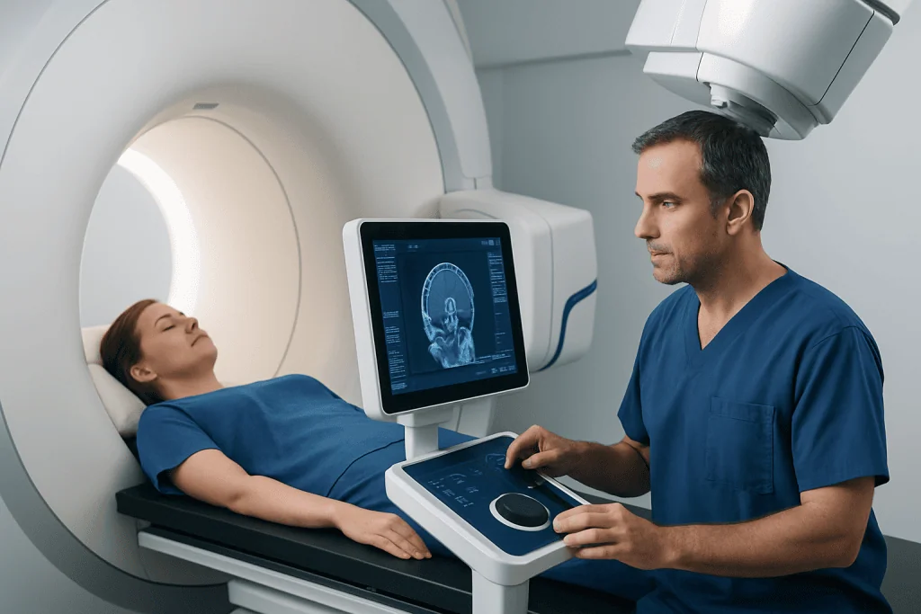
Traditional radiotherapy usually relies on a planning scan, often a CT scan, followed by daily positioning checks before each treatment. MR-guided radiotherapy adds MRI visualization directly in the treatment room. MRI is especially useful because it can show soft tissues such as the prostate, pancreas, liver, rectum, uterus, bladder, and nearby organs more clearly than some X-ray based imaging methods.
Before each session, the patient is positioned on the treatment table and an MRI scan is taken. The care team compares the day’s anatomy with the original treatment plan. If the tumor or nearby organs have shifted, the plan may be adjusted within safe clinical rules. This is called adaptive radiotherapy. In some cases, the system can also track tumor motion while the beam is on, pausing or adjusting delivery if the target moves outside the planned treatment area.
The process involves a multidisciplinary team. Radiation oncologists define the treatment target and organs at risk, medical physicists verify that the radiation dose is safe and accurate, radiation therapists position and support the patient, and imaging specialists help ensure high-quality images. Each step includes quality checks designed to make treatment precise and reproducible.
Who May Benefit From MR-Guided Radiotherapy?
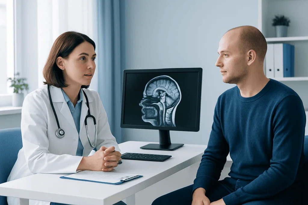
MR-guided radiotherapy may be considered when detailed soft-tissue imaging can meaningfully improve treatment accuracy. It is often discussed for tumors located near organs that are sensitive to radiation or that move from day to day. Examples may include selected cancers of the pancreas, liver, prostate, rectum, bladder, kidney, lung, gynecologic region, and certain metastatic tumors. Suitability depends on the individual case rather than the cancer name alone.
Patients receiving stereotactic body radiotherapy, also known as SBRT, may sometimes benefit from MR guidance because SBRT uses highly focused radiation doses over fewer sessions. When targets are small or close to important organs, seeing the anatomy clearly at each visit can be valuable. However, many patients are treated very effectively with other advanced techniques such as image-guided radiotherapy, intensity-modulated radiotherapy, proton therapy, or conventional radiotherapy.
MR-guided treatment is not appropriate for everyone. Some implanted medical devices, certain metal fragments, severe claustrophobia, or inability to lie still for the required time may affect eligibility. The radiation oncology team reviews MRI safety, general health, prior treatments, and treatment goals before recommending this approach.
What to Expect Before and During Treatment
The process begins with a consultation with a radiation oncologist. The doctor reviews the diagnosis, pathology results, imaging studies, previous treatments, current symptoms, and overall health. If MR-guided radiotherapy is suitable, a planning appointment is scheduled. During planning, imaging is performed in the treatment position, and customized supports may be used to help the patient remain comfortable and still.
The planning team creates a radiation plan that defines the target area, the dose, the number of treatment sessions, and the healthy organs to protect. Patients may receive instructions about bladder filling, fasting, bowel preparation, or breathing techniques depending on the treatment area. These instructions are important because they help make the body’s internal position more consistent from one session to the next.
During each treatment visit, the patient lies on the treatment table while MRI images are obtained. The team remains nearby and communicates through an intercom. The treatment itself is painless; the patient does not feel the radiation. A session may take longer than standard radiotherapy because imaging, plan adaptation, and safety checks are performed. The number of sessions varies according to cancer type, location, treatment purpose, and the prescribed radiation schedule.
Potential Benefits and Practical Limitations
The key potential benefit of MR-guided radiotherapy is improved visualization of soft tissues at the moment treatment is delivered. This may help the team target the tumor more confidently, adjust for daily anatomical changes, and reduce unnecessary exposure to nearby organs. In selected cases, this can support more personalized planning or shorter, highly focused treatment schedules.
Another advantage is motion management. Tumors in the abdomen and chest may move with breathing, while pelvic organs can shift with bladder and bowel changes. Real-time or near real-time MRI monitoring can help the team understand and respond to these movements. This does not remove all uncertainty, but it can add an important layer of image guidance.
There are also limitations. MR-guided systems are complex, and treatment sessions may be longer. Not all tumors require MRI guidance, and not all hospitals have this technology. Some patients cannot safely undergo MRI because of specific implants or metal fragments. In addition, the best treatment method depends on many factors, including the cancer diagnosis, stage, previous radiation exposure, and the expected balance between benefit and side effects.
Safety, Side Effects, and Aftercare
MR-guided radiotherapy uses noninvasive imaging and external-beam radiation. MRI itself does not use ionizing radiation, but the treatment beam does. Safety checks are performed before and during treatment to confirm patient identity, positioning, plan accuracy, and equipment performance. Patients are screened for MRI safety because the magnet can affect certain implants, devices, or metallic objects.
Side effects depend mainly on the part of the body being treated, the total dose, the number of sessions, and whether other treatments such as chemotherapy or immunotherapy are being given. Possible effects may include tiredness, skin sensitivity, nausea, bowel or bladder changes, swallowing discomfort, cough, or localized soreness, depending on the treatment area. Many side effects are temporary, but some may appear later, so follow-up is important.
Patients can support their treatment by following preparation instructions, keeping appointments, reporting new symptoms promptly, and discussing all medicines and supplements with the care team. Good hydration, balanced nutrition, gentle activity when approved, and adequate rest may help overall well-being. Any self-care plan should be adapted to the patient’s diagnosis and medical condition.
When to Speak With a Doctor
A patient may wish to ask about MR-guided radiotherapy if the tumor is located near sensitive organs, if previous imaging has shown movement or anatomical changes, or if stereotactic radiotherapy is being considered. It is also reasonable to ask whether MRI guidance offers a meaningful advantage over other modern image-guided techniques for the specific cancer type.
Medical advice is especially important if symptoms change during treatment. Patients should contact their care team for fever, uncontrolled pain, severe vomiting or diarrhea, bleeding, difficulty breathing, new neurological symptoms, or any side effect that feels unusual or concerning. The oncology team can assess whether symptoms are treatment-related and recommend safe management.
International patients considering advanced radiotherapy should seek a center that offers careful case review, MRI safety screening, radiation physics quality assurance, and coordinated oncology care. Acibadem International’s multidisciplinary specialists and JCI-accredited hospitals diagnose and treat cancer patients, including international patients, using individualized treatment planning where appropriate.
Frequently asked questions
Is MR-guided radiotherapy the same as regular radiotherapy?
It is a type of external-beam radiotherapy, but it uses MRI imaging in the treatment room to guide delivery. The radiation still comes from a treatment machine, while the MRI helps the team see soft tissues and check daily anatomy. This may allow more individualized targeting in selected patients.
Does MR-guided radiotherapy hurt?
The treatment itself is painless, and patients do not feel the radiation beam. Some people may feel discomfort from lying still or from the treatment position. The team can often use supports and communication during the session to help the patient remain comfortable.
How long does each session take?
Session length varies by treatment site and whether the plan is adapted that day. MR-guided sessions can take longer than standard radiotherapy because imaging, contour review, plan adjustment, and safety checks may be needed. The care team will explain the expected schedule before treatment begins.
Can every cancer be treated with MR-guided radiotherapy?
No. MR-guided radiotherapy is most useful when MRI visibility and daily adaptation are expected to improve treatment precision. Many cancers can be treated effectively with other modern radiation techniques. A radiation oncologist determines the best approach after reviewing imaging, diagnosis, stage, and patient-specific factors.
Is MRI guidance safe for patients with implants?
Some implants are MRI-safe or MRI-conditional, while others may not be compatible with MRI. Patients must tell the care team about pacemakers, neurostimulators, cochlear implants, surgical clips, metal fragments, joint replacements, and any implanted device. MRI safety screening is a routine part of the process.
What are the side effects of MR-guided radiotherapy?
Side effects depend on the treatment area and dose rather than the imaging guidance alone. They may include tiredness, skin irritation, nausea, bowel or bladder changes, cough, or discomfort in the treated region. The oncology team monitors symptoms and provides supportive care throughout treatment and follow-up.
References
- American Society for Radiation Oncology
- European Society for Radiotherapy and Oncology
- National Comprehensive Cancer Network
- International Atomic Energy Agency
- Radiological Society of North America
This article is for general information only and is not a substitute for professional medical advice. Please consult a qualified doctor about your individual situation.
Oncology care in Turkey — second opinion and treatment plan
JCI-accredited · board-certified surgeons · reply within 24h
Add us as a Preferred Source to see more of our trusted health content across Google Search, AI Overviews and Discover.

More from the Health Library



Related Specialists

Prof. Dr. Javad Parvizi
Orthopedic Surgery & Traumatology
Dr. Nesibe Sönmez Demiryoğuran
Emergency Service
Asst. Prof. Dr. Murat Tuğrul Eren
Urology
Dr. Aygun Babayeva
Pediatrics