MR Neurography: Advanced Imaging for Nerve Pain and Injury

MR neurography is a specialized MRI technique focused on nerves rather than bones or organs alone. It may help identify nerve compression, inflammation, injury, or changes in the tissues around a nerve.
Key Takeaways
- MR neurography is a specialized MRI technique focused on nerves rather than bones or organs alone.
- It may help identify nerve compression, inflammation, injury, or changes in the tissues around a nerve.
- The test is noninvasive and does not use ionizing radiation.
- Results are interpreted together with symptoms, physical examination, and sometimes nerve conduction studies or electromyography.
- MR neurography can be useful when standard MRI or other tests do not fully explain persistent nerve-related symptoms.
MR neurography is a specialized type of MRI designed to visualize peripheral nerves and surrounding tissues in greater detail than standard imaging. It can help doctors investigate unexplained nerve pain, numbness, weakness, or suspected nerve injury and plan the most appropriate treatment.
Overview of MR Neurography
MR neurography is an advanced imaging technique based on magnetic resonance imaging, or MRI. It is designed to make peripheral nerves easier to see and evaluate. While a standard MRI can show muscles, joints, bones, discs, and soft tissues, MR neurography uses specific sequences and protocols that better highlight nerves and detect changes in or around them.
This technique may be used when a person has ongoing nerve-related symptoms such as burning pain, tingling, numbness, weakness, or pain that travels along the course of a nerve. In some cases, symptoms continue even after routine imaging appears normal. MR neurography may offer more detail and help clarify whether a nerve is compressed, irritated, stretched, inflamed, or affected by scar tissue.
MR neurography is most often used to assess peripheral nerves outside the brain and spinal cord. Examples include nerves in the neck, shoulder, arm, pelvis, leg, and foot. It can also help evaluate nerve plexuses, such as the brachial plexus in the shoulder region or the lumbosacral plexus in the lower back and pelvis.
Importantly, MR neurography is a diagnostic tool rather than a treatment. Its value lies in helping doctors understand the source of symptoms more clearly, especially in complex cases. This information can support decisions about medication, rehabilitation, injections, or surgery when needed.
When MR Neurography May Be Recommended
Doctors may consider MR neurography when symptoms suggest a nerve problem but the exact location or cause is uncertain. It can be particularly helpful when pain, weakness, or sensory changes follow a nerve pathway and other tests have not provided a complete explanation. It may also be used after trauma or surgery if there is concern about nerve damage.
Situations in which MR neurography may be useful include suspected nerve entrapment, sports injuries, post-traumatic nerve injury, chronic sciatica-like symptoms, brachial plexus problems, pelvic nerve pain, or unexplained weakness in a limb. It may also help distinguish whether symptoms come from a peripheral nerve problem or another source, such as the spine or muscles.
Examples of conditions that may lead to MR neurography include carpal tunnel syndrome, nerve compression around the elbow or ankle, and symptoms related to the sciatic nerve. In selected cases, it may complement the evaluation of herniated disc or peripheral neuropathy when the clinical picture is complex.
MR neurography is not necessary for every person with nerve pain. Many common nerve conditions can be diagnosed through history, examination, and standard tests. A doctor usually recommends it when the additional detail is likely to influence diagnosis, treatment planning, or the decision to pursue further procedures.
What Symptoms and Conditions It Can Help Assess

MR neurography does not diagnose symptoms by itself, but it can help evaluate the structures that may be causing them. Nerve-related symptoms often include sharp or burning pain, electric shock-like sensations, tingling, numbness, muscle weakness, loss of coordination, or increased sensitivity in a specific area. These symptoms may be constant or may worsen with certain movements or positions.
The scan can sometimes show nerve swelling, signal changes suggesting irritation or injury, narrowing where a nerve passes through a tight space, or changes in nearby muscles that point to chronic nerve dysfunction. It may also reveal masses, cysts, scar tissue, inflammation, or anatomical variations affecting a nerve.
MR neurography may be considered in a range of clinical situations, such as:
- Entrapment neuropathies, where a nerve is compressed
- Traumatic nerve injury after an accident, fall, or sports injury
- Postoperative nerve symptoms or suspected scar-related tethering
- Plexus injuries involving the shoulder or pelvic nerves
- Persistent limb pain when standard imaging is inconclusive
- Selected cases of chronic pelvic pain or buttock-to-leg pain related to deep nerves
Even when MR neurography identifies an abnormality, the findings must match the person’s symptoms and examination. Some imaging changes may be mild or incidental, while clinically important nerve dysfunction may require confirmation with other tests. This is why interpretation by experienced radiologists and clinicians is important.
How MR Neurography Works and How It Differs From Standard MRI
MR neurography uses the same basic MRI technology as other scans: a strong magnet, radio waves, and advanced computer processing create detailed images without radiation. What makes it different is the use of specialized imaging sequences that suppress signals from surrounding fat and emphasize the appearance of nerves and tissue changes around them. This can make nerves stand out more clearly than on routine MRI.
In some cases, high-resolution images are obtained in several planes to trace the course of a nerve over a longer distance. Radiologists may assess the nerve’s size, shape, continuity, signal intensity, and relationship to nearby muscles, vessels, bones, discs, or scar tissue. Depending on the body area, the scan may focus on a small region or cover a broader pathway, such as from the lower back through the pelvis and into the leg.
Standard MRI remains very valuable and is often the first imaging study. It may identify common causes of nerve symptoms, such as a disc problem, joint disease, tumor, or soft tissue injury. MR neurography is usually reserved for cases that need more targeted nerve imaging. It is best viewed as a complementary tool rather than a replacement for conventional MRI.
Doctors may combine MR neurography with other assessments, such as neurological examination, ultrasound, blood tests, or electrodiagnostic studies. Depending on the findings, a patient may go on to treatments such as physical therapy and rehabilitation, image-guided pain management, or, in some situations, peripheral nerve surgery.
What to Expect During the Scan
Before the exam, the imaging team reviews the person’s symptoms, medical history, and any MRI safety concerns. Because MRI uses a strong magnet, it is important to mention implanted devices, prior surgeries, metal fragments, or any possibility of pregnancy. Many people can have MR neurography safely, but the care team must confirm that the scan is appropriate.
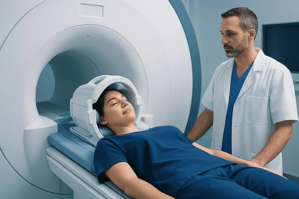
During the scan, the patient lies still on the MRI table while the area of interest is positioned carefully. The machine makes loud tapping or thumping sounds, so ear protection is usually provided. The test is painless, although remaining still can be uncomfortable for some people, especially if pain is already present. Scan time varies depending on the body region and protocol.
Some MR neurography studies are done without contrast, while others may require contrast material to help assess inflammation, masses, or postoperative changes. The radiologist and referring doctor decide whether contrast is useful based on the clinical question. Patients usually receive preparation instructions in advance.
People who feel claustrophobic should mention this beforehand, as practical solutions are often available. After the scan, most patients can return to their normal activities right away unless they received medication for relaxation. The images are then reviewed by a radiologist, and the results are discussed with the treating doctor in the context of symptoms and examination findings.
Benefits, Limitations, and Accuracy
One of the main benefits of MR neurography is that it provides a noninvasive way to visualize nerves and surrounding tissues in detail. It may help locate the site of nerve irritation, define the extent of an injury, and support treatment planning. It can also be useful in complex cases where symptoms do not fit a simple pattern or when surgery is being considered.
Another advantage is that the technique does not use ionizing radiation. For many patients, this makes it a suitable option when repeat evaluation is needed. In some cases, it may also help explain why symptoms persist despite previous treatments, allowing care to be directed more precisely.
At the same time, MR neurography has limitations. Not every nerve problem will be visible, and image quality can depend on the body region, equipment, protocol, and patient movement. Small fiber nerve disorders, functional pain syndromes, or very subtle abnormalities may not appear clearly on imaging. Availability may also be more limited than standard MRI because the technique requires specialized expertise.
Accuracy improves when MR neurography is interpreted as part of a full clinical assessment rather than in isolation. Doctors often compare the findings with physical examination, symptom pattern, and electrodiagnostic testing such as EMG testing. This combined approach usually provides the clearest picture of what is causing the patient’s symptoms.
Diagnosis, Treatment Planning, and Follow-Up
MR neurography is often most useful after an initial clinical assessment has already suggested a nerve-related problem. A doctor may first evaluate reflexes, strength, sensation, and the pattern of pain or numbness. Standard MRI, ultrasound, or nerve studies may come before or after MR neurography depending on the case. The goal is to build a complete and accurate diagnosis rather than rely on one test alone.
Once the source of symptoms is better defined, treatment planning becomes more focused. If imaging suggests nerve compression, treatment may involve activity modification, splinting, medication, rehabilitation, or targeted injections. If the scan shows a structural lesion, severe compression, or traumatic nerve disruption, referral to a surgeon may be appropriate. In other cases, the scan can reassure both patient and doctor by ruling out more serious structural causes.
Follow-up depends on the underlying problem, symptom severity, and response to treatment. Some patients improve with conservative care over time, while others need further specialist evaluation. It is important for patients to report any progression of weakness, worsening numbness, or loss of function, as these changes may alter the urgency of management.
For international patients seeking coordinated evaluation, Acibadem International’s multidisciplinary specialists and JCI-accredited hospitals diagnose and treat nerve-related conditions using advanced imaging and individualized care plans. The most appropriate next step always depends on the person’s symptoms, examination findings, and overall health.
Frequently asked questions
What is MR neurography used for?
MR neurography is used to examine peripheral nerves and the tissues around them in greater detail than standard MRI. Doctors may use it to investigate persistent nerve pain, numbness, tingling, weakness, or suspected nerve injury.
Is MR neurography the same as a regular MRI?
It uses the same MRI technology, but the imaging protocol is specialized to show nerves more clearly. A regular MRI often focuses more broadly on structures such as bones, discs, joints, and muscles.
Can MR neurography diagnose all causes of nerve pain?
No. It can be very helpful, but not every cause of nerve pain is visible on imaging. Doctors usually interpret the scan together with symptoms, examination findings, and sometimes nerve conduction studies or EMG.
Does MR neurography require contrast?
Not always. Many MR neurography studies are done without contrast, but contrast may be recommended in certain situations, such as suspected inflammation, masses, or postoperative changes.
Is MR neurography painful or unsafe?
The scan itself is noninvasive and painless, although lying still may be uncomfortable for some people. MRI is generally safe for many patients, but anyone with implants, metal fragments, or other MRI safety concerns should discuss this with the care team beforehand.
When might a doctor order MR neurography instead of other tests?
A doctor may order it when symptoms strongly suggest a nerve problem but routine imaging has not fully explained the cause. It may also be useful in complex injuries, persistent pain after trauma or surgery, or cases where treatment decisions depend on identifying the exact nerve involved.
References
- Radiological Society of North America
- American College of Radiology
- National Institute of Neurological Disorders and Stroke
- Mayo Clinic
- Cleveland Clinic
This article is for general information only and is not a substitute for professional medical advice. Please consult a qualified doctor about your individual situation.
Joint and spine care in Turkey — expert assessment & treatment
JCI-accredited · board-certified surgeons · reply within 24h
Add us as a Preferred Source to see more of our trusted health content across Google Search, AI Overviews and Discover.

More from the Health Library



Related Specialists
Fzt. Mustafa Özcan
Physical Medicine & Rehabilitation
Assoc. Prof. Dr. Selçuk Keskin
Urology
Prof. Dr. Nail Suat Ünver
Internal Medicine
Prof. Dr. Arzu Tiftikçi
Gastroenterology