Myelography Procedure: An Evidence-Based Patient Guide
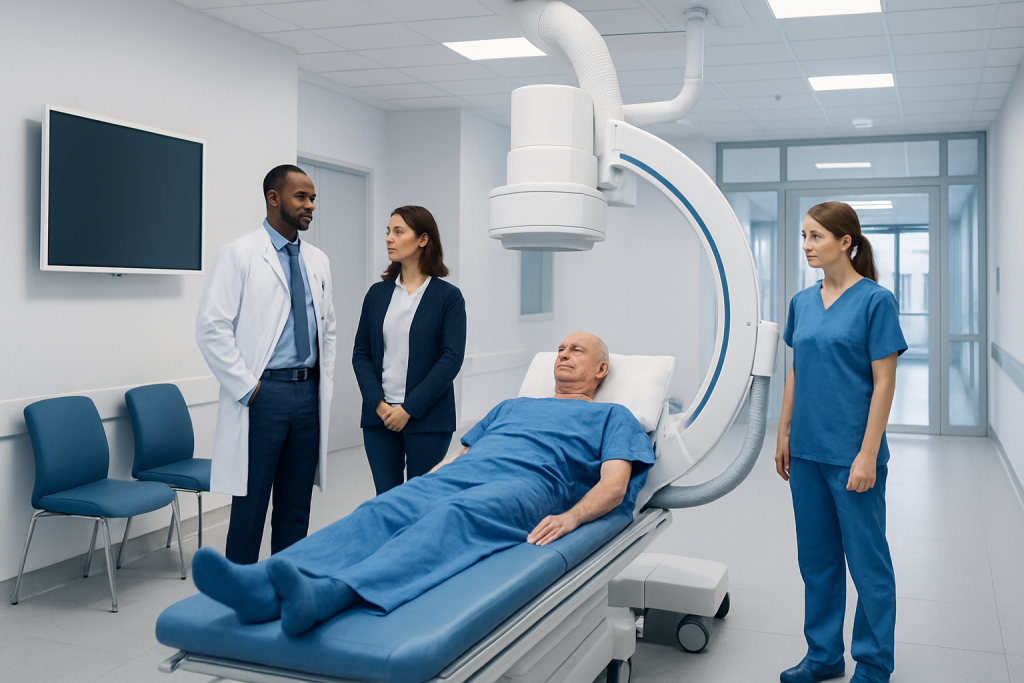
Myelography uses contrast dye placed in the fluid around the spinal cord, followed by X-ray or CT images. Most people are awake for a myelogram, with local anesthetic used at the injection site and sedation available in selected situations.
Key Takeaways
- Myelography uses contrast dye placed in the fluid around the spinal cord, followed by X-ray or CT images.
- Most people are awake for a myelogram, with local anesthetic used at the injection site and sedation available in selected situations.
- The test can provide detailed views of nerve-root compression and spinal canal narrowing, especially when MRI cannot be performed.
- Temporary headache, nausea and soreness are possible; serious complications are uncommon but require prompt medical attention.
- After the procedure, patients usually rest, drink fluids as advised and avoid strenuous activity for a short period.
- Individual preparation and recovery instructions can differ, so the imaging team's instructions should take priority.
A myelography procedure, also called a myelogram, is a specialized imaging test that combines contrast dye with X-ray or CT scanning to assess the spinal canal, spinal cord and nerve roots. It may help clinicians identify narrowing, disc-related pressure, cysts, or other causes of symptoms when MRI is not suitable or does not provide enough information.
Overview: What Is a Myelography Procedure?
A myelography procedure is an imaging examination of the spine. During the test, a clinician injects a small amount of contrast material into the cerebrospinal fluid, the clear fluid that surrounds the spinal cord and nerve roots. X-ray images, often followed by computed tomography (CT), show how the contrast moves through the spinal canal.
The examination may reveal areas where the normal flow of contrast is narrowed, displaced or blocked. These patterns can help assess spinal canal narrowing, pressure on nerve roots, some disc problems, postoperative changes, cysts, or other structural concerns. A myelogram is not usually the first imaging test for back or neck symptoms, but it remains useful when MRI is unsuitable, unclear, or affected by implanted devices or metal hardware.
This myelogram patient education guide describes common practice, but each hospital may use slightly different myelography instructions. The radiologist, neurologist, neurosurgeon or spine specialist will explain why the examination is being considered in the individual situation.
How Myelography Works and Who May Need It
Contrast material is visible on X-ray and CT images. Once it is introduced into the fluid space around the spinal cord, clinicians can observe the outline of the spinal canal and nerve roots. If a disc bulge, bone spur, scar tissue or another structure presses on this space, the contrast pattern may help show its location and extent.
A clinician may recommend myelography for persistent arm or leg pain, numbness, weakness, walking difficulty, or symptoms that suggest pressure on spinal nerves. It can be particularly valuable when evaluating suspected spinal stenosis, complex spinal anatomy after surgery, or symptoms that do not match findings from other scans.
Not everyone is a suitable candidate. The care team needs to know about pregnancy or a possible pregnancy, previous reactions to contrast agents, bleeding disorders, kidney disease, seizures, infection, and all medicines and supplements. Blood-thinning medicines may need individualized instructions before the test; patients should never stop prescribed medicines without guidance from the clinician managing them.
In many cases, MRI provides excellent spinal detail without an injection into the spinal fluid. Myelography is chosen selectively, based on symptoms, prior imaging, medical history and the clinical question that needs answering.
What Happens During the Test?

Before the examination, patients may be asked to follow specific eating, drinking and medication instructions. They should arrange for an adult to take them home if sedation is planned or if the local policy requires it. The team will review consent, allergies, current medicines and relevant health conditions, and may place an intravenous line if needed.
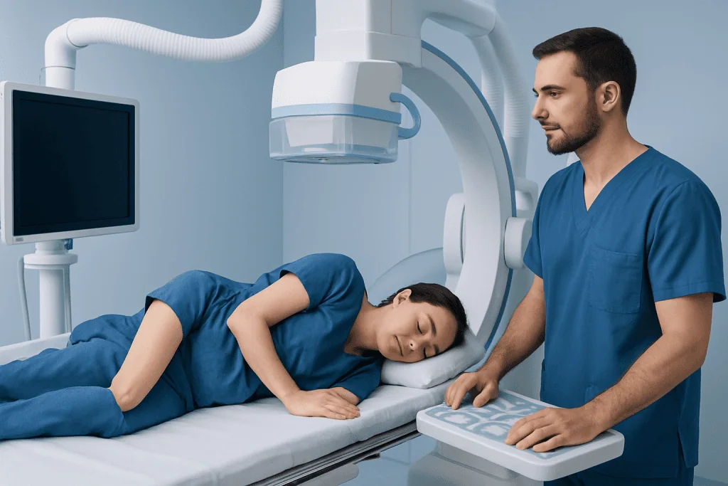
For the myelography procedure position, the person usually lies face down, on their side, or occasionally sits leaning forward, depending on the area being examined and the clinician’s approach. The lower back is cleaned and covered with sterile drapes. Local anesthetic numbs the skin and deeper tissues before a thin needle is guided into the fluid space in the lower spine, commonly using fluoroscopy, which is live X-ray guidance.
After confirming needle placement, the clinician injects contrast material and removes the needle. The table may be tilted gently so the contrast reaches the part of the spine of interest. X-ray images are taken, and many people then have a CT scan for more detailed cross-sectional images. The imaging portion often takes less than an hour, although preparation and observation add time.
Patients should tell the team immediately if they experience significant pain, dizziness, nausea, a new unusual sensation, or anxiety during the test. Staff monitor comfort and can explain each stage as it happens.
Are You Awake During a Myelogram? How Painful Is It?
Are you awake during a myelogram? Most people are awake. Local anesthetic is used to numb the injection site, and the clinical team communicates throughout the examination. Some people may receive medicine to help them relax, depending on their health needs and the facility’s protocol, but general anesthesia is not usually required.
How painful is a myelogram? Experiences vary. The local anesthetic may cause a brief sting or burning sensation, and there can be pressure as the needle is positioned. Some people feel temporary discomfort in the back, buttock or leg if a nearby nerve is irritated, but severe pain is not expected and should be reported immediately.
Afterward, mild tenderness at the injection site or a headache can occur. Clear communication helps the team tailor positioning, reassurance and symptom relief. People who are concerned about pain, claustrophobia, prior difficult procedures or anxiety should discuss this before the appointment so supportive options can be considered.
Recovery Timeline and What Not to Do After a Myelogram
Following the examination, patients are observed in a recovery area. Monitoring time varies by local practice, the type of contrast used, whether sedation was given, and how the person feels. Many people go home the same day, provided they are clinically well and have a safe plan for travel and support if needed.
How long do you lay flat after a myelogram? This differs between facilities and individual circumstances. Some centers ask patients to remain lying down or with the head elevated for a period of observation, while others permit gentle movement sooner. The care team’s written discharge instructions are the appropriate guide, as they reflect the technique used and the patient’s clinical needs.
What not to do after a myelogram? Patients are commonly advised to avoid driving if they received sedating medicine, avoid alcohol for the rest of the day if instructed, and postpone strenuous exercise, heavy lifting and bending for a short time. They should rest, change positions slowly, and drink fluids if the care team says this is appropriate. A responsible adult may be recommended to stay nearby overnight after sedation.
Minor soreness and tiredness generally improve over the next day or two. Headache may occur, especially when upright, and should be managed according to the discharge plan. Contacting the imaging department or treating clinician is appropriate when symptoms are persistent, worsening or concerning.
Benefits, Risks and Myelography Procedure Side Effects
The main benefit of myelography is targeted information about the spinal canal and nerve roots. It may clarify whether symptoms are related to nerve compression and can help specialists plan further care, including spine surgery when surgery is clinically appropriate. The result is interpreted alongside symptoms, physical examination findings and other tests; an imaging change alone does not always explain pain or require treatment.
Possible myelography procedure side effects include temporary headache, nausea, vomiting, dizziness, neck or back discomfort, and tenderness or minor bruising at the injection site. Some people notice symptoms later on the day of the test, which is one reason for following discharge instructions carefully.
More serious complications are uncommon but can include infection, bleeding, a significant contrast reaction, seizure, or a persistent spinal-fluid leak causing a severe positional headache. Radiation is used for fluoroscopy and CT; the imaging team applies the lowest radiation exposure reasonably achievable for the required diagnostic information. The expected benefit of the examination should outweigh these potential risks.
When results show a condition affecting the spine or nerves, care may involve radiology, neurology, pain medicine, rehabilitation and surgical specialists. Acibadem International’s multidisciplinary specialists and JCI-accredited hospitals assess and treat spinal conditions for international patients when coordinated care is needed.
When to Seek Medical Care
Patients should contact the care team promptly after a myelogram if they develop a severe or worsening headache, repeated vomiting, fever, increasing redness or drainage at the injection site, severe back pain, confusion, or a widespread rash. New weakness, numbness, trouble walking, loss of bladder or bowel control, or symptoms of a serious allergic reaction require urgent medical assessment.
Before the test, urgent assessment is also important for new or rapidly progressing limb weakness, numbness in the groin or saddle area, or new bladder or bowel changes. These symptoms may indicate significant nerve compression and should not wait for a routine imaging appointment.
For non-urgent questions about preparation, recovery or results, patients should contact the radiology department or the clinician who requested the test. They can explain the individual myelography instructions, when results will be available, and which next steps are appropriate.
Frequently asked questions
What is the difference between myelography and MRI?
MRI uses magnets and radio waves to create detailed images without ionizing radiation or injection into spinal fluid. Myelography uses contrast material plus X-ray or CT imaging and may be useful when MRI cannot be performed, is limited by metal hardware, or does not answer a specific question about the spinal canal or nerve roots.
How should a patient prepare for a myelography procedure?
Preparation varies, but the team commonly reviews medicines, allergies, pregnancy status and medical history before the appointment. Patients may receive instructions about food, drinks, blood-thinning medicines and arranging transportation home. The imaging center's own instructions should be followed because they are tailored to the planned procedure.
Can a person eat before a myelogram?
Some facilities allow light food, while others ask patients to avoid eating for a set period, especially if sedation may be used. Drinking clear fluids may also be handled differently between centers. Patients should confirm the instructions with the department performing the test rather than relying on general advice.
How long does it take to get myelogram results?
A radiologist reviews the images and sends a report to the clinician who ordered the examination. Timing depends on the facility and the clinical urgency. The requesting clinician explains what the findings mean in the context of the person's symptoms and other tests.
Can a myelogram cause a headache?
Yes. A headache can occur after the needle puncture, sometimes because a small amount of spinal fluid leaks from the puncture site. It may be worse when sitting or standing and improve when lying down; patients should follow their discharge plan and contact the care team if the headache is severe, persistent or accompanied by other concerning symptoms.
Is a myelogram safe for everyone?
Myelography is generally performed with careful screening, but it is not appropriate for every person. Pregnancy, certain infections, bleeding risks, past contrast reactions and particular neurological conditions may affect whether or how the test is done. A clinician weighs the expected diagnostic value against the individual's risks before recommending it.
References
- Radiological Society of North America
- American College of Radiology
- National Institute of Neurological Disorders and Stroke
- Mayo Clinic
This article is for general information only and is not a substitute for professional medical advice. Please consult a qualified doctor about your individual situation.
Joint and spine care in Turkey — expert assessment & treatment
JCI-accredited · board-certified surgeons · reply within 24h
Add us as a Preferred Source to see more of our trusted health content across Google Search, AI Overviews and Discover.