Necrosis: Diagnosis, Outlook, and Modern Treatment Approaches

Necrosis is not a disease itself but a sign of severe tissue injury. Common causes include reduced blood flow, infection, trauma, burns, chemicals, and some medical conditions.
Key Takeaways
- Necrosis is not a disease itself but a sign of severe tissue injury.
- Common causes include reduced blood flow, infection, trauma, burns, chemicals, and some medical conditions.
- Symptoms vary by location but may include pain, color change, swelling, numbness, skin breakdown, or foul-smelling discharge.
- Diagnosis usually combines a physical exam with imaging, blood tests, and sometimes tissue sampling.
- Treatment focuses on removing the cause, restoring blood supply when possible, treating infection, and managing damaged tissue.
- Early medical evaluation is important because untreated necrosis can spread and lead to serious complications.
Necrosis means the death of body tissue, usually because cells have been badly injured or deprived of blood supply. The outlook depends on the cause, location, and how quickly treatment begins, but prompt medical care can often limit further damage and help protect surrounding tissue.
Overview: what necrosis means
Necrosis is the irreversible death of cells or tissue in part of the body. It happens when tissue is damaged so severely that the cells can no longer survive. This may follow a loss of blood supply, a serious infection, trauma, extreme temperatures, radiation, chemicals, or inflammation linked to an underlying illness.
Unlike normal cell turnover, necrosis is not a controlled process. The affected tissue may become swollen, discolored, fragile, or infected, and nearby tissue can also be affected if the underlying cause continues. The term describes what has happened to the tissue, while the reason behind it still needs to be identified.
Necrosis can involve the skin, fat, muscle, bone, or internal organs. Examples include tissue death after a blocked blood vessel, severe pressure injury, complications of diabetes, or damage after major trauma. In some situations, necrosis overlaps with conditions such as gangrene, especially when poor blood flow and infection affect a limb or toe.
How necrosis develops and the main types
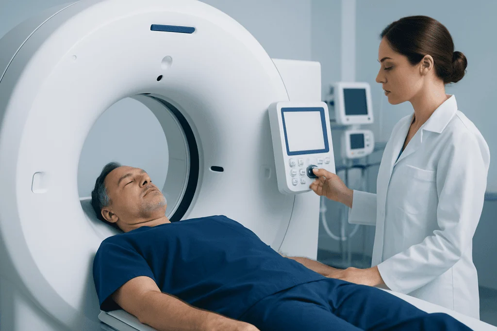
Doctors often describe necrosis by both its cause and its pattern under examination. In everyday care, what matters most is where it is happening, how deep it goes, and whether infection or loss of circulation is involved. These details guide urgency and treatment.
Several patterns of necrosis are recognized in medicine. Coagulative necrosis is often linked to reduced blood supply in solid organs. Liquefactive necrosis is more commonly seen in infections or certain brain injuries, where tissue becomes soft or liquid-like. Fat necrosis can occur after trauma or inflammation of the pancreas. Avascular necrosis refers specifically to bone tissue death caused by interrupted blood flow and is related to avascular necrosis.
Skin and soft-tissue necrosis may occur after pressure injuries, vascular disease, burns, severe wounds, or treatment complications. Some forms are limited and superficial, while others spread rapidly and require urgent surgery. Because the pattern alone does not explain the cause, doctors assess the whole clinical picture rather than relying on appearance alone.
Symptoms and possible complications

Symptoms depend on the area involved. Skin necrosis may begin with pain, tenderness, swelling, redness, blistering, or a patch that turns pale, blue, purple, brown, or black. As tissue dies, sensation may decrease, and the area may become dry and hard or wet and fragile. If infection is present, there may be warmth, pus, a bad smell, or fever.
Necrosis in deeper tissues or internal organs may cause less visible but still important symptoms. A person may notice severe persistent pain, reduced movement, numbness, weakness, non-healing wounds, or worsening function of the affected body part. Bone necrosis may cause joint pain and stiffness, especially with weight-bearing. Organ-related necrosis can cause symptoms specific to that organ and may quickly become serious.
Complications can include spreading infection, abscess formation, loss of function, delayed wound healing, scarring, and, in severe cases, sepsis or the need for surgery to remove dead tissue. Because dead tissue cannot recover, the aim is to stop progression, protect healthy tissue, and manage complications as early as possible.
- Increasing pain or sudden numbness
- Skin color change, especially black, blue, or gray areas
- Rapid swelling or blistering
- Fever, chills, or foul-smelling drainage
- A wound that is not healing or appears to be worsening
Causes and risk factors
Necrosis usually develops when tissue does not receive enough oxygen and nutrients or is directly injured beyond repair. Poor circulation is one of the most common reasons. This may happen because of blocked arteries, blood clots, severe swelling, pressure on tissue, or damage to blood vessels after injury. Diabetes, smoking, and peripheral artery disease can further reduce blood flow and slow healing.
Infection is another important cause. Bacteria can destroy tissue directly or trigger intense inflammation that cuts off local blood supply. Trauma, burns, frostbite, surgery, injections, radiation exposure, and certain chemicals or toxins can also lead to tissue death. Sometimes necrosis appears as a complication of an existing illness rather than a primary problem.
Risk factors vary by location but often include diabetes, vascular disease, immobility, advanced pressure injuries, autoimmune disease, pancreatitis, sickle cell disease, long-term corticosteroid use, heavy alcohol use, and previous major injury. Bone necrosis is more likely in people with joint trauma, steroid exposure, or blood supply problems affecting the hip, shoulder, or knee.
Diagnosis: how doctors confirm necrosis
Diagnosis starts with a careful history and physical examination. Doctors ask when symptoms began, whether there was an injury, burn, injection, surgery, infection, circulation problem, or chronic illness, and how quickly the area has changed. The skin color, temperature, sensation, pulse, swelling, and wound appearance can provide important clues about depth and severity.
Tests are chosen based on the suspected cause and location. Blood tests may look for signs of infection, inflammation, organ stress, or poor glucose control. Imaging such as ultrasound, X-ray, CT, or MRI can help assess blood flow, bone involvement, soft-tissue spread, or collections of fluid. In some cases, vascular imaging is needed to check for blocked arteries that may require urgent treatment.
If infection is suspected, doctors may take wound cultures or samples during a procedure, although treatment should not be delayed when a rapidly progressive infection is possible. A biopsy or tissue examination may help confirm the diagnosis or distinguish necrosis from other conditions. If poor circulation is central to the problem, specialists may use advanced imaging and angiography to plan care.
Modern treatment approaches
Treatment depends on the cause, location, extent of tissue damage, and whether infection is present. Dead tissue cannot be revived, so care focuses on stopping further injury, preserving healthy tissue, controlling pain, and supporting healing. This may include wound care, antibiotics when infection is confirmed or strongly suspected, improving blood sugar control, offloading pressure, and treating the underlying circulation problem.
When circulation is reduced, restoring blood flow can be essential. Depending on the situation, doctors may consider medication, endovascular procedures, or vascular surgery to improve perfusion. If infection or deep tissue damage is severe, urgent surgical removal of dead tissue, called debridement, may be necessary. Debridement can lower the bacterial burden and help healthy tissue heal more effectively.
Some patients benefit from reconstructive procedures after the damaged area is stable, especially if skin or soft tissue has been lost. This may involve advanced dressings, grafts, or carefully planned plastic and reconstructive surgery. For bone necrosis, management may range from activity modification and physical therapy to orthopedic procedures depending on stage and joint damage. If pancreatic inflammation leads to fat necrosis or if there is another digestive cause, related care may involve specialists in gastroenterology.
Because necrosis can involve more than one body system, treatment is often multidisciplinary. Surgeons, vascular specialists, infectious disease physicians, wound care teams, orthopedists, radiologists, and rehabilitation experts may all contribute to a safe plan. Near the end of treatment planning, some patients may choose centers such as Acibadem International, where multidisciplinary specialists in JCI-accredited hospitals diagnose and treat complex necrosis-related conditions for international patients.
Outlook, recovery, and self-care
The outlook for necrosis varies widely. Small, superficial areas may heal well once the cause is corrected and the tissue is properly managed. Deeper or more extensive necrosis can take longer to treat and may leave scarring or loss of function. Prognosis is generally better when treatment begins early, blood flow can be restored, and infection is controlled promptly.
Recovery often continues after the immediate problem is treated. Wound care instructions should be followed closely, including dressing changes, pressure relief, limb elevation if advised, and careful monitoring for new redness, drainage, odor, or pain. People with diabetes should aim for good glucose control, and those who smoke are strongly encouraged to stop because smoking further reduces tissue oxygen delivery.
Healthy self-care can support healing but should not replace medical treatment. Good nutrition, hydration, mobility within a doctor’s guidance, and management of long-term conditions all matter. Patients should avoid home removal of blackened or damaged tissue and should not apply harsh chemicals or unproven remedies to a wound, as these can worsen injury.
When to seek medical care
Medical assessment is important whenever a wound or painful area changes color, fails to heal, or seems to be worsening. Care should be sought promptly for black, blue, or gray tissue; sudden severe pain; loss of sensation; spreading redness; fever; foul-smelling drainage; or rapidly enlarging blisters. These signs may suggest progressing tissue death, infection, or poor circulation.
Urgent care is especially important for people with diabetes, vascular disease, immune suppression, recent surgery, or a major injury. Any limb that becomes cold, pale, numb, or weak should be assessed quickly because reduced blood flow can threaten tissue survival. Persistent joint pain after injury or steroid use should also be discussed with a doctor, particularly if movement becomes limited.
Even when symptoms seem mild, early evaluation may prevent a small problem from becoming a larger one. A qualified clinician can identify the cause, arrange the right tests, and recommend treatment tailored to the affected area and the person’s overall health.
Frequently asked questions
Is necrosis the same as gangrene?
Not exactly. Necrosis is a general term for tissue death, while gangrene usually refers to a form of necrosis that affects body tissue, often in a limb, and is commonly linked to poor blood flow and sometimes infection. A doctor can explain which term fits a specific case.
Can necrosis heal on its own?
Dead tissue itself does not recover. However, the surrounding healthy tissue may heal once the cause is treated and the damaged tissue is managed properly. Early treatment improves the chance of limiting spread and supporting recovery.
What does necrosis look like on the skin?
It may appear as an area that changes color from pale or purple to brown or black. The skin can become dry and firm or wet and broken down, sometimes with blisters or discharge. Appearance alone is not enough for diagnosis, so medical evaluation is important.
Is necrosis always an emergency?
Not every case is an immediate emergency, but all suspected necrosis deserves prompt medical attention. Some forms progress slowly, while others can spread quickly or become infected. Rapidly worsening pain, color change, fever, or foul odor should be assessed urgently.
How is necrosis diagnosed?
Doctors diagnose necrosis using the medical history, physical examination, and tests based on the affected area. These may include blood tests, imaging, vascular studies, and sometimes a tissue sample. The goal is to confirm tissue death and identify the cause.
Who is at higher risk of developing necrosis?
People with diabetes, smoking history, poor circulation, severe infections, pressure injuries, major trauma, or immune suppression are often at higher risk. Certain medicines and conditions that affect blood supply, such as long-term steroid use or vascular disease, can also increase risk.
References
- World Health Organization
- National Institute of Diabetes and Digestive and Kidney Diseases
- National Institute of Arthritis and Musculoskeletal and Skin Diseases
- Centers for Disease Control and Prevention
- Merck Manual Professional Edition
This article is for general information only and is not a substitute for professional medical advice. Please consult a qualified doctor about your individual situation.
Explore treatments in Turkey — costs, top hospitals & a free quote
JCI-accredited · board-certified surgeons · reply within 24h
Add us as a Preferred Source to see more of our trusted health content across Google Search, AI Overviews and Discover.