Nephroblastoma Ultrasound: Preparation, Procedure and Results

Ultrasound is painless, does not use ionizing radiation, and usually requires little or no preparation. Wilms tumor, also called nephroblastoma, often appears as a solid mass arising from one kidney, but ultrasound alone cannot confirm the diagnosis.
Key Takeaways
- Ultrasound is painless, does not use ionizing radiation, and usually requires little or no preparation.
- Wilms tumor, also called nephroblastoma, often appears as a solid mass arising from one kidney, but ultrasound alone cannot confirm the diagnosis.
- Doppler ultrasound can assess blood flow and look for possible extension into the renal vein or inferior vena cava.
- CT or MRI is commonly needed to define the tumor’s extent and support treatment planning.
- A new abdominal swelling, blood in the urine, unexplained fever, or persistent abdominal pain in a child should be assessed promptly by a doctor.
A nephroblastoma ultrasound is usually the first imaging test used when a child may have a kidney mass. It uses sound waves rather than radiation to examine the kidneys, assess blood flow, and help clinicians decide whether further imaging and specialist evaluation are needed.
Nephroblastoma Ultrasound: What It Shows
A nephroblastoma ultrasound is an imaging examination used to look at the kidneys and surrounding abdominal organs when a child may have a renal mass. Nephroblastoma, more commonly called Wilms tumor, is a childhood kidney cancer. Ultrasound can often show whether a lump is within the kidney, whether it is mainly solid or contains fluid-filled areas, and how it affects nearby structures.
The examination is commonly an early step after a parent, caregiver, or clinician notices an abdominal swelling, a firm area in the abdomen, blood in the urine, or other concerning symptoms. It may also be requested after an unexpected finding during another examination. Although ultrasound provides important information, it cannot by itself establish the exact type of tumor; further imaging and specialist assessment are usually required.
Ultrasound is especially useful in children because it is quick, painless, and does not expose the body to ionizing radiation. It can be performed in a hospital imaging department, outpatient center, or pediatric facility. A radiologist interprets the images and sends a report to the child’s care team.
How the Ultrasound Works and Who May Need It

Ultrasound works by sending high-frequency sound waves into the body through a handheld device called a transducer. The sound waves reflect differently from fluid, normal kidney tissue, blood vessels, and a mass. A computer converts these echoes into moving images visible on a screen.
A child may be referred for this test when there is a suspected kidney mass, unexplained abdominal enlargement, abdominal discomfort, high blood pressure, blood in the urine, or an abnormality detected on another test. It can also help distinguish a mass that seems to arise from the kidney from one originating in the adrenal gland, liver, bowel, or other nearby area.
Color and spectral Doppler techniques may be added during the examination. These assess the direction and pattern of blood flow, including flow in the renal vessels and the inferior vena cava. This information can be important if a tumor appears to extend into a blood vessel, although CT or MRI may still be needed for complete staging and surgical planning.
Ultrasound is appropriate for most children, including infants and young children. There is generally no radiation exposure and no injection is needed for a standard scan. The imaging team can adapt the approach to a child’s age, comfort, mobility, and ability to remain still.
Preparation, Step-by-Step Procedure and Recovery
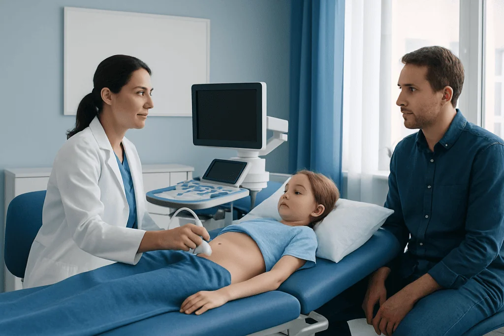
Preparation for a nephroblastoma ultrasound is usually simple. Families should follow the instructions provided by the imaging department, as requirements can differ by age and the area being scanned. Some abdominal ultrasound examinations ask a child not to eat or drink for a few hours beforehand, while others do not require fasting. Comfortable two-piece clothing may make access to the abdomen easier.
During the procedure, the child lies on an examination bed, usually on the back and sometimes briefly on one side. A sonographer applies a clear, water-based gel to the abdomen. The gel helps the transducer make good contact with the skin and is often cool but not painful. The transducer is moved gently over the abdomen and flanks to obtain views of both kidneys, the bladder, major blood vessels, and nearby organs.
The sonographer may ask an older child to take a breath in, hold still for a moment, or change position. Doppler images may be obtained to evaluate blood flow. The scan often takes about 20 to 45 minutes, depending on the child’s cooperation and whether a detailed vascular assessment is needed.
There is no recovery period after a standard ultrasound. The gel is wiped away, and the child can usually return to normal eating, drinking, school, and play right away. Sedation is not usually necessary. If a child needs additional imaging such as MRI, the care team will explain separate preparation and recovery instructions.
What Are the Sonographic Findings of Wilms Tumor?
On ultrasound, Wilms tumor commonly appears as a large, well-defined mass that arises from the kidney and changes its usual shape or architecture. It is often predominantly solid, but its internal appearance can be mixed. Areas of bleeding, tissue breakdown, or cyst-like change may make parts of the mass look darker or more fluid-like on the scan.
The affected kidney may be enlarged, while the other kidney is examined carefully for abnormalities. The radiologist also assesses whether the mass is displacing nearby organs, compressing the collecting system, or causing hydronephrosis, which is swelling of the kidney due to impaired urine drainage. Calcification is not a typical dominant feature in Wilms tumor, although it may occasionally be seen.
Doppler ultrasound can demonstrate blood flow within or around the mass and evaluate the renal vein and inferior vena cava for a possible tumor thrombus. A thrombus is material within a blood vessel that may contain tumor tissue. Detecting or suspecting this finding is important because it can affect the next imaging tests and the surgical approach.
These findings can raise concern for Wilms tumor, but they overlap with other childhood renal masses. The final diagnosis depends on the full clinical picture, cross-sectional imaging, and the treatment pathway determined by a pediatric oncology and pediatric urology team.
Would a Kidney Tumor Show Up on Ultrasound?
Many kidney tumors can be seen on ultrasound, particularly when they are large enough to change the kidney’s contour or differ clearly from normal kidney tissue. Ultrasound can identify a solid mass, a cystic lesion, a mixed mass, or an area that needs closer evaluation. It can also show whether a mass appears to originate within the kidney or next to it.
However, a normal or uncertain ultrasound does not always rule out a small lesion or explain every symptom. Image quality can be affected by bowel gas, body size, the position of the kidney, and a child’s ability to remain still. Some masses have features that cannot be fully characterized with ultrasound alone.
When a renal mass is suspected, clinicians commonly arrange contrast-enhanced CT or MRI of the abdomen and pelvis to evaluate its size, location, relation to blood vessels, and possible spread. A chest scan may also be considered as part of staging when Wilms tumor is diagnosed or strongly suspected. These tests help the multidisciplinary team plan safe, individualized care.
Families should avoid trying to interpret an ultrasound image or report in isolation. A radiology result is most helpful when reviewed alongside the child’s symptoms, examination findings, blood pressure, laboratory results, and any additional imaging.
What Is the “Rule of 10” for Wilms Tumors?
The “rule of 10” is an older teaching mnemonic that summarizes several approximate patterns associated with Wilms tumor. It has traditionally stated that about 10% of cases may involve both kidneys, around 10% may have a family history, and about 10% may be associated with congenital or genetic conditions. The exact proportions vary across studies, populations, and definitions.
This phrase is useful as a broad educational reminder, but it should not be used to predict an individual child’s diagnosis, outcome, or family risk. Most Wilms tumors occur in one kidney and are not linked to an inherited cancer syndrome. When clinicians identify features suggesting a genetic predisposition, they may recommend genetics assessment and tailored surveillance.
Some syndromes and developmental conditions can be associated with a higher likelihood of Wilms tumor, including certain overgrowth syndromes and conditions affecting the eyes, genital or urinary tract, or kidney development. The child’s specialist will consider family history and physical findings carefully, without assuming that every child requires genetic testing.
For a child with a suspected renal tumor, the priority is timely evaluation by an experienced pediatric team. Imaging findings, clinical assessment, and appropriate staging guide the next steps more reliably than any single mnemonic.
What Does Neuroblastoma Look Like on Ultrasound?
Neuroblastoma is a different childhood cancer that commonly begins in nerve tissue, often in or near an adrenal gland above the kidney. On ultrasound, it may appear as a solid, uneven-looking mass above or beside the kidney rather than arising from kidney tissue itself. It can be heterogeneous because of areas of bleeding, calcification, or tissue change.
Unlike a typical Wilms tumor, neuroblastoma may surround or encase nearby blood vessels rather than simply displacing them. It may also cross the middle of the abdomen or extend through openings near the spine in some cases. Calcifications are more commonly seen in neuroblastoma than in Wilms tumor, although ultrasound is not always the best test for identifying them.
There can be overlap between the appearance of adrenal and kidney masses, especially when a tumor is large. CT or MRI is therefore important to identify the organ of origin, assess the relationship with vessels and spinal structures, and plan further care. Other tests may be used when neuroblastoma is considered.
An ultrasound finding does not mean a child has cancer. Many abdominal findings have noncancerous explanations. Still, any suspicious mass should be evaluated promptly by pediatric specialists so that the diagnosis can be clarified and families can receive clear guidance.
Benefits, Limitations, Next Steps and When to Seek Medical Care
The main benefits of nephroblastoma ultrasound are that it is noninvasive, painless, widely available, and free from ionizing radiation. It can quickly provide useful details about the kidneys, a suspected mass, urine drainage, and blood vessels. Its limitations are that it cannot always determine the precise tumor type or fully assess the extent of disease, so CT or MRI is often necessary after an abnormal result.
If a tumor is suspected, care is typically coordinated among pediatric oncology, pediatric urology, radiology, pathology, anesthesia, and supportive-care professionals. Treatment for confirmed Wilms tumor may include surgery to remove part or all of the affected kidney, chemotherapy, and, in selected situations, radiotherapy. The appropriate sequence depends on the child’s imaging findings, tumor stage, kidney function, and overall health.
Parents or caregivers should seek medical care promptly if a child has a new or enlarging abdominal lump or swelling, blood in the urine, persistent abdominal pain, unexplained fever, reduced appetite with weight loss, or a noticeable change in energy. These symptoms can have many causes, but they deserve professional assessment. Urgent care is appropriate if severe pain, significant bleeding, breathing difficulty, fainting, or marked illness develops.
Acibadem International’s multidisciplinary specialists and JCI-accredited hospitals support international patients who need assessment and treatment for pediatric kidney tumors. Families should discuss imaging results and all treatment choices with a qualified pediatric oncology team.
Frequently asked questions
Is nephroblastoma ultrasound painful?
No. A standard ultrasound is painless and does not involve needles, radiation, or surgery. The gel may feel cool, and gentle pressure from the transducer may be felt over the abdomen.
Does a child need to fast before a kidney ultrasound?
Often, no special preparation is needed, but some imaging departments may ask for a short period without food or drink before an abdominal scan. Families should follow the instructions from the facility because preparation can vary by the child’s age and the type of ultrasound ordered.
Can ultrasound confirm Wilms tumor?
Ultrasound can strongly suggest a kidney mass and provide important information about its features, but it cannot confirm Wilms tumor on its own. CT or MRI and assessment by pediatric specialists are usually needed to clarify the diagnosis and plan treatment.
How long does a nephroblastoma ultrasound take?
The examination commonly takes about 20 to 45 minutes. The time may be longer if detailed images of blood vessels are needed or if breaks are required to help a young child stay comfortable.
Why is Doppler used during a Wilms tumor ultrasound?
Doppler ultrasound shows blood flow in and around the kidneys and major abdominal veins. It can help identify whether a mass may involve the renal vein or inferior vena cava, which is relevant to further imaging and treatment planning.
Can a kidney mass found on ultrasound be benign?
Yes. Not every kidney mass is cancerous, and ultrasound may also identify cysts, infections, congenital changes, or other noncancerous conditions. A specialist evaluates the imaging findings together with the child’s symptoms and additional tests to determine the cause.
References
- National Cancer Institute
- American Cancer Society
- Children’s Oncology Group
- Radiological Society of North America
- World Health Organization
This article is for general information only and is not a substitute for professional medical advice. Please consult a qualified doctor about your individual situation.
Ultrasound in Turkey — costs, top hospitals & a free quote
JCI-accredited · board-certified surgeons · reply within 24h
Add us as a Preferred Source to see more of our trusted health content across Google Search, AI Overviews and Discover.
Check your numbers in seconds
BMI, calories, due date, blood pressure and 30+ more clinical calculators — free, instant, doctor-reviewed ranges.

Check-up & Preventive Medicine
Comprehensive health screenings designed for early detection and prevention.
2 specialists in this unitMore from the Health Library



Related Specialists

Dr. Belinda İrem Ardal
Acibadem Life Clinical Service
Fzt. Faika Nur Erkol
Treatment of Lymphedema
Dt. Sıdıka Yaşar
Oral & Dental Health
Dr. İdris Berke Ceylan
Emergency Service