Nephrostomy Tube: An Evidence-Based Guide for Patients

A nephrostomy tube drains urine directly from the kidney when urine cannot flow normally to the bladder. Common reasons for placement include kidney stones, ureter blockage, infection with obstruction, and tumor-related compression.
Key Takeaways
- A nephrostomy tube drains urine directly from the kidney when urine cannot flow normally to the bladder.
- Common reasons for placement include kidney stones, ureter blockage, infection with obstruction, and tumor-related compression.
- Daily care focuses on keeping the tube secure, the dressing clean, and the drainage bag below kidney level.
- Fever, worsening pain, reduced drainage, leakage, or a tube that falls out needs prompt medical advice.
- Many people can walk, wash carefully, and continue light daily activities while the tube is in place.
A nephrostomy tube is a thin drainage tube placed through the skin into the kidney to help urine leave the body when the usual pathway is blocked or not working well. It is often a temporary but important treatment that can protect kidney function, relieve pressure, and help doctors manage the underlying cause safely.
Overview: what a nephrostomy tube is and why it is used
A nephrostomy tube is a soft, narrow tube placed through the skin of the back into the kidney so urine can drain into an external collection bag. Doctors use it when urine cannot pass normally from the kidney down the ureter and into the bladder. In simple terms, it is a way to bypass a blockage and protect the kidney.
This procedure is usually called a percutaneous nephrostomy. “Percutaneous” means through the skin. The tube may be needed for a short time, such as while swelling settles or a stone is treated, or for longer if the blockage is ongoing and needs staged treatment.
A nephrostomy tube does not treat the underlying cause by itself. Instead, it relieves pressure inside the kidney, allows urine to drain, and can reduce the risk of kidney damage. In some situations, it also helps doctors obtain urine samples directly from the kidney or creates a safer path for later procedures.
Because urine backing up into the kidney can lead to pain, infection, or loss of kidney function, timely drainage matters. For some patients, a nephrostomy tube is an urgent measure; for others, it is planned as part of treatment for a condition such as kidney stones or a narrowing in the urinary tract.
How it is placed and what to expect
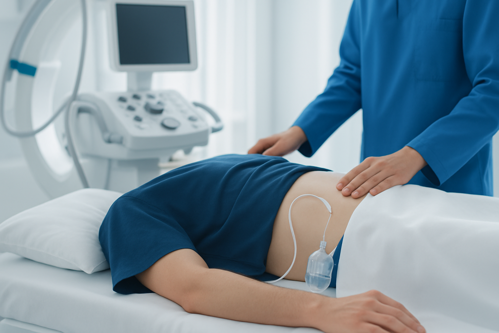
A nephrostomy tube is usually placed by an interventional radiologist or urologist using imaging guidance such as ultrasound or X-ray. The skin is cleaned carefully, the area is numbed with local anesthetic, and a small pathway is created into the kidney so the tube can be positioned correctly. Some patients also receive medicine to help them relax.
The procedure often takes less than an hour, although this can vary depending on the reason for the tube and the anatomy of the urinary tract. After placement, urine begins draining into a bag connected to the tube. The urine may look blood-tinged at first, which can be normal for a short time, but the care team watches for heavy bleeding or other concerns.
Many people stay in hospital for observation, especially if the tube was placed urgently, if there is an infection, or if other procedures are planned. Others may go home relatively soon with clear instructions on caring for the tube, dressing changes, bathing, activity, and follow-up appointments.
The tube is held in place with a locking mechanism, stitch, adhesive device, or dressing. It is important not to pull or twist it. If the nephrostomy is part of a broader treatment plan, the next steps may include imaging, urology evaluation, stone treatment, or relieving the blockage with another procedure such as a stent.
Common reasons a nephrostomy tube may be needed
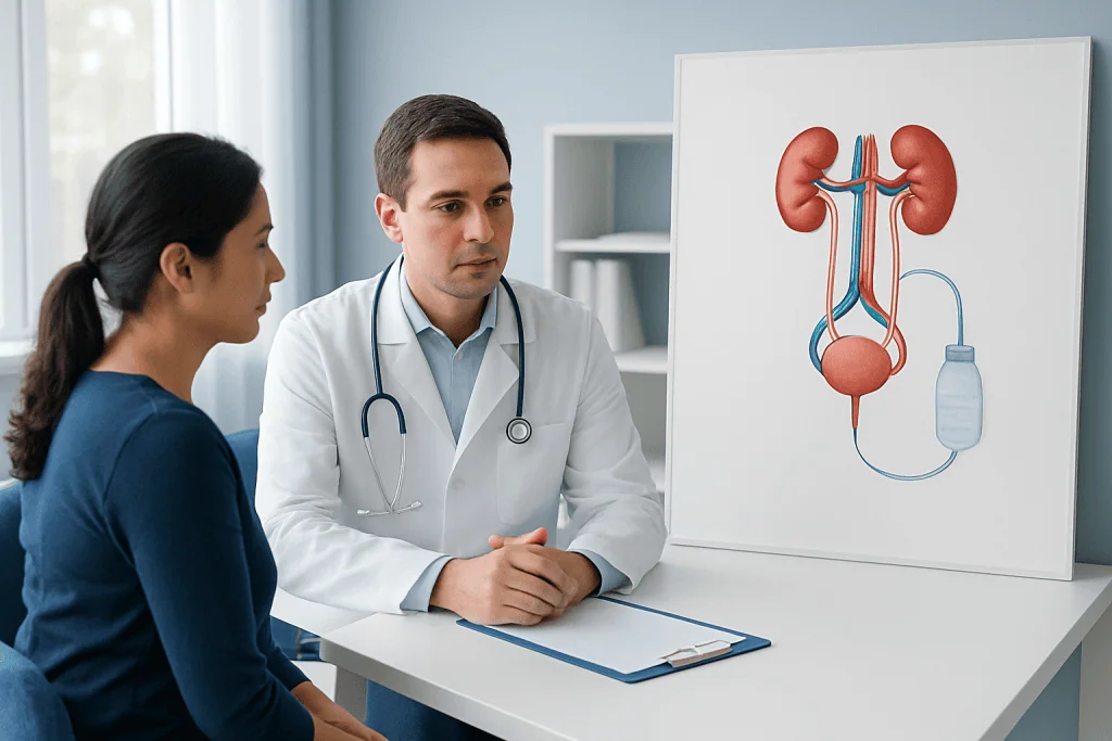
The most common reason for a nephrostomy tube is obstruction, meaning urine cannot drain from the kidney as it should. A blockage may happen suddenly or develop gradually. When pressure builds up in the kidney, it can cause swelling known as hydronephrosis, pain, infection, or declining kidney function.
Several conditions can lead to blockage. Kidney or ureter stones are a frequent cause, especially if a stone becomes lodged and urine cannot pass. In other cases, a ureter may be narrowed by scar tissue, inflammation, prior surgery, or congenital structural problems. Tumors in or near the urinary tract can also compress the ureter from the inside or outside.
A nephrostomy tube may also be used when there is infection together with obstruction, which can become serious because infected urine is trapped. In this situation, urgent drainage is often a key part of treatment along with antibiotics. Less commonly, the tube may be used after injury, after surgery, or to prepare for another procedure involving the kidney.
- Kidney stones blocking the ureter
- Ureter narrowing or scarring
- Tumor-related compression of the urinary tract
- Infected, obstructed urine flow
- Urinary tract injury or post-surgical drainage needs
Living with a nephrostomy tube: daily care and practical tips
Most patients can return to many everyday activities with some adjustments. The main goals are to keep the tube draining freely, prevent infection, and avoid accidental dislodgement. The drainage bag should usually stay below the level of the kidney so urine can flow downward rather than back up.
The dressing and skin around the tube need regular attention. Hands should be washed before touching the tube or bag. The care team may advise how often to change the dressing and when to replace drainage equipment. If the area becomes very red, swollen, increasingly painful, or develops a foul smell, medical advice is important.
Hydration advice varies depending on kidney function, heart health, and the reason for the tube, so patients should follow their clinician’s instructions rather than forcing fluids. Walking and gentle activity are often encouraged, but heavy lifting, sudden pulling, or contact sports may not be appropriate until the doctor says they are safe.
Daily life is usually easier when patients check the tube and bag routinely. Helpful habits include:
- Keep the tubing free from kinks or loops that block drainage.
- Secure the tube so it does not tug on the skin.
- Empty the collection bag as instructed and record output if asked.
- Do not clamp or flush the tube unless specifically told to do so.
- Ask before showering, swimming, or using lotions near the site.
Possible side effects and complications
Most nephrostomy tubes work well, but like any medical device they can have problems. A small amount of blood in the urine soon after placement may occur and often settles. Some soreness around the insertion site is also common for a short time. Persistent or increasing pain, however, should be assessed.
Infection is one of the most important complications to watch for. Warning signs can include fever, chills, feeling generally unwell, cloudy or foul-smelling urine, and new redness or drainage around the tube. Because the tube enters the urinary system directly, careful hygiene and proper bag handling matter.
The tube can also become blocked, kinked, or displaced. If urine suddenly stops draining, drains much less than usual, leaks around the tube, or the tube appears to have moved, the patient should contact the care team promptly. A tube that falls out completely is not something to replace at home; it needs medical attention.
Less commonly, there may be significant bleeding, injury to nearby structures, or problems related to the underlying disease rather than the tube itself. Follow-up imaging or tube exchange may be needed, particularly if the nephrostomy remains in place for an extended period. If ongoing treatment is required, specialists may discuss options through interventional radiology or related urinary tract procedures.
Diagnosis, follow-up, and treatment of the underlying cause
A nephrostomy tube is one part of care, not usually the end of the evaluation. Doctors still need to identify why urine flow was blocked. This often involves blood tests to assess kidney function, urine testing, and imaging such as ultrasound, CT, or contrast studies that show how urine moves through the kidney and ureter.
Follow-up appointments are important because the tube may need to be checked, exchanged, or removed at the right time. Some tubes stay in place only until swelling improves or definitive treatment is completed. Others are needed for longer if the blockage cannot yet be corrected or if treatment is being done in stages.
Definitive treatment depends on the cause. For example, a stone may require kidney stone treatment, while a narrowing of the urinary tract might be managed with dilation, a stent, or surgery. If a mass is compressing the ureter, treatment may focus on that condition while keeping the kidney drained safely.
Patients often want to know whether the tube will be removed. In many cases, yes, once the urinary tract is draining normally again and the doctor confirms that the kidney is safe. Removal timing varies, so individualized follow-up matters more than a fixed schedule.
When to seek medical care
Patients should seek medical care promptly if they have fever, chills, worsening flank or back pain, nausea with inability to keep fluids down, or feel suddenly much more unwell. These symptoms can suggest infection or another complication that should not wait.
Urgent advice is also needed if the nephrostomy tube stops draining, drains much less than usual, leaks heavily around the insertion site, or appears blocked or dislodged. Bright red bleeding that does not settle, large clots, or a tube that falls out completely also needs prompt assessment.
Less urgent but still important reasons to contact the care team include increasing redness of the skin, a loose dressing that cannot be replaced properly, persistent bad odor, or questions about bathing, activity, or supplies. Clear communication can prevent small issues from becoming larger ones.
Near the end of the care pathway, some patients may need coordinated review by urology, radiology, and nephrology teams. Acibadem International’s multidisciplinary specialists and JCI-accredited hospitals diagnose and treat urinary tract conditions for international patients when this type of coordinated care is needed.
Frequently asked questions
Is a nephrostomy tube permanent?
Usually not. Many nephrostomy tubes are temporary and stay in place only until the blockage is treated or urine flow returns to normal. Some patients need a longer-term tube if the underlying problem cannot be corrected right away.
Does having a nephrostomy tube hurt?
Some discomfort around the insertion site is common, especially soon after placement. Most patients describe soreness rather than severe pain once the tube is secured and draining well. New or worsening pain should be reported because it can signal blockage, infection, or displacement.
Can a person walk and sleep normally with a nephrostomy tube?
Many people can walk and do light daily activities with care. Sleeping is usually possible in a comfortable position that does not pull on the tube or compress the drainage bag. The care team may suggest ways to secure the tube and bag during rest and movement.
What should the urine look like in the drainage bag?
Urine is often yellow, but it may be lightly blood-tinged for a short time after placement. It should continue to drain rather than stopping suddenly. Cloudy urine, a strong odor, or a major change in color together with symptoms such as fever should be discussed with a doctor.
Can a person shower with a nephrostomy tube?
This depends on the clinician's instructions and the type of dressing used. Many patients can shower carefully after a certain period, but soaking the site or swimming may be restricted. The safest approach is to follow the local care instructions given after the procedure.
What happens if the nephrostomy tube stops draining?
A sudden drop in drainage can mean the tube is kinked, blocked, or displaced. The patient should check for obvious kinks and keep the bag below kidney level, but should not try to force a flush unless told to do so. Prompt contact with the medical team is important.
This article is for general information only and is not a substitute for professional medical advice. Please consult a qualified doctor about your individual situation.
Explore treatments in Turkey — costs, top hospitals & a free quote
JCI-accredited · board-certified surgeons · reply within 24h
Add us as a Preferred Source to see more of our trusted health content across Google Search, AI Overviews and Discover.

More from the Health Library

Bradycardia: A Complete Medical Overview

External Rotation: What Patients Need to Know

Pubisa: An Evidence-Based Guide for Patients

Renal: A Complete Medical Overview

Zyban: What Patients Need to Know