Nevus — Explained by Medical Evidence, Not Myths

Nevus is a broad medical term that includes many types of moles and some birthmarks. Most nevi are benign and stay stable over time.
Key Takeaways
- Nevus is a broad medical term that includes many types of moles and some birthmarks.
- Most nevi are benign and stay stable over time.
- Changes in size, color, shape, or symptoms such as bleeding or itching need medical evaluation.
- Doctors diagnose a nevus mainly by skin examination and sometimes dermoscopy or biopsy.
- Sun protection and regular skin self-checks help support early detection of concerning changes.
A nevus is the medical term for a mole or certain birthmarks formed by a cluster of skin cells, usually pigment-producing melanocytes. Most nevi are benign, but new, changing, irregular, or symptomatic lesions should be assessed by a qualified doctor to rule out skin cancer or other skin conditions.
What is a nevus?
A nevus is a common medical term for a mole or a localized skin mark caused by a cluster of cells. In many cases, these are melanocytic nevi, meaning they form from melanocytes, the cells that make pigment. A nevus may be present at birth or appear later in childhood, adolescence, or adulthood.
Most nevi are harmless. They can be flat or raised, smooth or rough, and vary in color from pink or tan to brown or nearly black. Some people have only a few, while others naturally develop many over their lifetime.
The word nevus does not describe a single condition. Instead, it covers different types of lesions with different appearances and levels of clinical importance. This is one reason a professional skin assessment can be helpful when a mark looks unusual or changes over time.
Medical evidence shows that most moles are benign, but a small number of pigmented lesions can resemble or develop into skin cancer. This is why doctors focus less on myths about “good” or “bad” moles and more on careful examination, pattern recognition, and monitoring for change.
Types of nevus and how they differ
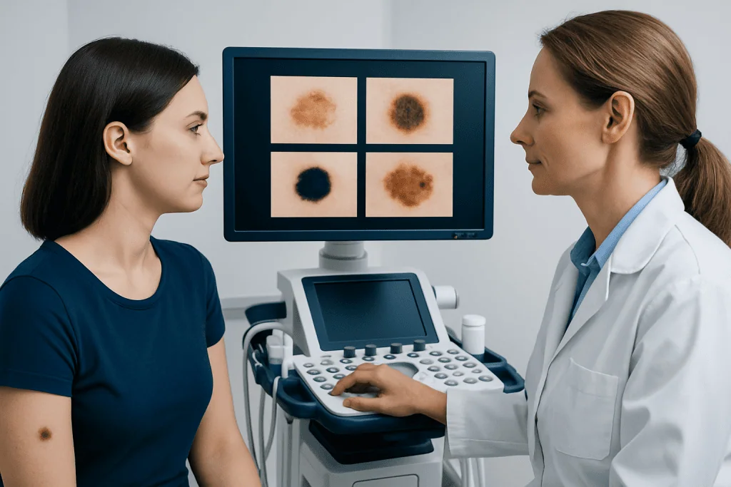
Nevi are often classified by when they appear, what cells they contain, and how they look under clinical examination. Congenital nevi are present at birth, while acquired nevi develop later. Small acquired moles are very common and are usually not dangerous.
Common melanocytic nevi may be junctional, compound, or intradermal, depending on where the nevus cells are located in the skin. Junctional nevi are often flat and darker. Compound nevi may be slightly raised. Intradermal nevi are often flesh-colored or soft and raised.
Some lesions are called atypical or dysplastic nevi. These may be larger, have less uniform color, or have somewhat irregular borders. They are not the same as melanoma, but they can increase clinical concern because they may resemble melanoma and can be associated with a higher overall risk in some individuals.
There are also specialized forms, such as blue nevus, Spitz nevus, and congenital melanocytic nevus. Not every pigmented spot is a nevus. Conditions such as freckles, seborrheic keratoses, and some forms of skin cancer can look similar, which is why diagnosis should not rely on appearance alone when a lesion is changing or unusual.
Signs, symptoms, and changes to watch for
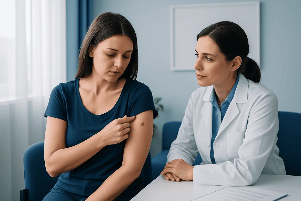
Most nevi do not cause symptoms. They are often discovered incidentally and remain stable for years. A typical benign nevus tends to be symmetrical, evenly colored, and consistent in shape and size.
Doctors often recommend paying attention to the ABCDE features of a mole: asymmetry, border irregularity, color variation, diameter becoming larger, and evolution over time. Evolution is especially important because change can be a key sign that a lesion needs expert review.
Other symptoms that deserve attention include itching, tenderness, crusting, spontaneous bleeding, persistent scaling, or a mole that suddenly becomes more raised. A new dark lesion in adulthood can also justify medical assessment, especially if it looks different from a person’s other moles.
Some people use the “ugly duckling” rule: a spot that stands out from the rest of the skin marks on the body may need closer evaluation. This approach does not diagnose cancer, but it can help identify lesions that should be checked with a skin examination and, when indicated, dermatology evaluation.
Causes and risk factors
Nevi develop when skin cells grow in clusters instead of spreading evenly through the skin. Genetics plays a major role, and many people inherit a tendency to develop more moles. Hormonal changes, especially during puberty and pregnancy, can also influence the appearance or darkening of nevi.
Sun exposure is another important factor, particularly for acquired melanocytic nevi. Ultraviolet radiation can affect pigment cells and may increase the number of nevi or promote changes in existing lesions. This does not mean every sun-exposed mole is dangerous, but it supports the importance of sun protection.
Risk factors that increase the need for careful skin monitoring include fair skin, a history of intense sun exposure or sunburns, many moles, atypical nevi, a personal history of skin cancer, or a family history of melanoma. Congenital large nevi also deserve specialist follow-up because their long-term risks differ from those of small common moles.
Not all changes are caused by cancer. Friction from clothing, shaving, or minor trauma can irritate a nevus and make it appear inflamed. Even so, persistent or unexplained changes should not be dismissed without clinical assessment.
How doctors diagnose a nevus
Diagnosis begins with a medical history and visual skin examination. A doctor will ask when the lesion appeared, whether it has changed, and whether there are symptoms such as bleeding, itching, or pain. Personal and family history of skin cancer also matters.
Many clinicians use dermoscopy, a noninvasive tool that magnifies the skin and helps reveal pigment patterns not visible to the naked eye. Dermoscopy can improve accuracy in distinguishing many benign lesions from those that are more suspicious.
If a nevus appears atypical or concerning, the doctor may recommend a biopsy or complete removal so the tissue can be examined under a microscope. Histopathology is the most reliable way to confirm whether a lesion is benign, atypical, or malignant.
Patients with many moles or atypical nevi may benefit from periodic skin mapping or follow-up photography. In selected cases, referral to specialists in dermatology or evaluation for melanoma risk may be appropriate. The aim is not to remove every mole, but to identify lesions that need closer attention while avoiding unnecessary procedures.
Treatment options and when removal is considered
Most benign nevi do not need treatment. If a mole is stable and has reassuring clinical features, doctors often recommend observation rather than removal. This approach is based on evidence and helps avoid unnecessary procedures and scarring.
Removal may be advised if the nevus shows suspicious changes, repeatedly becomes irritated, is difficult to monitor, or causes cosmetic concern. The decision depends on the lesion’s size, location, appearance, and the person’s medical history.
Common removal methods include shave removal, punch removal, or surgical excision. If cancer is a concern, excision that preserves the tissue for pathology is usually preferred over destructive methods, because the sample needs to be examined accurately. In some cases, related care may involve skin cancer treatment if a malignancy is diagnosed.
People should avoid trying to remove a nevus at home. Home treatments can cause infection, scarring, incomplete removal, and delayed diagnosis of serious conditions. Professional evaluation is the safest path whenever a mole appears unusual or troublesome.
Prevention, self-checks, and skin awareness
Nevi cannot always be prevented, especially when genetics plays a strong role. However, protecting the skin from ultraviolet exposure may help reduce preventable changes in moles and lowers the overall risk of skin damage and skin cancer.
Helpful habits include using broad-spectrum sunscreen, wearing protective clothing, seeking shade during peak sunlight hours, and avoiding tanning beds. These steps are useful for adults and children alike, particularly those with fair skin or many moles.
Regular self-examination can support early detection. A person can check the entire skin surface once a month, including the scalp, back, soles, and areas under nails when practical. Photos may help track lesions over time, especially for people with many moles.
Self-checks do not replace expert care. If there are multiple atypical lesions, a personal or family history of melanoma, or a mole in a hard-to-see area, scheduled reviews with a skin specialist may be recommended. For some patients, access to multidisciplinary care, including skin specialist care, can help with long-term monitoring.
When to seek medical care
Medical review is advisable if a nevus is new in adulthood, grows quickly, changes shape, develops more than one color, or looks noticeably different from other moles. It is also sensible to arrange an appointment if a lesion itches persistently, bleeds without clear injury, crusts, or does not heal as expected.
Urgent assessment may be needed if there is rapid evolution or strong concern for melanoma. Early evaluation helps doctors decide whether monitoring is enough or whether biopsy is the safer option. Prompt review is especially important for people with a personal or family history of skin cancer.
Children with congenital nevi should follow their doctor’s advice about routine observation, photography, and specialist review when needed. Large or unusual congenital lesions sometimes require assessment by more than one specialist.
Acibadem International’s multidisciplinary specialists in JCI-accredited hospitals diagnose and treat skin lesions, including suspicious nevi, for international patients. The most appropriate next step is always an in-person medical assessment rather than relying on myths, online images, or self-diagnosis.
Frequently asked questions
Is a nevus the same as a mole?
Often, yes. Nevus is the medical term commonly used for a mole, especially a melanocytic mole. However, the term can also include some other localized skin lesions or birthmarks, so doctors use it in a broader way than everyday language.
Are all nevi dangerous?
No. Most nevi are benign and never cause health problems. Doctors become more concerned when a lesion changes over time, has irregular features, or causes symptoms such as bleeding or persistent itching.
Can a benign nevus turn into melanoma?
Some melanomas arise in or near a pre-existing mole, but many develop as new lesions on previously normal skin. A benign nevus does not automatically become cancerous, yet any significant change should be assessed by a doctor.
Should a nevus be removed as a precaution?
Not usually. If a nevus looks benign and remains stable, observation is often the preferred approach. Removal is generally considered when there are suspicious changes, repeated irritation, diagnostic uncertainty, or cosmetic reasons.
What does an atypical nevus mean?
An atypical nevus is a mole with some unusual features, such as uneven color, larger size, or less regular borders. It is not the same as melanoma, but it may need closer monitoring or biopsy because it can resemble melanoma clinically.
How often should someone check their moles?
A monthly self-check is a practical routine for many people, especially those with multiple moles. Anyone with atypical nevi, a history of skin cancer, or a strong family history may also need regular professional skin examinations.
This article is for general information only and is not a substitute for professional medical advice. Please consult a qualified doctor about your individual situation.
Explore treatments in Turkey — costs, top hospitals & a free quote
JCI-accredited · board-certified surgeons · reply within 24h
Add us as a Preferred Source to see more of our trusted health content across Google Search, AI Overviews and Discover.

More from the Health Library

Endometrial Stripe: What Patients Need to Know

Diarrhea Light Headed: An Evidence-Based Guide for Patients

Juice Box: An Evidence-Based Guide for Patients

Lucid Dreaming: A Complete Medical Overview
Supraclavicular: An Evidence-Based Guide for Patients