Newborn Hip Ultrasound: Preparation, Procedure and Results

Newborn hip ultrasound uses sound waves, not radiation, to assess the developing hip joint. It is most informative in early infancy, before the hip bones have hardened enough to limit ultrasound views.
Key Takeaways
- Newborn hip ultrasound uses sound waves, not radiation, to assess the developing hip joint.
- It is most informative in early infancy, before the hip bones have hardened enough to limit ultrasound views.
- Babies may be referred after an abnormal hip examination or because of risk factors such as breech position or a family history of DDH.
- The scan usually takes about 15 to 30 minutes and requires no recovery time.
- An unusual ultrasound result does not always mean permanent hip dysplasia; follow-up imaging and specialist assessment may be needed.
A newborn hip ultrasound is a painless imaging test that uses sound waves to examine how an infant’s hip joints are shaped and positioned. It is commonly used when a physical examination suggests hip instability or when a baby has risk factors for developmental dysplasia of the hip (DDH).
Overview: why newborn hip ultrasound is performed
A newborn hip ultrasound is an imaging examination that shows the soft tissues and early bone structures of a baby’s hips. It helps clinicians assess whether the ball at the top of the thigh bone is well seated within the hip socket and whether the socket is developing appropriately. The examination is often part of early assessment for developmental dysplasia of the hip (DDH), a condition in which the hip joint is shallow, unstable or partly or fully displaced.
Unlike an X-ray, ultrasound does not use ionising radiation. This makes it particularly useful during the first months of life, when much of the hip joint is still cartilage and cannot be fully evaluated on an X-ray. A newborn hip ultrasound radiology examination may be arranged because of a finding during a routine newborn check, or as a screening test for a baby with recognised risk factors.
Early assessment can help identify hips that need observation or treatment while they are still developing. Many babies who have a slightly immature hip on an early scan simply need repeat assessment, while others benefit from prompt review by a paediatric orthopaedic specialist.
Who may need a baby hip ultrasound?
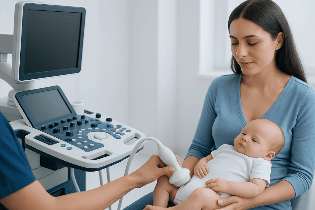
Not every newborn needs a hip ultrasound. Clinicians usually recommend it when the physical hip examination suggests instability, reduced movement or an asymmetrical finding. During these examinations, a clinician gently checks how the hips move; the test is designed to identify a hip that may be able to move out of, or back into, the socket.
A baby may also be referred even when the examination is normal if there are important risk factors for DDH. These commonly include being in a breech position late in pregnancy, a close family history of hip dysplasia, certain foot or neck positioning conditions, or other concerns identified by the paediatric team. Girls are affected more often than boys, but any baby with relevant findings can require assessment.
The timing depends on the reason for referral and local clinical guidance. Screening scans are often scheduled after the first few weeks of life because very early scans can show temporary immaturity that resolves naturally. If the doctor suspects a dislocated or significantly unstable hip, assessment may be arranged sooner.
How a newborn hip ultrasound works
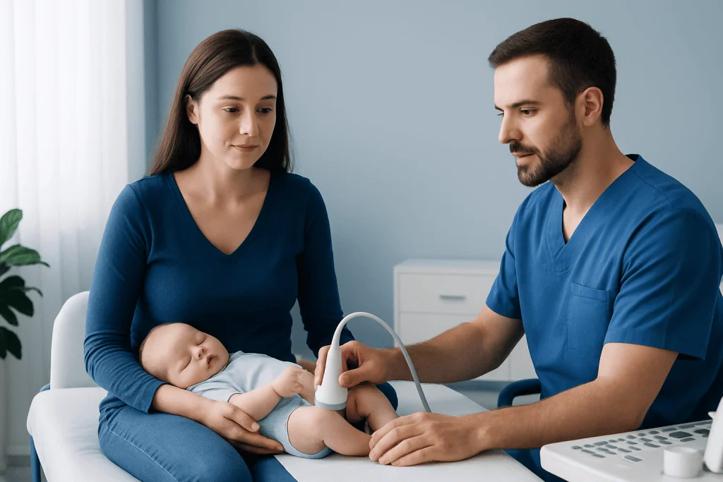
Ultrasound uses a handheld device called a transducer to send high-frequency sound waves into the body. The returning echoes are converted into real-time images on a screen. For hip imaging, the sonographer and reporting clinician examine the relationship between the rounded femoral head and the socket, as well as the shape and coverage of the socket.
The neonatal hip ultrasound protocol commonly includes images of each hip while the baby rests on their side. Depending on the clinical question, the examiner may also observe the hip during very gentle movement. This dynamic assessment can provide information about stability as well as joint shape. The baby hip ultrasound protocol is adapted to the infant’s age, comfort and referral reason.
Measurements and image features are interpreted using established methods, often including the Graf classification system. Results should be considered alongside the baby’s age, physical examination and risk factors. A scan is therefore one part of a wider clinical assessment rather than a stand-alone diagnosis.
What to expect at an infant hip ultrasound?
Parents or caregivers can usually remain with their baby throughout the appointment. There is no special preparation: the baby can generally feed as usual and continue taking prescribed medicines. It can be helpful to bring a feed, a pacifier if used, and familiar items that may help keep the baby calm.
For the scan, the baby is usually placed on their side in a supportive foam cradle or on an examination couch. The sonographer applies a small amount of clear gel to the skin over the hip and moves the transducer gently over the area. The gel may feel cool, but the examination itself should not hurt.
Babies may become unsettled if they are hungry, tired or uncomfortable, so allowing extra time for feeding and soothing can be useful. Parents should not try to hold the hips in a particular position unless the imaging professional asks them to. The team will guide positioning carefully and safely.
Step-by-step procedure, timing and recovery
How long does a newborn hip ultrasound take? The actual imaging commonly takes around 15 to 30 minutes. An appointment may take a little longer if the baby needs feeding or settling, if both hips need detailed dynamic images, or if additional review is required. The scan is performed while the baby is awake; sedation and anaesthesia are not normally needed.
The procedure starts with checking the referral information and confirming the baby’s identity. The sonographer positions the infant comfortably, obtains standard images of one hip and then the other, and may make gentle movements to see whether the joint remains stable. Images are saved for interpretation by an appropriately trained clinician or radiologist.
There is no recovery period. The gel is wiped away after the scan, and the baby can be fed, held and cared for normally straight away. No activity restrictions are needed. Results may be discussed on the day in some settings, while in others the referring paediatrician or family doctor receives a formal report and contacts the family.
Understanding results, including the 50% rule
Results may be reported as normal, physiologically immature, borderline, unstable, dysplastic or dislocated, depending on the images and measurements. A normal result means the hips appear appropriately formed and located for the baby’s age. An immature hip may be monitored with a repeat scan because early development can change quickly.
What does the 50% rule mean on a hip ultrasound for developmental dysplasia? The 50% rule is a descriptive guideline used in some ultrasound assessments: ideally, at least half of the femoral head should appear covered by the bony hip socket. Coverage below this level can suggest reduced containment of the femoral head and may support concern for dysplasia or instability. However, this observation is not interpreted alone; clinicians also consider hip shape, angles, stability, the baby’s age and the complete examination.
If the scan indicates a concern, the next step may be repeat ultrasound, referral to paediatric orthopaedics, or treatment designed to hold the hip in a stable position while it develops. Assessment and care for developmental dysplasia of the hip should be individualised, as some findings resolve with observation while others need earlier intervention.
Do babies outgrow hip dysplasia?
Do babies outgrow hip dysplasia? Some mild hip immaturity seen in early infancy improves as the baby grows, particularly when the hip is stable and the finding is identified in the first weeks of life. This is why clinicians may recommend a follow-up scan rather than immediate treatment in selected cases.
However, confirmed hip dysplasia, instability or dislocation should not be assumed to resolve without medical follow-up. A hip that remains shallow or unstable can affect normal joint development over time. Early monitoring allows clinicians to distinguish temporary immaturity from a condition that needs treatment.
When treatment is recommended, a soft positioning brace such as a Pavlik harness is often used in young infants to support healthy alignment while allowing controlled movement. The exact plan, duration and follow-up schedule depend on the ultrasound findings and specialist assessment. Parents should follow the care team’s instructions and attend all scheduled reviews.
Benefits, limitations and when to seek medical care
The main benefits of newborn hip ultrasound are that it is painless, does not use radiation and provides real-time images of developing joint structures. It can identify babies who need closer observation or early treatment, which may support normal hip development. Limitations include the fact that results can vary with the baby’s age, position and movement, and that early findings sometimes require repeat imaging for clarity.
When to seek medical care Parents should contact their baby’s doctor if they have been advised to arrange a hip ultrasound, if a baby’s legs appear to move differently, or if one hip seems less flexible during routine care. A difference in leg length, uneven thigh or buttock creases, or a noticeable limp later in infancy or childhood should also be assessed. These signs do not confirm dysplasia, but they deserve professional evaluation.
Families should not attempt to test hip stability at home or use positioning devices without clinical advice. For babies diagnosed with DDH, coordinated care may involve paediatricians, radiologists and paediatric orthopaedic specialists. Acibadem International’s multidisciplinary specialists and JCI-accredited hospitals support international patients requiring assessment and treatment for childhood hip conditions.
Frequently asked questions
Is newborn hip ultrasound safe?
Yes. Ultrasound uses sound waves rather than ionising radiation, and it is widely used to examine babies. The transducer is placed gently on the skin, and the test does not usually cause pain.
At what age is a hip ultrasound most useful for babies?
Hip ultrasound is most useful during the first several months of life, when the hip contains substantial cartilage that ultrasound can show clearly. As the baby grows and the bones harden, an X-ray may become more useful for follow-up assessment.
Does a normal newborn hip ultrasound rule out future hip problems?
A normal scan is reassuring and indicates that the hips looked appropriate at the time of imaging. However, parents should still attend routine child health checks and seek medical advice if they later notice limited hip movement, leg-length differences or changes in walking.
What happens if a baby’s hip ultrasound is abnormal?
The next step depends on the type and severity of the finding. A clinician may recommend repeat ultrasound, referral to a paediatric orthopaedic specialist, or early treatment to support stable hip development.
Can a baby wear a diaper during a hip ultrasound?
The diaper may need to be loosened or removed briefly so the hips can be positioned and scanned properly. Parents can bring a spare diaper and wipes for after the examination.
Why might a doctor repeat a newborn hip ultrasound?
A repeat scan may be recommended when an early hip appears immature, when images are not fully clear, or when the clinician wants to monitor development after an abnormal finding. Repeat imaging helps determine whether the hip is maturing normally or needs further care.
References
- American Academy of Pediatrics
- International Hip Dysplasia Institute
- American College of Radiology
- Radiopaedia
- National Institute for Health and Care Excellence
This article is for general information only and is not a substitute for professional medical advice. Please consult a qualified doctor about your individual situation.
Ultrasound in Turkey — costs, top hospitals & a free quote
JCI-accredited · board-certified surgeons · reply within 24h
Add us as a Preferred Source to see more of our trusted health content across Google Search, AI Overviews and Discover.

Check-up & Preventive Medicine
Comprehensive health screenings designed for early detection and prevention.
3 specialists in this unit