Nuclear Medicine Injection: How It Works, Results and What to Expect

A nuclear medicine injection contains a radiotracer that helps imaging equipment assess body function, not only anatomy. The scan type determines the tracer, preparation instructions, timing, and how long the appointment lasts.
Key Takeaways
- A nuclear medicine injection contains a radiotracer that helps imaging equipment assess body function, not only anatomy.
- The scan type determines the tracer, preparation instructions, timing, and how long the appointment lasts.
- Most people have no significant side effects; serious reactions are uncommon, but staff should be told about pregnancy, breastfeeding, allergies, and medicines.
- Radiation exposure is generally low and is carefully selected to provide useful diagnostic information.
- Drinking fluids and following the imaging team's instructions can help the tracer leave the body after many scans.
A nuclear medicine injection delivers a small amount of a radioactive tracer, called a radiotracer, that allows special cameras to show how organs, bones, or tissues are functioning. It is commonly used for diagnosis, and the tracer usually leaves the body naturally over time.
Overview: What Is a Nuclear Medicine Injection?
A nuclear medicine injection is a diagnostic procedure in which a healthcare professional injects a very small amount of a radiotracer into a vein. The radiotracer travels through the body and collects in specific organs, tissues, or cells. A special camera then detects the energy it gives off, creating images that show how the body is functioning.
Unlike an X-ray, CT scan, or MRI, which mainly show structure, nuclear medicine can reveal processes such as blood flow, bone repair, heart muscle activity, thyroid activity, or how tissues use glucose. Depending on the reason for testing, the scan may be a PET scan, SPECT scan, bone scan, thyroid scan, renal scan, or another type of nuclear imaging.
The injection is most often used to investigate a symptom, clarify an uncertain finding from another test, assess the extent of a known condition, or monitor response to treatment. It is a diagnostic tool rather than a treatment in most situations, although some nuclear medicine treatments also use radioactive substances at different doses.
How a Nuclear Medicine Injection Works
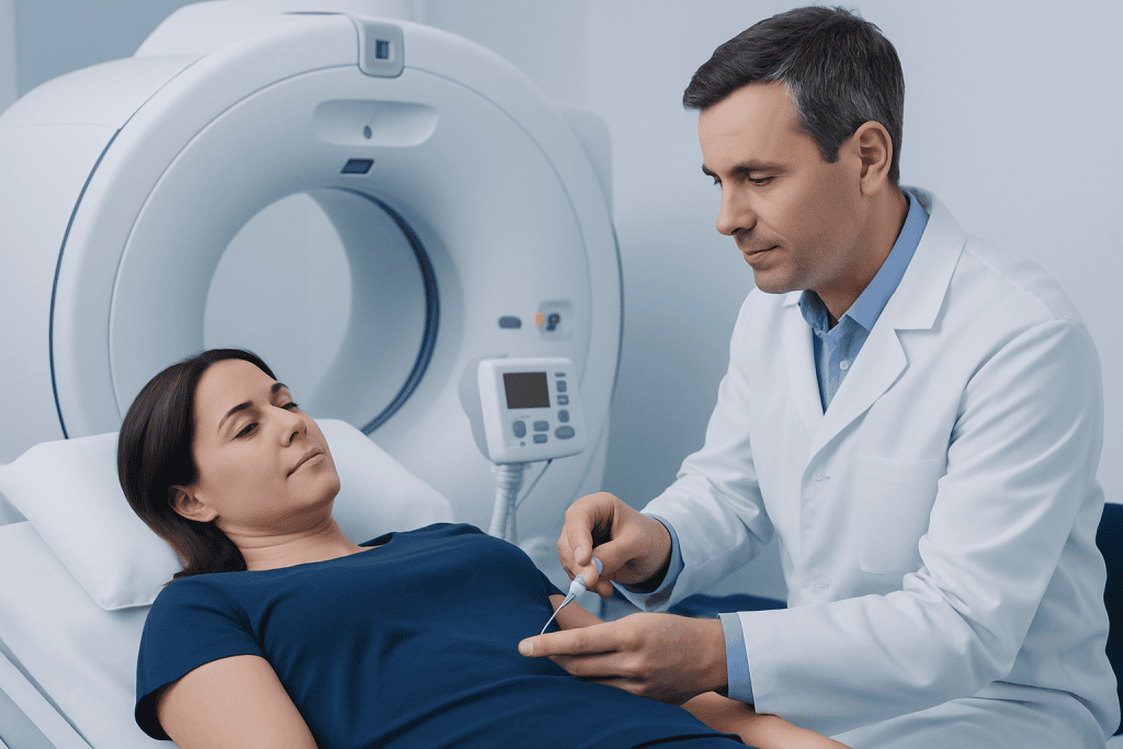
Radiotracers are medicines linked to a radioactive atom. They are designed to behave in a particular way in the body. For example, a bone tracer is taken up in areas of bone remodeling, while a glucose-like tracer used in many PET scans may gather in cells with higher glucose use.
After the injection, the radiotracer needs time to circulate and reach the area being studied. This waiting period may be short or may last several hours, depending on the examination. During imaging, a gamma camera or PET scanner records signals from the tracer and a specialist interprets the pattern of uptake.
Areas with greater or lower tracer uptake are not a diagnosis by themselves. They can reflect normal body processes, inflammation, infection, injury, healing, or cancer, depending on the scan and clinical context. The nuclear medicine physician considers the images alongside symptoms, medical history, laboratory results, and other imaging tests.
Candidacy and Preparation

A doctor may recommend a nuclear medicine injection when information about organ function could guide care. Examples include assessing unexplained bone pain, evaluating heart blood flow, investigating thyroid disorders, examining kidney drainage, locating some infections, or staging and monitoring certain cancers.
People should tell the imaging team if they are pregnant, might be pregnant, are breastfeeding, have kidney problems, have diabetes, are taking medicines or supplements, or have had recent imaging tests. Pregnancy and breastfeeding do not always prevent testing, but the team may adjust the plan, delay a non-urgent scan, or provide specific instructions to reduce exposure to a baby.
Preparation varies. Some scans require fasting, avoiding strenuous exercise, drinking extra water, temporarily changing certain medicines, or avoiding iodine-containing substances. Others need little preparation. Patients should follow the instructions from their own imaging center, since preparation for a PET scan differs from preparation for a thyroid or cardiac scan.
- Wear comfortable clothing and remove metal items if requested.
- Bring a list of medicines, allergies, and relevant previous test results.
- Ask in advance about eating, drinking, exercise, diabetes medicines, and breastfeeding guidance.
Step by Step: During and After the Procedure
At the appointment, staff confirm the reason for the scan, review safety questions, and may check blood glucose or other relevant measurements. The radiotracer is then injected through a small needle, similar to a routine blood test or intravenous injection. Most people feel only a brief pinch.
There may be a waiting period before imaging. Patients may be asked to rest quietly, drink water, empty their bladder, or avoid close contact with others in a waiting area, depending on the tracer. The imaging itself is painless. It usually involves lying still on a table while a camera moves around or near the body.
After the images are complete, most people can go home the same day and return to usual activities unless given different instructions. The images are reviewed by a nuclear medicine physician or radiologist, and the referring clinician explains what the results mean and whether any follow-up testing or care is needed.
Acibadem International’s multidisciplinary specialists and JCI-accredited hospitals provide nuclear medicine assessment and coordinated care for international patients when this type of imaging is clinically appropriate.
Benefits, Risks and Recovery Timeline
The main benefit of a nuclear medicine injection is that it can provide functional information that may not be visible on structural imaging alone. It can help clinicians identify abnormal activity, choose further tests, plan treatment, or evaluate how well treatment is working. In many cases, it can reduce uncertainty without the need for more invasive procedures.
The amount of radiation used is generally small and is selected according to the test and the person’s circumstances. The risk from diagnostic radiation is considered low, while the expected benefit is the medically useful information gained. The tracer itself is usually cleared through urine, stool, breathing, or natural radioactive decay.
Side effects are uncommon. Mild bruising, soreness, or redness at the injection site can occur. Allergic reactions are rare, but any new rash, swelling, breathing difficulty, dizziness, or feeling unwell should be reported immediately to staff or assessed urgently. For most diagnostic nuclear medicine scans, there is no incision, sedation, or extended recovery period.
Recovery is typically immediate. Many people are advised to drink fluids and urinate regularly for the remainder of the day, unless they have been told to limit fluids for another medical reason. Individual aftercare guidance takes priority because it depends on the specific radiotracer and scan.
How Long Does Nuclear Medicine Last in Your Body?
How long nuclear medicine lasts in the body depends on the radiotracer used, the scan performed, kidney function, hydration, and the body’s natural metabolism. Many commonly used diagnostic tracers lose most of their radioactivity and are eliminated within hours to a few days. The imaging team can explain the expected timeframe for the exact tracer received.
Radiotracers have a physical half-life, meaning the radioactivity decreases naturally over time, and a biological clearance process, meaning the body removes the substance. These processes occur together. Drinking fluids and emptying the bladder regularly may help remove some tracers more quickly when this is appropriate.
For some exams, temporary precautions may be advised around pregnant people, infants, or breastfeeding. These precautions are usually limited in duration and are tailored to the tracer. Patients should not rely on general internet advice when they have been given tracer-specific instructions by their care team.
What Not to Do After a Nuclear Medicine Scan
After a nuclear medicine scan, patients should not ignore the discharge instructions given for their particular test. They should not assume that all scans have identical rules, because recommendations differ for PET, bone, thyroid, cardiac, and other studies. If sedation was used, driving, alcohol, and important decisions should be avoided until the effects have fully worn off.
Unless the clinical team says otherwise, it is often helpful to avoid dehydration after the scan. Patients may be asked to drink more water, use the toilet frequently, wash their hands carefully, and flush the toilet thoroughly. These practical measures reduce the time a tracer remains in the body and limit unnecessary exposure to others.
Patients should not delay asking for advice if they are breastfeeding, caring closely for a baby or young child, or sharing a home with someone who is pregnant. The nuclear medicine department can provide clear, individualized guidance on contact, feeding, and hygiene. Usual meals, work, and gentle daily activities are commonly possible after diagnostic imaging.
Is Nuclear Medicine Better Than MRI?
Nuclear medicine is not universally better than MRI; the tests answer different clinical questions. MRI produces detailed images of anatomy and soft tissues without ionizing radiation. It is often particularly useful for the brain, spine, joints, muscles, pelvic organs, and many other structural concerns.
Nuclear medicine imaging is especially valuable when clinicians need to understand function, metabolism, blood flow, or molecular activity. For example, a nuclear medicine scan may help assess heart perfusion, bone turnover, thyroid function, or the activity of some cancers. In certain situations, PET combined with CT or MRI provides both functional and anatomical information.
The best examination depends on the symptom, suspected diagnosis, previous test results, medical history, and urgency. A referring doctor and imaging specialist select the test that is most likely to answer the specific clinical question while avoiding unnecessary testing.
What Diseases Can Nuclear Medicine Detect?
Nuclear medicine can help detect or assess a wide range of conditions, but the scan does not diagnose every disease on its own. In oncology, PET and other nuclear medicine studies may help evaluate some cancers, determine whether disease has spread, guide biopsy planning, or monitor treatment response. Results must always be interpreted carefully because inflammation and infection can also cause increased uptake.
Other uses include evaluating coronary artery disease and heart muscle function, thyroid overactivity or nodules, certain kidney and urinary drainage problems, bone fractures that may not show on early X-rays, bone infection, inflammatory disorders, and selected neurological conditions. Some scans are also used to investigate gastrointestinal bleeding or problems with gallbladder function.
Abnormal uptake does not automatically mean cancer or another serious illness. A finding may be benign, related to recent injury or treatment, or require correlation with CT, MRI, ultrasound, laboratory tests, or follow-up imaging. The clinician who requested the scan can explain how the result applies to the person’s health situation.
When to Seek Medical Care
Patients should contact the imaging department or their doctor if they develop concerning symptoms after an injection, such as hives, swelling of the face or throat, breathing difficulty, fainting, or severe persistent pain at the injection site. Severe breathing difficulty or signs of a serious allergic reaction require emergency medical care.
Medical advice is also important if a person discovers they may have been pregnant at the time of the test, has questions about breastfeeding, or did not receive clear discharge instructions. These concerns can usually be addressed promptly with tracer-specific guidance.
A scan result should be discussed with the referring clinician, particularly if symptoms persist, worsen, or new symptoms develop. Nuclear medicine results are one part of a broader clinical assessment and should not be used alone to self-diagnose or to make treatment decisions.
Frequently asked questions
Does a nuclear medicine injection hurt?
The injection usually feels similar to a routine blood draw or intravenous injection: a brief pinch followed by mild, temporary discomfort at most. The scan itself is painless, although lying still may be uncomfortable for some people. Staff can help with positioning and explain what to expect.
Can a nuclear medicine scan make someone radioactive?
A radiotracer temporarily gives off a small amount of radiation while it is in the body. It does not make a person permanently radioactive. The tracer naturally loses radioactivity and is eliminated over a short period, with timing depending on the examination.
Can people eat before a nuclear medicine injection?
It depends on the type of scan. Some tests require fasting or dietary changes, while others allow normal meals and medicines. The imaging center's instructions should always be followed because preparation affects image quality and accuracy.
How quickly are nuclear medicine scan results available?
The scan images need to be reviewed by a trained nuclear medicine physician or radiologist, often alongside other medical information. Timing varies by facility and by test complexity. The referring doctor usually discusses the final report and any next steps with the patient.
Can nuclear medicine detect cancer?
Some nuclear medicine examinations can help find, stage, or monitor certain cancers by showing areas of unusual cellular activity or by targeting particular biological features. However, increased tracer uptake can also occur with infection, inflammation, or healing. A diagnosis may require correlation with other tests and sometimes a biopsy.
Is it safe to be around family after a nuclear medicine injection?
Most diagnostic scans allow normal contact with family after the appointment, although some tracers require short-term precautions. Extra advice may be needed around infants, young children, pregnant people, or during breastfeeding. The nuclear medicine team should provide guidance specific to the tracer used.
References
- International Atomic Energy Agency
- Radiological Society of North America
- Society of Nuclear Medicine and Molecular Imaging
- National Cancer Institute
- U.S. Food and Drug Administration
This article is for general information only and is not a substitute for professional medical advice. Please consult a qualified doctor about your individual situation.
Explore treatments in Turkey — costs, top hospitals & a free quote
JCI-accredited · board-certified surgeons · reply within 24h
Add us as a Preferred Source to see more of our trusted health content across Google Search, AI Overviews and Discover.

More from the Health Library



Related Specialists

Prof. Dr. Gamze Erfan
Dermatology
Dyt. Bilgesu Pekcan
Nutrition & Diet
Asst. Prof. Dr. Mehmet Karaarslan
Internal Medicine
Dr. Mustafa Asan
Anesthesiology