Nuclear Radiation Technologist: How It Works, Results and What to Expect

Nuclear medicine technologists prepare patients, administer radiotracers when appropriate, operate imaging equipment, and support safe examinations. Nuclear medicine scans can assess organ function and detect changes that may not be visible on standard X-rays or some other imaging tests.
Key Takeaways
- Nuclear medicine technologists prepare patients, administer radiotracers when appropriate, operate imaging equipment, and support safe examinations.
- Nuclear medicine scans can assess organ function and detect changes that may not be visible on standard X-rays or some other imaging tests.
- Most radiotracers leave the body naturally over time, and the radiation exposure is carefully planned to be as low as reasonably achievable.
- Training includes patient care, anatomy, radiation safety, imaging technology, and quality control.
- Salary, responsibilities, and IV-starting practices vary by country, employer, certification, and local scope-of-practice rules.
A nuclear radiation technologist, more commonly called a nuclear medicine technologist, is a trained imaging professional who performs scans using radiotracers to show how organs, tissues, and bones are functioning. They work closely with radiologists, nuclear medicine physicians, nurses, and other specialists to support diagnosis, treatment planning, and follow-up care.
Overview: What Does a Nuclear Radiation Technologist Do?
A nuclear radiation technologist is usually referred to as a nuclear medicine technologist. This healthcare professional performs diagnostic imaging procedures that use carefully selected radioactive materials, called radiotracers, to show how parts of the body are functioning. The images can help clinicians evaluate the heart, bones, kidneys, lungs, thyroid, brain, and other organs.
Unlike a standard X-ray, which mainly shows anatomy, nuclear medicine can provide functional information. For example, it may show how well blood reaches heart muscle, how active bone tissue is, or how an organ processes a tracer. The technologist is responsible for preparing the patient, following radiation-safety procedures, operating the scanner, checking image quality, and communicating important observations to the supervising physician.
Nuclear medicine technologists do not independently diagnose a condition or prescribe treatment. A nuclear medicine physician or radiologist interprets the completed examination alongside the person’s symptoms, medical history, laboratory results, and other imaging findings.
How Nuclear Medicine Imaging Works
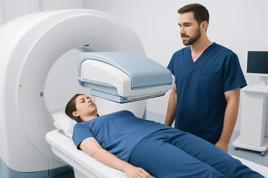
Nuclear medicine examinations begin with a radiotracer. This is a small amount of radioactive material linked to a substance that behaves in a particular way in the body. Depending on the test, it may be injected into a vein, swallowed, or inhaled. The chosen tracer travels to the tissue or organ being assessed and releases small amounts of energy that can be detected by a special camera.
A gamma camera, SPECT scanner, PET scanner, or combined system such as PET/CT or SPECT/CT records this energy and creates detailed images. The technologist positions the patient, explains when it is important to remain still, and monitors the equipment throughout the scan. Some tests take place shortly after tracer administration, while others require images several hours later or on a different day.
These studies may be used when clinicians are investigating symptoms or monitoring known conditions. Examples include heart perfusion imaging, bone scans, thyroid uptake tests, renal scans, and PET imaging in selected cancer, neurological, or cardiac settings. A nuclear medicine study is one part of a wider diagnostic process and is selected only when it is likely to provide useful information.
Who May Need a Nuclear Medicine Scan?

A doctor may recommend a nuclear medicine scan when a functional assessment could help answer a clinical question. For example, a scan may be considered for unexplained bone pain, suspected infection or inflammation, assessment of heart blood flow, evaluation of thyroid activity, or follow-up of certain cancers. The precise examination depends on the person’s symptoms and health history.
Not everyone is a suitable candidate for every type of scan. Pregnancy, possible pregnancy, breastfeeding, kidney disease, allergies, current medicines, recent imaging tests, and difficulties lying flat may affect the choice or timing of an examination. Patients should tell the care team about all medicines and supplements, including thyroid medicines, diabetes medicines, and any recent contrast imaging.
Before the appointment, the imaging department provides individual preparation instructions. Some examinations require fasting, temporary dietary changes, extra fluids, or avoiding specific medicines for a defined period. It is important not to stop prescribed medication unless the referring clinician or nuclear medicine team specifically advises this.
What to Expect: Step by Step
At the appointment, the nuclear medicine technologist confirms the patient’s identity, reviews relevant safety questions, explains the examination, and checks whether preparation instructions were followed. They may ask about pregnancy or breastfeeding, allergies, medical conditions, and previous procedures. Patients have an opportunity to ask questions before the scan begins.
The radiotracer is then administered in the form required for the test. If an injection is needed, the technologist or another qualified member of the team places a small needle into a vein. There may be a short waiting period while the tracer reaches the target organ or tissue. This can range from minutes to several hours, depending on the scan.
During imaging, the patient lies or sits in a position that allows the camera to obtain clear pictures. The scanner itself is usually painless. Some machines move close to the body, which can feel unfamiliar, but the technologist remains in contact and can provide reassurance. The complete visit may be longer than the scanning time because of preparation and waiting intervals.
After the images are checked for quality, the patient can usually return home the same day. A specialist reviews the images and sends a report to the referring doctor, who discusses the results in the context of the patient’s overall care.
Benefits, Risks and Recovery Timeline
The main benefit of nuclear medicine is that it can reveal functional changes that may appear before structural changes are clear on other tests. This may help clinicians make a diagnosis, determine the extent of a condition, plan treatment, or evaluate whether treatment is working. It can also help avoid unnecessary procedures when results provide reassuring information.
Radiation exposure from diagnostic nuclear medicine is carefully calculated and kept as low as reasonably achievable while still producing useful images. The tracer typically loses radioactivity naturally and leaves the body through urine, stool, breath, or normal metabolism. The care team may advise drinking fluids and emptying the bladder regularly after some tests.
Side effects are uncommon, although temporary discomfort, bruising, or soreness can occur at an injection site. Allergic-type reactions to radiotracers are rare. Some scans involve CT imaging as well, which adds a separate source of radiation. The imaging team weighs expected benefits against potential risks and tailors the examination when possible.
Recovery is generally immediate. Most people resume ordinary activities the same day unless they received sedation or have test-specific instructions. Patients should follow any guidance about contact with infants, young children, or pregnant people, as recommendations vary according to the radiotracer used.
How Much Do Nuclear Radiation Techs Make?
Income for a nuclear radiation technologist varies considerably by country, city, workplace, education level, certification, shift patterns, and years of experience. Technologists working in large hospitals, specialist imaging centers, academic institutions, or roles requiring advanced skills may have different pay structures from those in smaller facilities.
Because wages change over time, the most reliable source is an official labor-market or professional-registry resource in the country where the person plans to work. In the United States, employment and wage information is commonly published through government labor statistics. Local job postings and professional organizations can also provide a more current picture of regional opportunities.
When comparing roles, it is useful to look beyond base salary. Benefits, overtime arrangements, on-call duties, pension contributions, licensing requirements, workplace safety policies, and opportunities for continuing education can all influence the overall employment package.
Is Nuclear Med Tech a Hard Job?
Nuclear medicine technology can be demanding, but many people find it rewarding because it combines science, technology, patient care, and teamwork. The work requires strong attention to detail: correct patient identification, accurate preparation, radiation-safety procedures, image quality checks, and documentation are essential parts of every examination.
The role also requires communication skills. Patients may be worried about a scan, uncomfortable because of pain, or unable to remain still for long periods. A technologist needs to explain the process clearly, respond calmly to concerns, and work respectfully with people of different ages and health needs.
Physical demands can include standing for extended periods, helping patients transfer safely, and maintaining careful positioning during imaging. Work may include early, evening, weekend, or on-call shifts in some hospitals. Education and supervised clinical training prepare technologists for these responsibilities, while ongoing learning helps them stay current with equipment and safety standards.
Who Makes More, RN or Nuclear Medicine Technologist?
There is no universal answer because registered nurse and nuclear medicine technologist salaries differ by location, experience, specialty, employer, scheduling, and professional credentials. In some regions, nurses may earn more because of overtime, shift differentials, leadership positions, or high-demand specialties. In other settings, nuclear medicine technologists may have competitive compensation because of their specialized imaging training.
The two careers also have different day-to-day responsibilities. Registered nurses provide broad clinical care, medication administration, patient education, coordination, and ongoing assessment across many care settings. Nuclear medicine technologists focus on radiotracer-based imaging procedures, radiation safety, scanner operation, and image acquisition.
Someone choosing between these careers may wish to consider training pathways, preferred work environment, patient-contact style, job availability, and long-term career goals rather than salary alone. Speaking with accredited training programs and licensed professionals can help clarify local requirements.
Do Nuclear Med Techs Start IVs?
In many healthcare settings, nuclear medicine technologists are trained and authorized to start intravenous lines when an examination requires an injected radiotracer. However, this depends on local laws, professional licensing standards, employer policies, clinical training, and the technologist’s documented competency.
Where a technologist is not authorized to place an IV, a nurse, physician, or another qualified clinician may do so. Some patients may already have a suitable IV line in place. The team follows infection-prevention and medication-safety procedures regardless of who performs the task.
Patients who have difficult veins, a history of fainting during injections, a vascular access device, or concerns about needles should tell the imaging team before the appointment. This allows the staff to plan for comfort, safety, and appropriate support.
When to Seek Medical Care
People should seek medical advice for new, severe, persistent, or worsening symptoms rather than relying on an imaging test alone. Urgent assessment is appropriate for symptoms such as chest pain, significant trouble breathing, sudden weakness or difficulty speaking, fainting, severe allergic symptoms, or uncontrolled bleeding. Emergency services should be contacted when symptoms are severe or rapidly progressing.
After a nuclear medicine procedure, patients should contact the imaging department or their doctor if they develop increasing redness, swelling, pain, or drainage at an injection site, or if they have symptoms that may suggest an allergic reaction, such as widespread hives, facial swelling, wheezing, or difficulty breathing. Serious reactions are uncommon, but prompt assessment is important.
For people seeking diagnostic evaluation or treatment planning, Acibadem International’s multidisciplinary specialists and JCI-accredited hospitals support international patients across imaging and clinical services. The referring doctor and nuclear medicine team can explain whether a particular scan is appropriate and how its results may guide next steps.
Frequently asked questions
What is the difference between a nuclear radiation technologist and a radiologic technologist?
A nuclear medicine technologist performs studies using radiotracers that show body function. A radiologic technologist commonly performs X-rays and may work with other imaging modalities depending on training. Both roles use important safety procedures and work as part of an imaging care team.
Is radiation from a nuclear medicine scan dangerous?
Diagnostic nuclear medicine uses carefully selected amounts of radiotracer, with exposure kept as low as reasonably achievable. The expected benefit of obtaining clinically useful information is considered before the test is requested. Patients can ask the imaging team about the tracer, radiation exposure, and any aftercare instructions specific to their scan.
How long does a nuclear medicine scan take?
The imaging portion may take from minutes to over an hour, depending on the examination. The full appointment can be longer because the body may need time to absorb or distribute the radiotracer. The imaging department can provide the most accurate estimate before the visit.
Can a person drive after a nuclear medicine scan?
Most people can drive and return to normal activities after a routine diagnostic scan. Driving may not be appropriate if sedation was used or if the patient feels unwell for another reason. The care team provides instructions based on the specific procedure.
Should pregnant or breastfeeding patients have nuclear medicine imaging?
Pregnant patients or those who may be pregnant should inform the medical team before the test, as the scan may need to be postponed, modified, or replaced with another method. Breastfeeding guidance depends on the specific radiotracer and may involve a temporary interruption. Individual advice from the nuclear medicine team is essential.
When will nuclear medicine scan results be available?
A nuclear medicine physician or radiologist reviews the images and prepares a report for the referring clinician. Timing varies by facility, scan type, and clinical urgency. The referring doctor is usually best placed to explain what the result means for the individual patient.
References
- International Atomic Energy Agency
- Society of Nuclear Medicine and Molecular Imaging
- Radiological Society of North America
- U.S. Bureau of Labor Statistics
- World Health Organization
This article is for general information only and is not a substitute for professional medical advice. Please consult a qualified doctor about your individual situation.
Explore treatments in Turkey — costs, top hospitals & a free quote
JCI-accredited · board-certified surgeons · reply within 24h
Add us as a Preferred Source to see more of our trusted health content across Google Search, AI Overviews and Discover.