Papilledema Treatment: How It Works, Results and What to Expect

Papilledema is optic nerve swelling caused by raised pressure inside the skull and needs prompt medical assessment. Treatment is directed at the underlying cause, not at eye swelling alone.
Key Takeaways
- Papilledema is optic nerve swelling caused by raised pressure inside the skull and needs prompt medical assessment.
- Treatment is directed at the underlying cause, not at eye swelling alone.
- Many people improve when the cause is treated early, but visual monitoring remains important.
- Medicines, lifestyle measures and procedures may be used depending on severity and cause.
- Sudden or worsening visual symptoms, severe headache, confusion or weakness require urgent medical care.
Papilledema treatment focuses on urgently identifying and reducing raised pressure inside the skull, while protecting the optic nerve and vision. The right approach depends on the underlying cause and may include medicines, weight management, treatment of a triggering condition, or procedures when vision is at risk.
Overview: How papilledema treatment works
Papilledema treatment aims to lower raised pressure within the skull, known as intracranial pressure, and to prevent permanent injury to the optic nerves. Papilledema is not a diagnosis by itself; it is a sign that pressure around the brain may be elevated. Because some causes can be serious, assessment should not be delayed.
A care team first confirms that optic disc swelling is truly papilledema and then looks for the cause. This usually involves an eye examination, brain imaging and, when appropriate, a lumbar puncture to measure cerebrospinal fluid pressure and test the fluid. Treatment then addresses the cause, such as idiopathic intracranial hypertension, a blood-clotting problem, infection, inflammation, medication effect, or a mass affecting fluid flow.
The main goals are to preserve vision, ease pressure-related symptoms such as headache, and manage the condition causing the pressure rise. Eye specialists, neurologists, neuroradiologists and neurosurgeons may work together when care is complex.
Symptoms and why visual monitoring matters
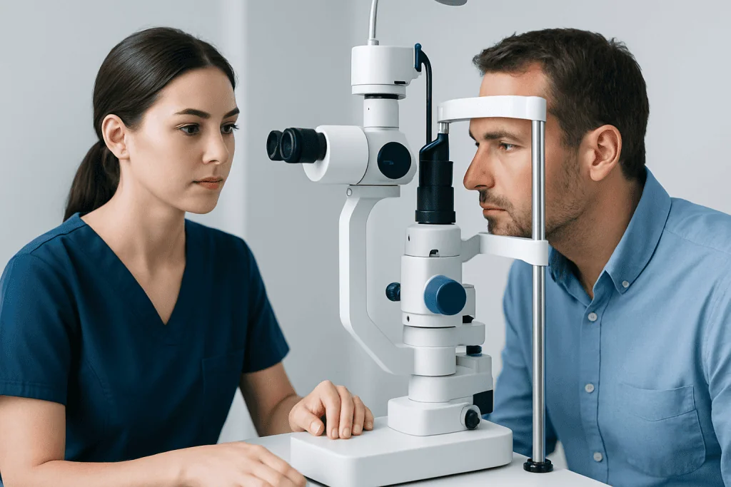
Some people have few symptoms at first, while others develop headaches, brief episodes of dimmed or graying vision, double vision, nausea, vomiting or a pulse-like sound in the ears. Visual blurring may become more noticeable as pressure remains elevated. Symptoms can vary, so their absence does not rule out papilledema.
Vision testing is central to follow-up. Clinicians may check visual acuity, colour vision, pupil responses, eye movements and visual fields. Retinal imaging, including optical coherence tomography, can help document optic nerve swelling over time. These tests provide a clearer picture of whether treatment is protecting the optic nerve.
Not all optic nerve swelling is papilledema. Other eye or nerve conditions can create a similar appearance, including optic neuritis and an anatomical variation called optic disc drusen. Accurate diagnosis is important because the investigations and treatments differ.
Causes, risk factors and candidacy for treatment

Anyone with confirmed papilledema is a candidate for prompt specialist assessment because the treatment plan depends on why pressure is raised. A common cause is idiopathic intracranial hypertension, a condition in which intracranial pressure is elevated without a brain mass or another clear structural cause. It is more common in people with obesity, but it can occur in people of different body sizes and ages.
Other possible causes include a brain mass, hydrocephalus, cerebral venous sinus thrombosis, meningitis or other infections, bleeding, severe uncontrolled blood pressure, inflammatory disorders, and certain medicines or vitamin-related exposures. A clinician will review recent illnesses, medical history, pregnancy status, medicines, supplements and risk factors for clotting.
Vitamin A in excessive amounts can contribute to raised intracranial pressure in susceptible people. This may occur with high-dose supplements, certain acne medicines related to vitamin A, or other retinoid medications. People should not stop prescribed medicine without advice, but they should tell their clinician about all prescriptions, over-the-counter products and supplements.
People with rapidly declining vision, severe papilledema or a cause requiring urgent intervention may need hospital-based treatment and close monitoring. Those with milder, stable findings may be treated as outpatients, provided follow-up is reliable and timely.
Papilledema treatment: medicines, procedures and step-by-step care
The first step is usually urgent evaluation. After examining the eyes and nervous system, clinicians commonly arrange magnetic resonance imaging or computed tomography to exclude a mass, bleeding or hydrocephalus. Imaging of the brain’s veins may be needed when a venous sinus clot is suspected. If imaging shows it is safe, a lumbar puncture may measure pressure and identify signs of infection or inflammation.
For idiopathic intracranial hypertension, treatment may include a medicine that reduces cerebrospinal fluid production, support for gradual, clinically appropriate weight reduction when relevant, and regular visual-field monitoring. Managing associated headaches may require an individualized plan. If a clot, infection, inflammatory condition or medicine effect is identified, the underlying problem is treated directly.
When vision is worsening despite medical therapy, or when pressure must be relieved quickly, procedures may be considered. These can include optic nerve sheath fenestration to protect vision in selected cases, a cerebrospinal fluid shunt to divert fluid, or venous sinus stenting for carefully selected patients with relevant venous narrowing and pressure findings. The choice depends on symptoms, imaging, visual risk and the team’s assessment of benefits and risks.
Procedural care includes pre-treatment testing, discussion of alternatives and anaesthesia planning where needed. During the procedure, the specialist performs the planned intervention to reduce pressure or protect the optic nerve. Follow-up then includes eye examinations, symptom review and imaging or pressure assessment when clinically indicated.
Benefits, risks and recovery timeline
The potential benefit of papilledema treatment is preservation of vision by reducing sustained pressure on the optic nerves. Headache and other pressure-related symptoms may also improve, although headache response does not always match changes in optic nerve swelling. Follow-up is necessary because improvement in symptoms alone does not confirm that the optic nerves are safe.
Recovery varies greatly with the cause and severity. When an underlying trigger is corrected promptly, optic nerve swelling may begin to improve over days to weeks, while full stabilization can take longer. With idiopathic intracranial hypertension, treatment and monitoring may continue for months or longer. Existing visual field loss may improve, remain stable or, if nerve damage has occurred, be permanent.
Medicines may have side effects and need monitoring. Procedures also have specific risks, including infection, bleeding, anaesthesia-related complications, low-pressure headaches, blockage or revision of a shunt, and possible visual complications depending on the intervention. A specialist explains the expected benefit, alternatives and individual risks before recommending a procedure.
At home, patients should take medicines as prescribed, attend scheduled visual testing, and report new visual symptoms promptly. It is helpful to bring an updated list of medicines and supplements to every appointment.
How long does it take to cure papilledema?
There is no single timeline for curing papilledema because it is a sign of raised intracranial pressure rather than one disease. If the cause is temporary and treated quickly, swelling may start to settle within days or weeks. In other situations, especially idiopathic intracranial hypertension or conditions requiring surgery, treatment and monitoring may continue for months or longer.
The most important measure of progress is not only whether headache improves, but whether eye examinations and visual fields show that the optic nerves are stabilizing. Some people need ongoing care to prevent recurrence, and any residual vision changes should be discussed with an eye specialist or neurologist.
How serious is papilledema?
Papilledema should always be taken seriously because it can signal raised pressure inside the skull and may threaten vision if untreated. The degree of urgency depends on the cause, the speed of symptom onset and whether vision is changing. Many causes can be treated effectively, particularly when assessment occurs early.
It is reassuring that papilledema does not automatically mean a life-threatening condition. However, it should not be monitored at home or attributed to migraine without a medical evaluation. Prompt investigation helps clinicians identify urgent causes and choose the safest treatment plan.
What vitamin causes papilledema?
Excess vitamin A can contribute to raised intracranial pressure and papilledema in some people. This risk is linked to high-dose vitamin A supplements and to medicines related to vitamin A, known as retinoids. It is not usually caused by eating normal amounts of vitamin-A-containing foods as part of a balanced diet.
Anyone being assessed for papilledema should tell the clinical team about supplements, acne treatments, skin medicines and other prescriptions. A clinician can advise whether a product may be contributing and how it should be safely adjusted or stopped.
Does papilledema mean brain tumor? When to seek medical care
No. Papilledema does not necessarily mean a brain tumor. A tumor is one possible cause of raised intracranial pressure, but there are many other causes, including idiopathic intracranial hypertension, blood clots in brain veins, infection, medication effects and problems with cerebrospinal fluid circulation. Brain imaging is used to investigate these possibilities safely.
Medical care should be sought urgently for new or worsening vision loss, persistent severe headache, repeated vomiting, double vision, confusion, fainting, seizures, weakness, difficulty speaking or severe drowsiness. People who have been told they have optic nerve swelling should follow the recommended referral and testing plan without delay.
Acibadem International’s multidisciplinary specialists and JCI-accredited hospitals assess and treat conditions associated with papilledema for international patients, using coordinated eye, neurology and neurosurgical care when needed.
Frequently asked questions
Can papilledema go away on its own?
Papilledema may improve when the underlying cause is removed or treated, but it should not be assumed to resolve without medical care. Because ongoing raised pressure can damage vision, a clinician should investigate and monitor it promptly.
Is papilledema painful?
The optic nerve swelling itself is not usually painful. However, the raised pressure causing it can be associated with headache, nausea, neck discomfort or pain with eye movement in some people. Symptoms vary according to the underlying cause.
Can weight loss help papilledema?
For people with idiopathic intracranial hypertension and excess body weight, gradual weight reduction can help lower intracranial pressure and may improve papilledema. It is only one part of care and should be planned with a healthcare professional, particularly when symptoms or vision changes are significant.
Will a lumbar puncture treat papilledema?
A lumbar puncture is mainly used to measure cerebrospinal fluid pressure and examine the fluid after imaging has shown it is safe. Removing some fluid can sometimes provide temporary symptom relief, but it is usually not a long-term treatment for the cause of papilledema.
Can papilledema cause permanent blindness?
Untreated or severe papilledema can lead to permanent visual field loss and, in advanced cases, major vision loss. Early diagnosis, regular eye testing and treatment directed at the cause can reduce this risk.
What specialist treats papilledema?
Care commonly involves an ophthalmologist, often a neuro-ophthalmologist, and a neurologist. Depending on the cause, neuroradiologists, neurosurgeons, infectious disease specialists or other clinicians may also be involved.
References
- American Academy of Ophthalmology
- National Eye Institute
- National Institute of Neurological Disorders and Stroke
- Mayo Clinic
- Merck Manual Professional Edition
This article is for general information only and is not a substitute for professional medical advice. Please consult a qualified doctor about your individual situation.
Explore treatments in Turkey — costs, top hospitals & a free quote
JCI-accredited · board-certified surgeons · reply within 24h
Add us as a Preferred Source to see more of our trusted health content across Google Search, AI Overviews and Discover.
Check your numbers in seconds
BMI, calories, due date, blood pressure and 30+ more clinical calculators — free, instant, doctor-reviewed ranges.