

Perfusion MRI: Procedure, Recovery and Results

Perfusion MRI evaluates how blood moves through an organ or area of tissue rather than showing anatomy alone. It is commonly used for brain conditions, stroke assessment, tumors and selected heart blood-flow evaluations.
Key Takeaways
- Perfusion MRI evaluates how blood moves through an organ or area of tissue rather than showing anatomy alone.
- It is commonly used for brain conditions, stroke assessment, tumors and selected heart blood-flow evaluations.
- Many examinations use an intravenous contrast agent; patients should tell the imaging team about kidney disease, pregnancy, allergies or implanted devices.
- The scan itself is painless, but remaining still in the MRI scanner and receiving contrast can be uncomfortable for some people.
- Results are interpreted by a radiologist and shared with the referring clinician, who explains what they mean in the individual clinical context.
Perfusion MRI is a noninvasive imaging examination that maps blood flow through tissue, most commonly in the brain or heart. It can help clinicians identify areas receiving reduced or unusually increased blood supply and interpret these findings alongside symptoms, other scans and laboratory tests.
Overview: what perfusion MRI shows
Perfusion MRI is a magnetic resonance imaging technique that measures blood flow through tissue. Unlike a standard MRI, which mainly shows the structure of organs, perfusion imaging adds information about how well blood is reaching an area and how it moves through small blood vessels. It may be used in the brain, heart and, less commonly, other organs.
In brain imaging, perfusion MRI can help doctors assess tissue affected by reduced circulation, such as after a suspected stroke, or evaluate the blood supply of a mass. In cardiac imaging, myocardial perfusion MRI examines blood flow to the heart muscle, often at rest and during medication-induced stress. The examination does not by itself confirm a diagnosis; it is one part of a broader clinical assessment.
MRI does not use ionizing radiation, unlike CT and many nuclear medicine examinations. However, some perfusion MRI methods require a contrast injection, and MRI is not suitable for every person or every implanted medical device.
How does perfusion MRI work?
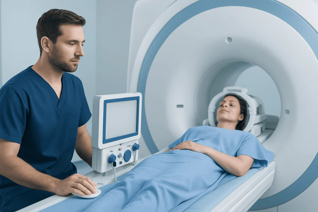
MRI uses a strong magnetic field and radiofrequency waves to create detailed images. For many perfusion studies, a gadolinium-based contrast agent is injected into a vein while the scanner rapidly obtains a series of images. Computer software analyzes how the contrast passes through tissue and produces color-coded maps of blood-flow-related measurements.
In the brain, these maps may describe cerebral blood volume, blood flow and the time it takes blood or contrast to pass through an area. They can help identify tissue with altered circulation. Different approaches are available, including dynamic susceptibility contrast imaging and arterial spin labeling, which may measure perfusion without injected contrast in selected situations.
For the heart, images are usually taken during the first pass of contrast through the heart muscle. A medication may temporarily make the heart work as if during exercise, helping reveal blood-flow differences that are not apparent at rest. This may be considered alongside other evaluations for coronary artery disease when clinically appropriate.
Who may need a perfusion MRI?

A doctor may request perfusion MRI when information about tissue blood flow could change diagnosis or management. Brain perfusion imaging may be considered in the evaluation of a suspected acute stroke, certain brain tumors, seizures, cognitive concerns or other neurological findings. It may also help distinguish active tumor tissue from treatment-related changes in some circumstances.
Cardiac perfusion MRI may be used for people with chest symptoms, suspected reduced blood flow to the heart muscle, or unclear findings from other cardiac tests. It can support assessment of myocardial ischemia, meaning an imbalance between the heart muscle’s oxygen needs and its blood supply.
Before the examination, the referring clinician and MRI team review medical history and safety factors. Important information includes a pacemaker, neurostimulator, cochlear implant, aneurysm clip, metal fragments, prior surgery, kidney problems, previous contrast reactions, pregnancy or possible pregnancy, and claustrophobia. Many modern implanted devices can be scanned under specific conditions, but this must be verified in advance.
- People with significant kidney impairment may need blood tests or a different imaging approach before gadolinium contrast is used.
- People who are pregnant should discuss the expected benefit and alternatives with their care team.
- Those with anxiety in enclosed spaces may benefit from preparation, an open discussion with the imaging department, or clinician-prescribed support when suitable.
What happens during a perfusion MRI procedure?
Preparation instructions vary by the body area being scanned. Patients may be asked to remove jewelry, watches, hearing aids, removable dental work and clothing with metal. For cardiac stress perfusion imaging, the team may ask patients to avoid caffeine for a specified period and may review medications beforehand. They should follow the instructions provided by their own imaging center rather than stopping medication without medical advice.
At the appointment, a radiographer confirms safety information and places an intravenous line if contrast is planned. The patient lies on a padded table, and a specialized receiver coil is positioned around the head or chest. Ear protection is provided because MRI produces loud tapping and knocking sounds. The table moves into the scanner, while staff monitor and communicate with the patient from an adjacent room.
During contrast perfusion imaging, the injection may cause a brief cool sensation in the arm or a temporary unusual taste. For a cardiac stress study, a medication is administered under close monitoring to increase blood flow in the heart. Temporary warmth, flushing, shortness of breath, chest discomfort, headache or a faster heartbeat can occur; the team watches closely and can respond if symptoms arise.
Clear images require staying still, and the radiographer may ask for short breath-holds during a heart MRI. The scan is painless. If discomfort, anxiety or new symptoms develop, the patient can use the call device to alert the team.
How long is a perfusion MRI?
A perfusion MRI commonly takes about 30 to 60 minutes in the scanner, although the total visit may be longer because of screening, changing, intravenous access, preparation and post-scan observation. A brain perfusion sequence may take only several minutes as part of a longer brain MRI examination.
Myocardial perfusion MRI often takes around 45 to 90 minutes for the full appointment, depending on whether rest and stress images are needed, the patient’s heart rhythm, preparation requirements and whether additional heart MRI sequences are performed. The imaging team can provide a more precise estimate before the visit.
Allowing extra time is sensible, especially for a first MRI appointment or if translation, device checks or kidney-function testing is needed. Patients should not feel concerned if the examination takes longer than expected, as image quality and safety checks are important.
Recovery, benefits and possible risks
Most people can return to normal activities shortly after a perfusion MRI. The intravenous line is removed before leaving, and mild soreness or bruising at the injection site can occur. Drinking fluids after contrast is often reasonable unless a clinician has advised fluid restriction for a medical reason.
The main benefit is detailed, radiation-free information about blood flow that can complement structural MRI images. In the brain, this may assist urgent decision-making and treatment planning. In the heart, it can help clinicians assess whether reduced blood supply may be contributing to symptoms and determine whether further testing or treatment is appropriate.
Gadolinium-based contrast agents are generally well tolerated, but mild effects such as nausea, headache, dizziness or temporary injection-site discomfort may occur. Serious allergic-like reactions are uncommon but can happen, which is why trained staff and emergency equipment are available. In people with severe kidney disease, certain contrast agents may carry additional risks; the care team weighs these carefully.
MRI safety screening is essential because the magnetic field can affect some metal objects and electronic implants. The scan may also be difficult for people with severe claustrophobia or those unable to lie flat. In such cases, the team can discuss suitable alternatives or supportive measures.
What do perfusion MRI results mean?
A radiologist reviews the images and compares perfusion maps with routine MRI sequences, previous scans and the clinical question. Results may be described as normal, reduced, delayed or increased perfusion. The wording can sound technical, but it does not automatically indicate a particular condition or predict an outcome on its own.
Reduced brain perfusion can occur when blood supply is impaired, while increased perfusion may be seen in some tumors or areas of inflammation. In cardiac imaging, a stress-related perfusion defect can suggest reduced blood flow to part of the heart muscle. Further testing may or may not be needed depending on the person’s symptoms, risk factors and all available results.
The formal report is usually sent to the clinician who ordered the scan. That clinician explains the findings, whether any follow-up is needed, and how they relate to the person’s health. If brain tumor assessment is part of the reason for testing, perfusion results are commonly reviewed with other imaging and specialist input, including brain tumor treatment planning.
When to seek medical care
People should seek urgent medical care for sudden facial drooping, arm weakness, speech difficulty, severe new confusion, loss of vision, a sudden severe headache, or new trouble walking. These symptoms can be signs of a stroke and should not wait for an outpatient MRI appointment.
Urgent assessment is also important for new or worsening chest pressure, chest pain with shortness of breath, fainting, sweating, nausea, or pain spreading to the arm, jaw, back or shoulder. These symptoms may have several causes, but prompt evaluation is needed to rule out a heart emergency.
After an MRI, immediate medical attention is appropriate for breathing difficulty, swelling of the face or throat, widespread hives, severe dizziness or other signs of a serious allergic reaction. For non-urgent questions about scan preparation or results, patients should contact the ordering clinician or imaging department. Acibadem International’s multidisciplinary specialists and JCI-accredited hospitals diagnose and treat neurological and cardiovascular conditions for international patients.
Frequently asked questions
What does MRI with perfusion mean?
MRI with perfusion means that the MRI examination includes sequences designed to assess blood flow through tissue. This is often done by tracking an injected contrast agent, although some techniques can estimate perfusion without contrast. It provides functional information in addition to standard anatomical MRI images.
How long is a perfusion MRI?
The time in the scanner is often about 30 to 60 minutes, but total appointment time can be longer. A cardiac stress perfusion MRI may take 45 to 90 minutes or more when preparation, monitoring and additional images are needed. The imaging center can give instructions based on the exact study ordered.
Is a myocardial perfusion serious?
Myocardial perfusion refers to blood flow to the heart muscle; it is not a diagnosis by itself. An abnormal myocardial perfusion result can suggest that part of the heart may receive less blood during stress, which deserves review by a clinician. The seriousness depends on the pattern of findings, symptoms, medical history and other test results.
Can I drive after a myocardial perfusion scan?
Many people can drive after a myocardial perfusion MRI if they feel well and have not received a sedative. However, driving may not be appropriate if medication used for the test causes lingering symptoms, if the person feels unwell, or if sedation was given. Patients should follow the instructions from their imaging and cardiology teams and arrange transport when advised.
Does perfusion MRI always require contrast?
No. Many perfusion MRI examinations use gadolinium-based contrast because it provides detailed dynamic information about blood flow. Some methods, particularly arterial spin labeling in brain imaging, can assess perfusion without injected contrast. The choice depends on the clinical question, the body area and individual safety considerations.
How soon are perfusion MRI results available?
The images are reviewed by a radiologist, and the report is commonly sent to the referring clinician within days, although timing varies by facility and clinical urgency. In emergency situations, preliminary interpretation may be much faster. The clinician who ordered the test is best placed to explain the result and next steps.
References
- American College of Radiology
- Radiological Society of North America
- American Heart Association
- National Institute of Neurological Disorders and Stroke
- European Society of Cardiology
This article is for general information only and is not a substitute for professional medical advice. Please consult a qualified doctor about your individual situation.
Explore treatments in Turkey — costs, top hospitals & a free quote
JCI-accredited · board-certified surgeons · reply within 24h
Add us as a Preferred Source to see more of our trusted health content across Google Search, AI Overviews and Discover.

More from the Health Library



Related Specialists

Assoc. Prof. Dr. Burcu Bulum Akbulut
Pediatric Nephrology
Dr. Erdem Kaplan
Emergency Service
Dr. Serkan Tokgönül
Aesthetic Plastic & Reconstructive Surgery
Dr. Havva Gölalan
Oral & Dental Health