PET-CT in Cancer Care: Uses, Preparation, and Result Meaning
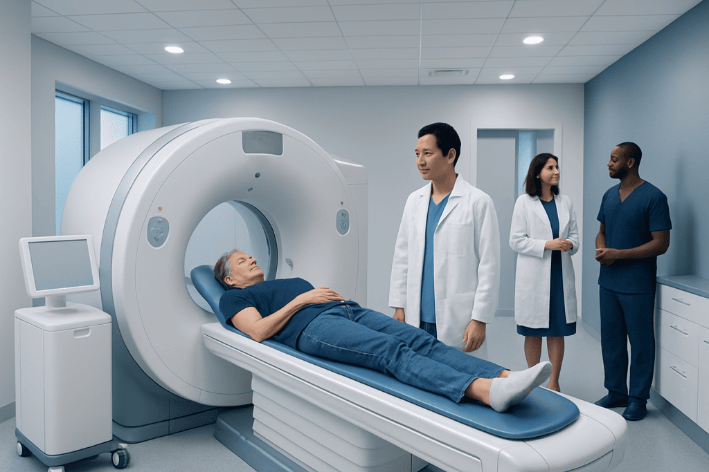
PET-CT combines PET, which shows areas of increased cell activity, with CT, which shows detailed body structure. In cancer care, PET-CT is often used for staging, checking treatment response, and looking for recurrence, but it is not needed for every cancer or every patient.
Key Takeaways
- PET-CT combines PET, which shows areas of increased cell activity, with CT, which shows detailed body structure.
- In cancer care, PET-CT is often used for staging, checking treatment response, and looking for recurrence, but it is not needed for every cancer or every patient.
- Preparation usually includes fasting, avoiding strenuous exercise, and carefully following diabetes or medication instructions provided by the imaging team.
- PET-CT results must be interpreted by specialists because inflammation, infection, recent surgery, and normal body processes can sometimes mimic cancer activity.
- Patients should discuss what the scan result means for their specific diagnosis and treatment plan with their oncology team.
PET-CT is an advanced imaging test that combines metabolic and anatomical information to help doctors understand cancer more clearly. It can support diagnosis, staging, treatment planning, and follow-up when used alongside clinical examination, laboratory tests, biopsy, and other imaging.
Overview: What Is PET-CT?
PET-CT is a medical imaging test that combines two techniques in one examination: positron emission tomography, known as PET, and computed tomography, known as CT. PET shows how active cells are by tracking a small amount of radioactive tracer, most commonly a glucose-related tracer called FDG. CT provides detailed images of the body’s organs, bones, lymph nodes, and tissues. Together, the two images help doctors see both where an abnormal area is located and how active it appears.
In cancer care, this combination can be especially useful because many cancer cells use more glucose than normal cells. Areas that take up more tracer may appear as “hot spots” on the PET image. The CT portion helps identify whether that activity corresponds to a tumor, lymph node, organ, scar tissue, or another structure. This makes PET-CT different from a standard CT or MRI, which mainly show anatomy rather than metabolic activity.
PET-CT is not a screening test for the general population, and it is not required for every type of cancer. It is most helpful when the result can guide a clear medical decision, such as choosing treatment, determining the stage of disease, assessing response after therapy, or investigating a suspected recurrence. The decision to request PET-CT is made by the treating doctor based on the cancer type, clinical situation, and previous test results.
Common Uses of PET-CT in Cancer Care
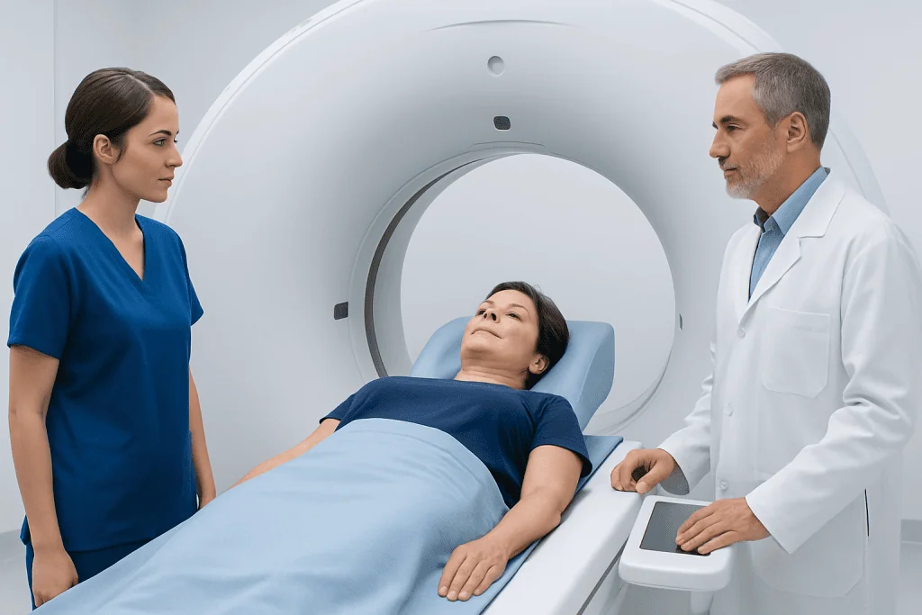
PET-CT may be used at different stages of the cancer pathway. Before treatment begins, it can help doctors determine the extent of disease, a process called staging. Accurate staging is important because treatment choices may differ depending on whether cancer is limited to one area, has reached nearby lymph nodes, or has spread to distant organs.
During or after treatment, PET-CT can help assess whether cancer activity has decreased, remained stable, or progressed. This can be useful after chemotherapy, immunotherapy, radiotherapy, targeted therapy, or combined treatments. In some cancers, PET-CT can help distinguish active disease from scar tissue or treated tissue that still looks abnormal on CT alone.
Doctors may also recommend PET-CT when symptoms, blood tests, tumor markers, or other scans raise concern for recurrence. It can sometimes identify areas of active disease that are difficult to characterize with other imaging methods. However, PET-CT is always interpreted in context, and suspicious findings may need confirmation with MRI, ultrasound, follow-up imaging, endoscopy, or biopsy.
- Staging certain cancers before treatment
- Planning radiotherapy fields or surgical approaches in selected cases
- Evaluating response to cancer treatment
- Investigating possible recurrence after treatment
- Helping select the best site for biopsy when multiple abnormal areas are present
Which Cancers May Be Evaluated with PET-CT?
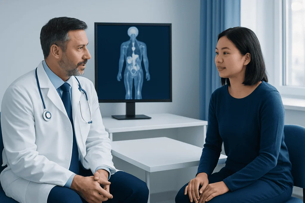
PET-CT is commonly used in several cancer types, including many lymphomas, lung cancers, head and neck cancers, esophageal cancers, colorectal cancers, melanomas, and some gynecologic cancers. In these settings, PET-CT can provide information that may affect staging or treatment planning. It may also be used in selected cases of breast, thyroid, pancreatic, and other cancers, depending on the clinical question.
Not all cancers show strong uptake of FDG, the most common PET tracer. Some tumors grow slowly or have biology that makes them less visible on FDG PET-CT. In other situations, a different tracer may be considered, such as prostate-specific membrane antigen imaging for prostate cancer in appropriate settings. The best imaging method depends on tumor type, stage, previous treatments, and what the doctor needs to know.
Because PET-CT is powerful but not universal, patients should not worry if their doctor does not recommend it. For some cancers, MRI, contrast-enhanced CT, ultrasound, mammography, endoscopy, or tissue biopsy may provide more useful information. The oncology team chooses imaging based on evidence-based guidelines and each patient’s individual circumstances.
How to Prepare for a PET-CT Scan
Preparation helps improve image quality and reduce the chance of unclear results. Most patients are asked not to eat for several hours before an FDG PET-CT scan, although drinking plain water is usually encouraged. The imaging center will provide specific instructions about fasting, medications, and arrival time. Patients should follow the instructions they receive, as protocols may vary depending on the tracer, scan purpose, and medical history.
Strenuous exercise is often avoided for at least the day before the scan because active muscles can take up more tracer and affect image interpretation. Patients are usually advised to stay warm and relaxed before the scan, since shivering and muscle tension can also increase tracer uptake. Comfortable clothing without metal parts is helpful, and jewelry or removable metal objects may need to be taken off.
People with diabetes need special instructions because blood glucose levels can influence FDG PET-CT results. They should not change diabetes medication or insulin schedules without guidance from their doctor or imaging team. Patients who are pregnant, might be pregnant, or are breastfeeding should inform the medical team before the appointment. Those with kidney problems, contrast allergies, or prior reactions to imaging contrast should also mention this, especially if the CT portion will use intravenous contrast.
What Happens During the Procedure?
On arrival, the patient is usually checked in, asked about medical history, and may have blood glucose measured. The radioactive tracer is then injected into a vein. The amount of radioactivity is small and selected for diagnostic imaging. After injection, the patient rests quietly for a period of time, often in a comfortable room, while the tracer circulates through the body and is taken up by tissues.
During the scan, the patient lies on a table that moves slowly through the PET-CT scanner. The test is painless, although lying still can be uncomfortable for some people. The scanner does not fully enclose the body in the same way as some MRI scanners, and it does not usually create loud knocking sounds. The imaging team can communicate with the patient during the examination.
The CT part is performed quickly and may be done with or without contrast, depending on the purpose of the scan. If contrast is used, a warm sensation or metallic taste may occur briefly. The PET images take longer to acquire. After the scan, most patients can return to usual activities unless they have received sedating medication, which is uncommon. Drinking fluids may help the body clear the tracer more quickly.
Understanding PET-CT Results
PET-CT results are reviewed by specialists trained in nuclear medicine and radiology. The report describes areas of tracer uptake, CT findings, comparison with previous scans, and the impression or conclusion. One term patients may see is SUV, or standardized uptake value. SUV is a measurement of tracer uptake, but it is only one part of interpretation. A higher SUV does not automatically mean cancer, and a lower SUV does not always rule it out.
Increased tracer uptake can occur for reasons other than cancer. Infection, inflammation, recent surgery, radiation changes, healing fractures, and some benign conditions may show activity. Normal organs such as the brain, heart, kidneys, bladder, liver, and bowel can also show expected tracer uptake. This is why doctors interpret the scan together with symptoms, physical examination, laboratory results, pathology, and previous imaging.
A PET-CT report may suggest that findings are consistent with active disease, treatment response, stable disease, or no clear evidence of abnormal metabolic activity. Sometimes the report recommends further evaluation, such as targeted MRI, ultrasound, endoscopy, short-interval follow-up, or biopsy. Patients should review the report with their oncology team, who can explain what the findings mean for the overall treatment plan rather than focusing on individual words or numbers alone.
Benefits, Limitations, and Safety
The main benefit of PET-CT is that it combines functional and structural information in one test. This can help identify active disease, clarify the significance of findings on other scans, and support more personalized treatment planning. For some patients, PET-CT may prevent unnecessary procedures by showing that a suspicious area is inactive, or it may guide doctors to a more appropriate biopsy site.
Like all medical tests, PET-CT has limitations. Very small lesions may be below the scan’s resolution. Some cancers may not take up enough tracer to be clearly seen. False-positive results can occur when inflammation or infection looks active. False-negative results can occur when disease is small, slow-growing, or affected by blood glucose levels or recent treatment timing. This is why PET-CT is best understood as one part of a complete diagnostic picture.
PET-CT involves exposure to ionizing radiation from both the tracer and CT scan. The dose is carefully controlled and considered acceptable when the test is medically justified. The benefit of obtaining important cancer information should be weighed against radiation exposure, especially in children, younger adults, and people who require repeated imaging. Patients should tell their doctors about recent scans and any possibility of pregnancy so the safest imaging plan can be chosen.
When to Speak with a Doctor
Patients should speak with their doctor if they have questions about why PET-CT is being recommended, how it may influence treatment, or whether another imaging test might be more appropriate. It is also important to ask about timing, especially after surgery, chemotherapy, immunotherapy, or radiotherapy, because scans performed too soon after treatment may be harder to interpret.
Before the appointment, patients should inform the imaging team about diabetes, pregnancy, breastfeeding, kidney disease, contrast allergies, severe claustrophobia, or difficulty lying flat. They should also bring relevant previous scans and reports if these are not already available to the medical team. Comparing current and earlier images often improves accuracy.
After the scan, patients should arrange a follow-up discussion with their oncology team rather than interpreting the report alone. A clear explanation can reduce uncertainty and help patients understand the next steps. Acibadem International’s multidisciplinary specialists and JCI-accredited hospitals diagnose and treat cancer for international patients, including coordinating advanced imaging such as PET-CT when clinically appropriate.
Frequently asked questions
Is PET-CT used to diagnose cancer?
PET-CT can support the diagnosis of cancer by showing areas of abnormal metabolic activity and their location. However, a definite cancer diagnosis usually requires tissue confirmation with a biopsy or pathology report. PET-CT is most often used together with clinical evaluation, laboratory tests, and other imaging.
Does a positive PET-CT always mean cancer?
No. Areas of increased tracer uptake can also be caused by infection, inflammation, healing after surgery, radiation changes, or certain benign conditions. The scan must be interpreted by specialists and reviewed by the treating doctor in the context of the patient’s full medical history.
How long does a PET-CT scan take?
The scan itself may take a relatively short time, but the full appointment usually lasts longer because the tracer needs time to circulate before imaging begins. Many patients spend a few hours at the imaging center from arrival to completion. The exact timing depends on the protocol used by the center.
Can people with diabetes have PET-CT?
Yes, many people with diabetes can have PET-CT, but they need individualized preparation instructions. Blood glucose levels can affect FDG tracer uptake and image quality. Patients should ask the imaging team how to manage meals, insulin, and diabetes medicines before the scan.
Is PET-CT painful or uncomfortable?
The test is generally not painful, aside from the small needle injection used for the tracer. Some people may feel uncomfortable lying still during the scan. Patients who have pain, anxiety, or difficulty remaining flat should tell the imaging team in advance so supportive measures can be discussed.
Will PET-CT show whether treatment is working?
In many cancers, PET-CT can help show whether metabolic activity has decreased after treatment, which may suggest response. The timing of the scan is important because inflammation from recent therapy can affect results. The oncology team will interpret the scan alongside symptoms, examination findings, blood tests, and other imaging.
What should patients do after receiving PET-CT results?
Patients should review the results with their oncologist or referring doctor. The report may contain technical terms that are easy to misunderstand without medical context. The doctor can explain whether the findings change the stage, treatment plan, follow-up schedule, or need for further testing.
References
- European Association of Nuclear Medicine
- American College of Radiology
- National Comprehensive Cancer Network
- American Cancer Society
- International Atomic Energy Agency
This article is for general information only and is not a substitute for professional medical advice. Please consult a qualified doctor about your individual situation.
Oncology care in Turkey — second opinion and treatment plan
JCI-accredited · board-certified surgeons · reply within 24h
Add us as a Preferred Source to see more of our trusted health content across Google Search, AI Overviews and Discover.

Medical Oncology Department
Medical treatment of cancer with chemotherapy, immunotherapy and targeted therapies under a multidisciplinary tumor board.
60 specialists in this unit