PET Scan for Neuroendocrine Tumor: Preparation, Procedure and Results
Most neuroendocrine PET scans use a tracer that attaches to somatostatin receptors commonly found on well-differentiated neuroendocrine tumors. Preparation varies by tracer and medication use, so patients should follow the imaging center's individual instructions.
Key Takeaways
- Most neuroendocrine PET scans use a tracer that attaches to somatostatin receptors commonly found on well-differentiated neuroendocrine tumors.
- Preparation varies by tracer and medication use, so patients should follow the imaging center's individual instructions.
- The injection, uptake period and scan commonly take several hours in total, although the camera scan itself is often shorter.
- A PET scan can show suspicious activity but cannot by itself prove that a tumor is cancerous.
- After the scan, drinking fluids and following radiation-safety instructions helps the tracer leave the body naturally.
A PET scan for neuroendocrine tumor is a specialized imaging test that uses a small amount of radioactive tracer to show neuroendocrine tumor cells throughout the body. It can help confirm the extent of known disease, identify suitable treatments and monitor response, but the scan result is interpreted alongside pathology, blood tests and other imaging.
PET Scan for Neuroendocrine Tumor: an answer-first guide
A PET scan for neuroendocrine tumor is an imaging examination that can map neuroendocrine tumor cells in the body by using a tracer designed to bind to particular receptors on those cells. It is commonly combined with CT, and sometimes MRI, to show both the location of abnormal cells and the surrounding anatomy.
For many well-differentiated neuroendocrine tumors (NETs), the preferred examination is somatostatin receptor PET imaging, often called a Ga-68 DOTATATE, DOTATOC or Cu-64 DOTATATE PET scan. The result can help a multidisciplinary team stage a tumor, evaluate whether it has spread, select treatment and assess changes over time.
Neuroendocrine tumors can arise in several organs, including the digestive tract, pancreas and lungs. Their behavior varies widely, so imaging is tailored to the individual tumor type, grade, symptoms and previous test results. A PET result is valuable information, but it is one part of a complete clinical assessment.
How neuroendocrine PET imaging works and who may need it
Positron emission tomography (PET) uses a radiotracer, a substance that gives off a very small amount of radiation. In somatostatin receptor PET, the tracer attaches to somatostatin receptors, which are often present in high numbers on well-differentiated NET cells. A scanner detects the tracer and creates detailed images of where it has collected.
The PET images are usually paired with CT images. CT helps the radiologist identify the precise anatomical location of tracer uptake, such as a lymph node, liver lesion or bone lesion. In selected situations, MRI may also be useful, especially for evaluating the liver.
Clinicians may recommend this scan when a NET has been diagnosed or strongly suspected, when conventional scans leave uncertainty, when staging is needed before treatment, or when monitoring a known tumor. It can also help determine whether a patient may be suitable for receptor-targeted radionuclide treatment, such as peptide receptor radionuclide therapy (PRRT).
Not every neuroendocrine tumor shows somatostatin receptors. Higher-grade or rapidly growing tumors may be better assessed with FDG PET, which measures glucose use, or with a different combination of scans. The care team selects the tracer most likely to answer the clinical question.
Preparation and candidacy before the scan

Preparation instructions depend on the specific tracer, the scan center and a person’s health conditions. Patients should tell the team about pregnancy, possible pregnancy, breastfeeding, kidney problems, diabetes, allergies, medications and any difficulty lying still or being in enclosed spaces. The imaging team can explain whether any additional precautions are needed.
For somatostatin receptor PET, patients may be asked to adjust the timing of somatostatin analogue medicines, such as long-acting injections or short-acting medicines. This should never be done independently. The oncology or endocrinology team will provide individualized advice because pausing treatment can worsen hormone-related symptoms for some people.
Many centers encourage good hydration before and after the examination. Fasting requirements differ: some somatostatin receptor PET scans do not require fasting, while FDG PET scans usually require dietary preparation and blood glucose checks. Comfortable clothing without metal is helpful, and patients may need to remove jewelry or other metal objects before imaging.
A person who has claustrophobia, significant pain, mobility limitations or anxiety should mention this before the appointment. The team can discuss practical measures to make the examination more manageable and safe.
What should you not do before a PET scan?
Patients should not make assumptions about fasting, exercise or medication changes before a PET scan because the correct instructions depend on the tracer being used. They should not stop prescribed medicines, including injections used for neuroendocrine tumor symptoms, unless the clinician who ordered the scan has specifically advised them to do so.
For an FDG PET scan, strenuous exercise is commonly avoided for at least a day beforehand because active muscles can take up the tracer and make interpretation more difficult. Patients are often asked to avoid food for several hours before FDG PET, while drinking plain water may be permitted. In contrast, the instructions for a somatostatin receptor PET scan may be less restrictive.
Patients should avoid arriving dehydrated and should inform the center if they are ill, have recently had surgery, received an injection or vaccination, or have an active infection. Inflammation can sometimes cause tracer uptake and may affect interpretation. The imaging department’s written instructions take priority over general guidance.
Step by step: what happens during a neuroendocrine PET scan
On arrival, a technologist confirms identity, medical history and preparation. A small intravenous cannula is placed, and the radiotracer is injected into a vein. Most people feel only the brief needle sensation associated with the injection, and the tracer itself does not usually cause noticeable effects.
There is then an uptake period, commonly around 45 to 90 minutes depending on the tracer and local protocol. During this time, patients may be asked to rest quietly and drink water. They may also be asked to empty their bladder before the scan because tracer in urine can obscure structures in the pelvis.
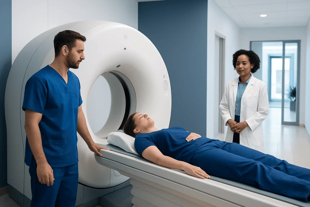
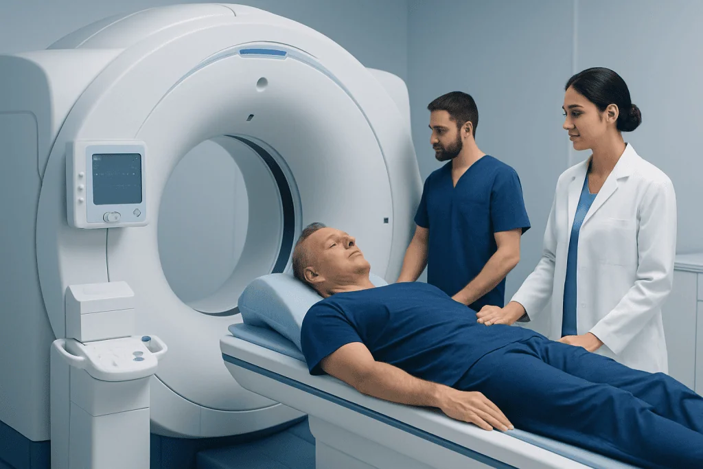
For the imaging portion, the patient lies on a padded table that moves slowly through the scanner. It is important to remain still, but staff can communicate throughout the examination. A CT scan may be performed at the same visit, sometimes with contrast if appropriate.
Afterward, a nuclear medicine physician or radiologist reviews the images in the context of previous scans and medical history. The referring specialist then discusses what the findings mean for diagnosis, monitoring or treatment planning.
How long does a neuroendocrine PET scan take?
A neuroendocrine PET scan appointment commonly takes about two to three hours from check-in to completion, although the total time can be longer depending on the tracer, imaging protocol and whether contrast CT is included. Much of this time is the quiet uptake period after the tracer injection.
The actual time on the scanner is often approximately 20 to 45 minutes. Some patients need additional images of a specific area, which can extend the appointment. The imaging team can provide a more accurate estimate when scheduling the scan.
Results may not be available immediately because the images require expert review and comparison with earlier tests. If the scan was ordered to make a time-sensitive treatment decision, patients can ask their referring clinician when and how results will be communicated.
Results, benefits and limitations
On a somatostatin receptor PET scan, areas with increased tracer uptake may represent NET tissue with somatostatin receptors. The report describes where uptake is seen, how intense it is, and whether there are findings in organs such as the liver, lymph nodes, bones or lungs. The report may also identify non-tumor findings that need correlation with other tests.
The main benefit is highly sensitive whole-body imaging that may reveal disease not seen clearly on conventional CT or MRI. This can improve staging and help guide an individualized care plan, including surgery, medicines, liver-directed treatment, observation or receptor-targeted therapy. Related evaluation and care may be part of neuroendocrine tumor management.
However, uptake is not always cancer. Inflammation, infection, healing tissue and some normal organs may show tracer activity. Small lesions may be difficult to detect, and tumors with few or no somatostatin receptors may not appear strongly. For these reasons, specialists interpret PET findings together with biopsy results, laboratory data, symptoms and other imaging.
When a tissue diagnosis is needed, a biopsy remains the usual way to identify tumor type and grade. Imaging can guide where to biopsy, but it does not replace pathology.
Can a PET scan tell if a tumor is cancerous?
A PET scan can identify areas that are suspicious for tumor activity, but it cannot independently confirm that a tumor is cancerous. Some noncancerous conditions, including inflammation and certain benign growths, can produce increased tracer uptake. Conversely, some cancers may have low or absent uptake with a particular tracer.
For neuroendocrine tumors, somatostatin receptor uptake suggests that cells in an area have receptors targeted by the tracer. This information can strongly support a diagnosis in the right clinical setting, but confirmation may require biopsy and laboratory assessment, especially for a new or uncertain lesion.
The report should be discussed with the specialist who knows the full medical context. A multidisciplinary team can determine whether observation, further imaging, biopsy or treatment is the most appropriate next step.
Recovery, risks and radiation-safety precautions
Most people can return to normal activities after a PET scan, including eating and drinking as usual unless they received separate instructions for CT contrast or sedation. Drinking extra water over the next day, if medically appropriate, and urinating regularly can help clear the tracer through the kidneys.
The radiation exposure from a PET scan is generally limited and is carefully planned to provide useful diagnostic information. The tracer loses radioactivity naturally over time. Allergic reactions to PET tracers are uncommon; if CT contrast is used, it has its own small risk of reactions and may require precautions in people with impaired kidney function.
Patients are often advised to limit prolonged close contact with pregnant people, babies and young children for a short period after the scan. Exact guidance differs by tracer and local regulations, so patients should follow the instructions given by their nuclear medicine department.
Acibadem International’s multidisciplinary specialists and JCI-accredited hospitals support international patients needing neuroendocrine tumor assessment and treatment planning.
Why do you have to flush the toilet twice after a PET scan?
Some centers advise patients to flush the toilet twice after a PET scan because a small amount of the radiotracer leaves the body in urine. Double flushing reduces the chance that trace radioactive material remains in the toilet bowl and is a simple radiation-safety measure.
This instruction is temporary and does not mean the person is dangerous to others. Good handwashing, regular toileting and following the department’s advice about contact with others are usually sufficient while the tracer clears. Patients with questions about household precautions should contact the imaging center that performed the scan.
If a patient is breastfeeding, pregnant or caring closely for a baby or young child, they should ask for tracer-specific instructions before leaving the department. Recommendations may differ according to the radiotracer used.
When to seek medical care
Patients should contact the imaging center or their healthcare professional promptly if they develop symptoms of a possible allergic reaction after contrast or an injection, such as trouble breathing, swelling of the face or throat, widespread hives, severe dizziness or fainting. These reactions are unusual, but urgent assessment is important if they occur.
They should also contact their doctor if they are unable to follow the preparation instructions, discover a possible pregnancy, have uncontrolled diabetes before an FDG PET scan, or experience new or worsening symptoms related to a known neuroendocrine tumor. Examples may include persistent severe abdominal pain, significant flushing or wheezing, fainting, or symptoms of bowel obstruction.
A PET scan is most useful when its findings are integrated into ongoing specialist care. Patients should attend follow-up appointments to review results and discuss whether further testing or treatment is needed.
Frequently asked questions
Is a PET scan painful?
The scan itself is not painful. Patients may feel a brief pinch when the intravenous line is placed and may find it uncomfortable to lie still for the imaging portion. Staff can help with positioning and can discuss concerns such as pain or claustrophobia before the test.
Do all neuroendocrine tumors show up on a DOTATATE PET scan?
No. DOTATATE and similar scans work best when tumor cells have somatostatin receptors, which is common in many well-differentiated NETs. Some higher-grade or poorly differentiated neuroendocrine cancers may show little uptake and may require FDG PET or other imaging instead.
Can patients eat before a neuroendocrine PET scan?
It depends on the radiotracer and local protocol. Some somatostatin receptor PET scans allow normal eating, while FDG PET scans commonly require fasting. Patients should follow the instructions from their imaging center rather than using general advice.
When can patients be near family after a PET scan?
Most patients can be around others after leaving the department, but they may be asked to limit lengthy, close contact with infants, young children and pregnant people for a short time. The exact recommendation depends on the tracer used. The nuclear medicine team provides individualized safety instructions.
What happens if the PET scan shows new areas of uptake?
New uptake does not automatically mean that a tumor has spread or grown. The care team compares the images with prior scans, symptoms, laboratory results and other tests to determine the likely cause. Additional imaging, biopsy or follow-up may be recommended when a finding is uncertain.
Can a neuroendocrine PET scan be used to plan treatment?
Yes. It can show the distribution of receptor-positive disease and may help clinicians decide whether surgery, observation, systemic treatment or receptor-targeted radionuclide therapy is appropriate. Treatment decisions also depend on tumor grade, location, symptoms, overall health and prior therapies.
References
- National Cancer Institute
- Society of Nuclear Medicine and Molecular Imaging
- European Association of Nuclear Medicine
- North American Neuroendocrine Tumor Society
- American Cancer Society
This article is for general information only and is not a substitute for professional medical advice. Please consult a qualified doctor about your individual situation.
Oncology care in Turkey — second opinion and treatment plan
JCI-accredited · board-certified surgeons · reply within 24h
Add us as a Preferred Source to see more of our trusted health content across Google Search, AI Overviews and Discover.