

Posterior Fossa Surgery: Procedure, Recovery and Results

Posterior fossa surgery is brain surgery because it involves structures within the skull, including the cerebellum and brainstem. A posterior fossa decompression enlarges space at the base of the skull and is commonly used for selected cases of Chiari malformation.
Key Takeaways
- Posterior fossa surgery is brain surgery because it involves structures within the skull, including the cerebellum and brainstem.
- A posterior fossa decompression enlarges space at the base of the skull and is commonly used for selected cases of Chiari malformation.
- Surgical positioning, including prone, lateral, or carefully selected sitting positions, is individualized for safety and surgical access.
- Recovery often begins in a monitored setting and can take weeks to months, depending on the procedure and neurological condition.
- Potential benefits must be balanced with risks such as bleeding, infection, cerebrospinal fluid leakage, and temporary or lasting neurological changes.
- New or worsening severe headache, weakness, confusion, swallowing difficulty, fever, or wound drainage needs urgent medical assessment.
Posterior fossa surgery is a type of neurosurgery performed in the lower back part of the skull, where the cerebellum and brainstem are located. It may relieve pressure, remove a tumor or other lesion, improve cerebrospinal fluid flow, or treat a vascular or structural condition; recovery and expected results depend on the reason for surgery and the approach used.
Overview: What Is Posterior Fossa Surgery?
Posterior fossa surgery refers to operations performed in the small space at the lower back of the skull. This area contains the cerebellum, which helps coordinate movement and balance, and the brainstem, which supports essential functions such as breathing, alertness, swallowing, and eye movements. Because these structures are closely packed, planning and monitoring are especially important.
There are several posterior fossa procedures. A surgeon may create an opening in the skull, called a craniotomy, or remove a small portion of bone near the base of the skull, called a craniectomy. The precise technique depends on whether the goal is to relieve crowding, remove a tumor, treat bleeding or a vascular abnormality, drain a cyst, obtain a biopsy, or restore the circulation of cerebrospinal fluid.
Posterior fossa surgery may be recommended for adults or children after careful evaluation by a neurosurgical team. The expected benefit is based on the underlying diagnosis, symptoms, imaging findings, and the likelihood that surgery can safely address the cause of pressure or neurological dysfunction.
How It Works and Who May Be a Candidate
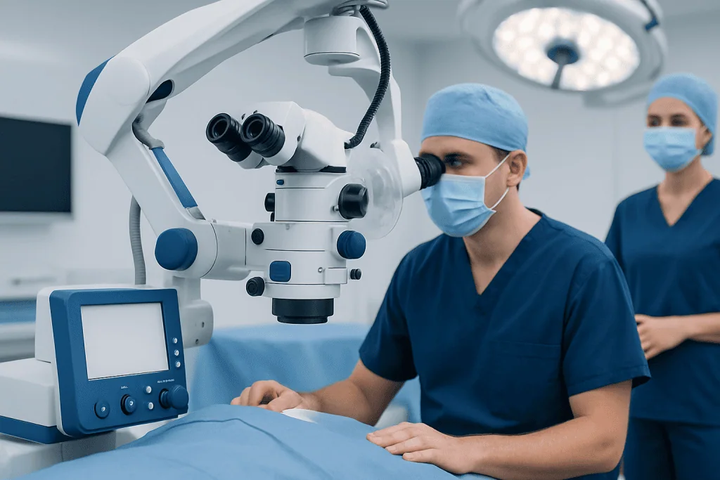
The posterior fossa is surrounded by bone and has limited room to accommodate swelling, a mass, or altered fluid flow. Surgery can create more space, provide access to a lesion, or reduce pressure on sensitive tissue. For example, posterior fossa decompression may be considered for selected people with symptomatic Chiari malformation, in which lower parts of the cerebellum extend downward through the opening at the base of the skull.
Candidacy is individualized. A neurosurgeon considers symptoms such as headache brought on by coughing or straining, balance problems, weakness, numbness, swallowing changes, double vision, or evidence of raised pressure. MRI, and sometimes CT scans, help define the anatomy, identify tumors or vascular changes, and assess cerebrospinal fluid pathways. Not every imaging finding requires surgery; some people are best managed with observation and follow-up imaging.
People being considered for surgery may also need neurological examination, blood tests, anesthesia assessment, and additional tests tailored to the suspected condition. When symptoms are related to structural crowding, evaluation for Chiari malformation can help clarify whether decompression is likely to be useful.
- Symptoms that significantly affect daily life or are progressing
- Imaging showing compression, blockage of fluid flow, or a lesion requiring treatment
- Concern for neurological deterioration, bleeding, hydrocephalus, or brainstem compression
- An overall health status that allows anesthesia and postoperative recovery
Step by Step: Posterior Fossa Surgical Approaches

Posterior fossa surgery is performed under general anesthesia. The patient is positioned to give the surgical team safe access to the back or side of the head. Hair may be clipped only where needed, and the head is supported in a fixed position. The surgeon makes an incision, typically behind the ear or at the back of the head and upper neck, depending on the target area.
After opening or removing a small section of skull, the surgeon uses magnification, specialized instruments, and often real-time neurological monitoring to work around delicate nerves and blood vessels. In decompression surgery, bone at the base of the skull may be removed, and the covering of the brain, called the dura, may be expanded with a patch to improve space and cerebrospinal fluid flow. For a tumor or other lesion, the procedure may involve removal, biopsy, drainage, or treatment of abnormal blood vessels.
The bone opening may be replaced after a craniotomy, while a decompressive craniectomy intentionally leaves more room. The layers of tissue are then closed, and some patients temporarily need a drain or monitoring device. The operation length varies substantially according to the diagnosis, anatomy, surgical approach, and whether additional procedures are required. A planned Chiari malformation surgery pathway should include discussion of the intended technique and realistic treatment goals.
What Is the Recommended Sitting Position for Posterior Fossa Surgery?
There is no single recommended position for every posterior fossa operation. Common options include prone positioning, with the patient face down; lateral or park-bench positioning, with the patient on one side; and, in selected cases, a semi-sitting or sitting position. The best choice depends on the location of the surgical target, the patient’s anatomy, the planned approach, anesthesia considerations, and the experience of the operating team.
The sitting position can provide a direct downward surgical view and may allow blood and fluid to drain away from the operative field. However, it also has specific risks, including venous air embolism, where air enters the bloodstream through open veins. For that reason, it is used selectively and requires meticulous anesthesia monitoring, preventive measures, and a team familiar with this approach.
Patients should not try to determine the position themselves. Before surgery, the anesthesiologist and neurosurgeon explain the planned position, steps taken to protect pressure points and the neck, and any individual factors that may make one position safer than another.
Posterior Fossa Surgery Recovery Timeline and Expected Results
Posterior fossa surgery recovery usually begins in a recovery room or intensive monitoring setting. Staff check alertness, limb movement, speech, swallowing, breathing, pain, blood pressure, and the surgical wound. Depending on the procedure and condition, a hospital stay may range from several days to longer when complex neurological monitoring or rehabilitation is needed.
In the first days, fatigue, headache, neck stiffness, nausea, and incision discomfort are common and are managed by the clinical team. Walking, breathing exercises, and nutrition are restarted gradually when appropriate. Some people need physical therapy, occupational therapy, speech and swallowing therapy, or balance rehabilitation before leaving hospital or after discharge.
At home, recovery commonly continues over several weeks, while energy, neck mobility, and activity tolerance improve gradually. Follow-up appointments assess wound healing, symptoms, neurological function, and imaging when needed. The outcome may include relief of pressure-related symptoms, prevention of further neurological damage, or treatment of the underlying lesion, but existing nerve or brain tissue injury may not fully reverse.
For people traveling for care, Acibadem International’s multidisciplinary specialists and JCI-accredited hospitals can coordinate diagnosis, neurosurgical treatment, and follow-up planning for international patients.
How Long Does It Take to Recover From Posterior Fossa Decompression Surgery?
Recovery from posterior fossa decompression surgery varies, but many people need several weeks before returning to lighter daily activities and may need several months for fuller recovery. The timeline depends on age, overall health, the extent of bone and dural work, preoperative symptoms, complications, and whether associated conditions such as syringomyelia or hydrocephalus are present.
During the first few weeks, patients are generally advised to protect the incision, avoid heavy lifting and strenuous activity, and follow the surgeon’s instructions about driving, work, bathing, and exercise. Tiredness can persist even after the wound has healed. A gradual return to activity is usually safer than trying to resume all normal tasks quickly.
Headaches or neck discomfort may improve gradually rather than immediately. Symptoms caused by long-standing nerve compression, such as altered sensation, balance issues, or weakness, can take longer to change and may require rehabilitation. Follow-up MRI may be used to assess decompression and cerebrospinal fluid flow when clinically appropriate.
Risks, Benefits, and Posterior Fossa Syndrome
The potential benefits of posterior fossa surgery include relieving compression, improving cerebrospinal fluid flow, removing or diagnosing a lesion, preserving neurological function, and reducing symptoms linked to the underlying disorder. However, surgery in this area has important risks. These include bleeding, infection, blood clots, anesthetic complications, cerebrospinal fluid leakage, wound problems, swelling, stroke, and the need for further surgery.
Because the posterior fossa contains pathways involved in coordination, cranial nerve function, and vital functions, some people may have changes in balance, speech, facial movement, hearing, vision, swallowing, sensation, or strength. The likelihood and permanence of these changes depend strongly on the condition being treated and the location of surgery. The surgeon should explain the risks most relevant to the individual procedure.
Posterior fossa syndrome, also called cerebellar mutism syndrome, is most often discussed after surgery for certain childhood tumors in the posterior fossa. It can involve reduced or absent speech after surgery, emotional or behavioral changes, low muscle tone, swallowing difficulty, and coordination problems. It is not a typical outcome of every posterior fossa operation.
What Is the Recovery Rate for Posterior Fossa Syndrome?
There is no single recovery rate for posterior fossa syndrome because its severity, timing, treatment needs, and long-term effects differ between children. Many affected children regain speech and make meaningful functional improvements over time, often with coordinated rehabilitation. Recovery can take months or longer, and some may continue to have difficulties with speech, coordination, behavior, learning, or emotional regulation.
Early assessment by neurology, rehabilitation, speech and language therapy, occupational therapy, physiotherapy, psychology, and education specialists can support recovery and daily functioning. Families benefit from clear follow-up plans that address both physical and cognitive-emotional needs.
Before an operation for a posterior fossa tumor, the surgical team should discuss this syndrome when relevant to the child’s diagnosis and planned approach. This conversation should include the potential for rehabilitation and the uncertainty inherent in predicting an individual child’s recovery.
Is Posterior Fossa Decompression Considered Brain Surgery?
Yes. Posterior fossa decompression is considered brain surgery, or neurosurgery, because it is performed inside the skull near the cerebellum and brainstem. Although the main surgical work may focus on bone at the base of the skull, the dura, and the upper spinal canal rather than removing brain tissue, it involves structures that protect and surround the brain.
This does not mean that every decompression procedure is the same as surgery to remove a brain tumor. The technique, risks, expected recovery, and follow-up differ according to the purpose of surgery. A neurosurgeon can explain whether the planned operation includes bone removal alone, dural expansion, treatment of associated spinal findings, or another procedure.
Understanding the exact operation can help patients and families prepare. It is reasonable to ask about the surgical goal, incision location, expected hospital stay, activity restrictions, pain management, warning signs, and the role of rehabilitation in posterior fossa surgery recovery.
When to Seek Medical Care
Urgent medical assessment is important for a sudden severe headache, new weakness or numbness, difficulty speaking, new confusion, fainting, seizures, severe unsteadiness, repeated vomiting, trouble swallowing, or changes in breathing. These symptoms can have many causes, but they require prompt evaluation, particularly when they are new, sudden, or worsening.
After posterior fossa surgery, patients should contact their surgical team promptly for fever, increasing redness or swelling at the incision, fluid leaking from the wound, persistent vomiting, worsening headache, new neck stiffness, increasing drowsiness, vision changes, weakness, or new trouble with coordination, speech, or swallowing. Emergency care is appropriate for severe or rapidly progressing symptoms.
Routine follow-up remains essential even when recovery is going well. The care team can adjust rehabilitation, manage symptoms, review pathology or imaging results, and guide a safe return to everyday activities.
Frequently asked questions
What is posterior fossa surgery used for?
Posterior fossa surgery is used to treat conditions affecting the cerebellum, brainstem, nearby cranial nerves, and cerebrospinal fluid pathways. Reasons can include Chiari malformation, brain tumors, cysts, hydrocephalus, bleeding, vascular abnormalities, or other causes of pressure in this area. The exact goal may be decompression, removal, biopsy, drainage, or restoration of fluid flow.
How long does it take to recover from posterior fossa decompression surgery?
Many people need several weeks to return to lighter activities after posterior fossa decompression, while fuller recovery may take several months. Recovery depends on the extent of surgery, the original symptoms, overall health, and whether rehabilitation is needed. The surgeon’s activity instructions should guide the individual timeline.
What is the recovery rate for posterior fossa syndrome?
A single recovery rate cannot accurately describe posterior fossa syndrome because recovery differs widely between children. Many children regain speech and improve with rehabilitation, although recovery may take months or longer and some effects can persist. Ongoing support for speech, movement, learning, and emotional health is important.
What is the recommended sitting position for posterior fossa surgery?
There is no universally recommended position. Surgeons may use prone, lateral, or selected sitting or semi-sitting positions depending on the target area and patient-specific safety factors. The sitting position has potential advantages but requires careful monitoring because of risks such as venous air embolism.
Is posterior fossa decompression considered brain surgery?
Yes. It is a neurosurgical procedure performed within the skull near the cerebellum and brainstem. Even when surgery primarily removes bone or expands the dura, it is considered brain surgery because of its location and the structures involved.
What should patients avoid after posterior fossa surgery?
Patients are usually advised to avoid heavy lifting, strenuous exercise, and activities that could strain the neck or affect wound healing until cleared by their surgeon. Driving, work, travel, and bathing restrictions vary by procedure and recovery progress. It is important to follow the surgical team’s specific instructions rather than relying on a standard timeline.
References
- National Institute of Neurological Disorders and Stroke
- American Association of Neurological Surgeons
- National Cancer Institute
- Children's Oncology Group
- Mayo Clinic
This article is for general information only and is not a substitute for professional medical advice. Please consult a qualified doctor about your individual situation.
Oncology care in Turkey — second opinion and treatment plan
JCI-accredited · board-certified surgeons · reply within 24h
Add us as a Preferred Source to see more of our trusted health content across Google Search, AI Overviews and Discover.

More from the Health Library


Related Specialists

Dr. Mustafa Burak Seven
Pediatrics
Dr. Mehriban Allahverdiyeva
Inpatient Clinic Physicians Clinical Service
Prof. Dr. Bekir Sıtki Cebeci
Cardiology
Dyt. Duygu Gençel
Nutrition & Diet