Pregnancy Ultrasound: Preparation, Procedure and Results

Pregnancy ultrasound does not use ionizing radiation and is considered safe when performed for a medical reason by trained professionals. Most early abdominal scans require a comfortably full bladder, while later scans usually do not.
Key Takeaways
- Pregnancy ultrasound does not use ionizing radiation and is considered safe when performed for a medical reason by trained professionals.
- Most early abdominal scans require a comfortably full bladder, while later scans usually do not.
- The examination commonly takes 15 to 45 minutes, depending on gestational age and the information needed.
- A sonographer may share limited observations during the scan, but a clinician reviews the final findings in clinical context.
- Urgent symptoms such as heavy bleeding, severe pain or fainting require medical assessment rather than waiting for a routine scan.
A pregnancy ultrasound is a non-invasive imaging examination that uses sound waves to create pictures of the developing fetus, placenta and uterus. It helps the clinical team confirm important details about the pregnancy, while the type and timing of the scan determine how a person should prepare.
Pregnancy Ultrasound: What It Is and How It Works
A pregnancy ultrasound, also called a prenatal or obstetric ultrasound, uses high-frequency sound waves to produce moving images of structures inside the pelvis and abdomen. It can show the gestational sac, embryo or fetus, heartbeat, placenta, amniotic fluid, uterus and, later in pregnancy, many aspects of fetal anatomy. The scan is commonly part of routine antenatal care, but it may also be arranged when symptoms or clinical findings need further assessment.
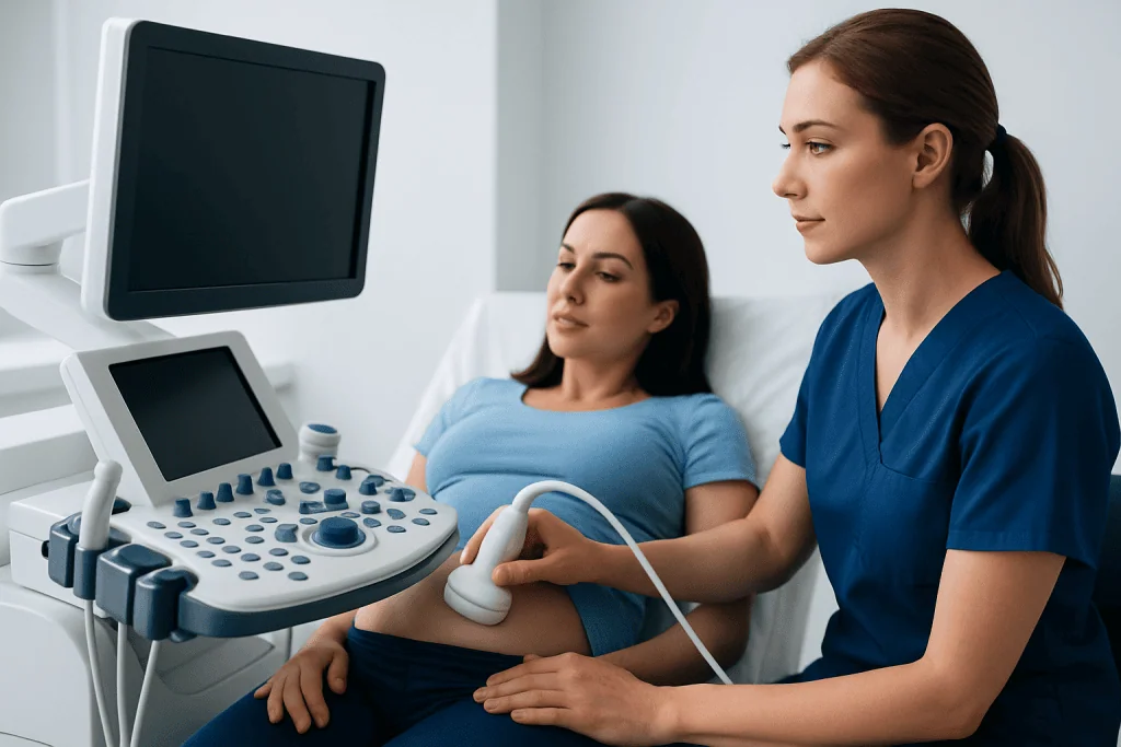
During an abdominal ultrasound, a clinician or sonographer places a small handheld device called a transducer on the abdomen after applying gel. The transducer sends sound waves into the body and receives returning echoes, which a computer converts into images. In early pregnancy, a transvaginal ultrasound may provide a clearer view because the probe is placed gently into the vagina and sits closer to the uterus.
Ultrasound does not use X-rays or other ionizing radiation. Decades of clinical use support its safety when it is medically indicated and performed by appropriately trained professionals using the lowest exposure settings and shortest scanning time needed to obtain useful images. It is not intended as a non-medical keepsake examination.
Why Pregnancy Ultrasounds Are Offered
The purpose of a pregnancy ultrasound changes as pregnancy progresses. An early scan may confirm that the pregnancy is located in the uterus, estimate gestational age, identify the number of embryos and check for cardiac activity when expected. Dating information can help the maternity team plan screening and monitor growth appropriately.
A mid-pregnancy anatomy scan examines fetal development in more detail. It commonly evaluates the brain, spine, heart, abdomen, kidneys, limbs, placenta, umbilical cord and amniotic fluid. Images may not always show every structure clearly at one appointment because fetal position, gestational age, body tissue and other technical factors can affect visibility. A repeat scan may simply be needed to complete views.
Later scans may assess growth, fetal position, placental location, fluid levels or blood flow in the umbilical cord and fetal vessels. Doppler ultrasound is a specialized method that evaluates blood movement. It may be helpful in selected pregnancies, particularly where clinicians are monitoring concerns such as suspected growth restriction or placental function.
Preparation and Candidacy

Most pregnant people can have an ultrasound, and there are no injections, anesthesia or recovery restrictions for a standard examination. The maternity team chooses the most appropriate type of scan based on gestational age, symptoms, prior test results and the clinical question. A transvaginal scan can be particularly useful early in pregnancy and is generally safe; it does not cause miscarriage.
Preparation instructions vary. For an early abdominal scan, a full bladder can help lift the uterus and improve the view through the abdomen. The imaging center may ask the patient to drink water before the appointment and avoid urinating until after the scan. By the second and third trimesters, a full bladder is often unnecessary because the uterus is larger.
Patients should wear comfortable clothing that allows access to the lower abdomen, bring relevant pregnancy records if requested, and tell the team about pain, bleeding, prior ultrasound findings or any concerns. They should follow the specific instructions from their obstetrician or imaging department, as local protocols can differ.
What not to do before a pregnancy ultrasound?
Patients should not assume that preparation is the same for every pregnancy ultrasound. They should avoid emptying their bladder if they have been specifically instructed to arrive with a full bladder, and they should not deliberately drink excessive amounts of water, as this can cause discomfort. There is usually no need to fast unless the clinic has given a separate instruction for another examination being performed at the same visit.
Can I eat anything 2 hours before my ultrasound?
Yes. For most pregnancy ultrasounds, a person can eat normally, including during the two hours before the appointment. Eating does not generally interfere with an obstetric scan. If the appointment includes an abdominal, gallbladder or other non-pregnancy ultrasound, fasting instructions may apply, so the patient should confirm the plan with the imaging center.
What Happens During the Procedure
On arrival, the patient may be asked to confirm pregnancy dates, symptoms and relevant medical history. The sonographer explains the examination and positions the patient on an examination bed, usually lying on their back with a slight tilt if needed for comfort. Gel is placed on the abdomen to help transmit sound waves and prevent air pockets between the skin and transducer.
The sonographer moves the transducer across the abdomen and takes still images, measurements and video clips. Gentle pressure may be used to obtain clearer views, which can be uncomfortable if the bladder is very full or the abdomen is tender, but it should not be painful. The patient can let the sonographer know if they need a break or are uncomfortable.
For a transvaginal ultrasound, the patient undresses from the waist down in private and is covered with a drape. A narrow, covered and lubricated probe is inserted a short distance into the vagina. This usually provides detailed early-pregnancy images and is often completed within a short time. It does not require a full bladder.
The length of the appointment varies, but many scans take about 15 to 45 minutes. Detailed anatomy scans can take longer, especially if the fetus is in a position that limits views. After the examination, the gel is wiped away and the patient can resume usual daily activities immediately.
Results, Follow-Up and Recovery Timeline
There is no physical recovery period after a routine pregnancy ultrasound. Patients can eat, drink, travel, work and exercise as usual unless they have been given separate advice because of symptoms or another health condition. Mild temporary discomfort from bladder fullness, probe pressure or a transvaginal examination generally settles quickly.
How long do ultrasound results take for a pregnant woman?
Some findings may be discussed during or immediately after the scan, but the timing of a formal result varies by clinic and healthcare system. A sonographer often acquires the images, while a radiologist, obstetrician or maternal-fetal medicine specialist reviews them and issues a report. Results may be available the same day or within several days, and the maternity clinician explains what they mean for the individual pregnancy.
An unclear or incomplete scan does not automatically mean that there is a problem. Fetal position, early gestational age, maternal anatomy and image quality can limit what is visible. The care team may recommend another ultrasound, a more detailed assessment, laboratory testing or referral to a specialist depending on the finding. Ultrasound results are interpreted alongside medical history, examination findings and other prenatal screening results.
Acibadem International’s multidisciplinary specialists and JCI-accredited hospitals support international patients who need obstetric assessment, follow-up imaging and coordinated pregnancy care.
Benefits, Limitations and Possible Risks
The main benefit of pregnancy ultrasound is that it provides real-time information without surgery or radiation. It can help confirm pregnancy location and viability, date the pregnancy, evaluate fetal development, identify placental position and guide appropriate maternity care. It can also offer reassurance when findings are consistent with expected development.
However, ultrasound has limitations. It cannot detect every fetal condition, and a normal scan cannot guarantee that a baby has no health concerns. Conversely, an unexpected image finding may need repeat imaging or additional tests before conclusions can be drawn. Screening ultrasound and diagnostic testing are different; the clinical team can explain which type of information a particular scan can and cannot provide.
No significant harmful effects have been established from diagnostic obstetric ultrasound used appropriately. Professional guidance supports using ultrasound only when there is a valid clinical purpose and avoiding unnecessarily prolonged exposure. If Doppler or more detailed imaging is recommended, it is because the team believes the additional information may help guide care.
What happens if I can’t hold my pee before an ultrasound?
Patients should tell the sonographer or receptionist rather than becoming very uncomfortable. Depending on the stage of pregnancy and the reason for the scan, the examination may still be possible, the patient may be asked to drink some more water and wait briefly, or the team may use a transvaginal approach for clearer early-pregnancy images. Emptying part of the bladder may also be sufficient in some situations, but this should be done only after speaking with the imaging team.
When to Seek Medical Care
A routine ultrasound is not a substitute for urgent medical assessment. Pregnant people should contact their maternity provider promptly for vaginal bleeding, persistent or worsening pelvic or abdominal pain, fever, severe vomiting with inability to keep fluids down, dizziness, fainting, fluid leaking from the vagina, or a sudden change in how they feel. The appropriate urgency depends on the stage of pregnancy and the severity of symptoms.
Later in pregnancy, reduced or changed fetal movements should be discussed urgently with the maternity team, following the guidance provided for that pregnancy. Severe headache, visual changes, sudden swelling of the face or hands, chest pain, shortness of breath or seizures need immediate medical attention. These symptoms can have several causes and require assessment rather than self-diagnosis.
If an ultrasound identifies an issue requiring closer monitoring, the obstetrician may recommend repeat imaging or referral to maternal-fetal medicine, genetics, cardiology or neonatal specialists. Patients should ask what the finding means, whether further tests are needed and when results will be reviewed. Clear follow-up plans can make the next steps more manageable.
Frequently asked questions
Does a pregnancy ultrasound hurt?
A standard abdominal pregnancy ultrasound should not be painful, although pressure from the probe may feel uncomfortable, especially with a full bladder or a tender abdomen. A transvaginal scan can cause mild pressure but is usually well tolerated. Patients should tell the sonographer if they have significant discomfort.
Do I need a full bladder for every pregnancy ultrasound?
No. A full bladder is most often useful for early abdominal pregnancy scans, when the uterus is still low in the pelvis. It is commonly not needed for scans later in pregnancy. The imaging center's instructions should be followed because the required preparation depends on the examination.
Can an ultrasound confirm how far along a pregnancy is?
Yes, ultrasound measurements can help estimate gestational age, particularly in early pregnancy. The clinician compares measurements with the reported first day of the last menstrual period and other available information. Dating is most accurate when performed early in pregnancy.
Can a pregnancy ultrasound show the baby's sex?
The fetal sex may be visible during a mid-pregnancy scan, depending on fetal position, gestational age and image quality. It may not be possible to determine with certainty at every scan. The main purpose of medical ultrasound is assessment of pregnancy health and development.
Is transvaginal ultrasound safe in early pregnancy?
Yes, transvaginal ultrasound is widely used in early pregnancy and is considered safe when clinically indicated. It can provide detailed images of the uterus, gestational sac, embryo and ovaries. The probe does not reach the uterus and does not cause miscarriage.
What does it mean if the sonographer cannot give results right away?
It often means the images need formal review by a radiologist, obstetrician or another qualified clinician. This is a normal part of many imaging services and does not by itself indicate an abnormal result. The patient should ask when and how the report will be discussed.
References
- American College of Obstetricians and Gynecologists
- International Society of Ultrasound in Obstetrics and Gynecology
- U.S. Food and Drug Administration
- National Health Service
This article is for general information only and is not a substitute for professional medical advice. Please consult a qualified doctor about your individual situation.
Ultrasound in Turkey — costs, top hospitals & a free quote
JCI-accredited · board-certified surgeons · reply within 24h
Add us as a Preferred Source to see more of our trusted health content across Google Search, AI Overviews and Discover.

Check-up & Preventive Medicine
Comprehensive health screenings designed for early detection and prevention.
2 specialists in this unitMore from the Health Library



Related Specialists

Prof. Dr. Cevdet Düger
Anesthesiology
Asst. Prof. Dr. Zeynep Tuğçe Sarıkaya
Intensive Care
Dr. Mehmet İbrahim Cansunar
General Surgery
Prof. Dr. Reha Baran
Pulmonary Medicine