Private mri scan Before & After: What Realistic Results Look Like
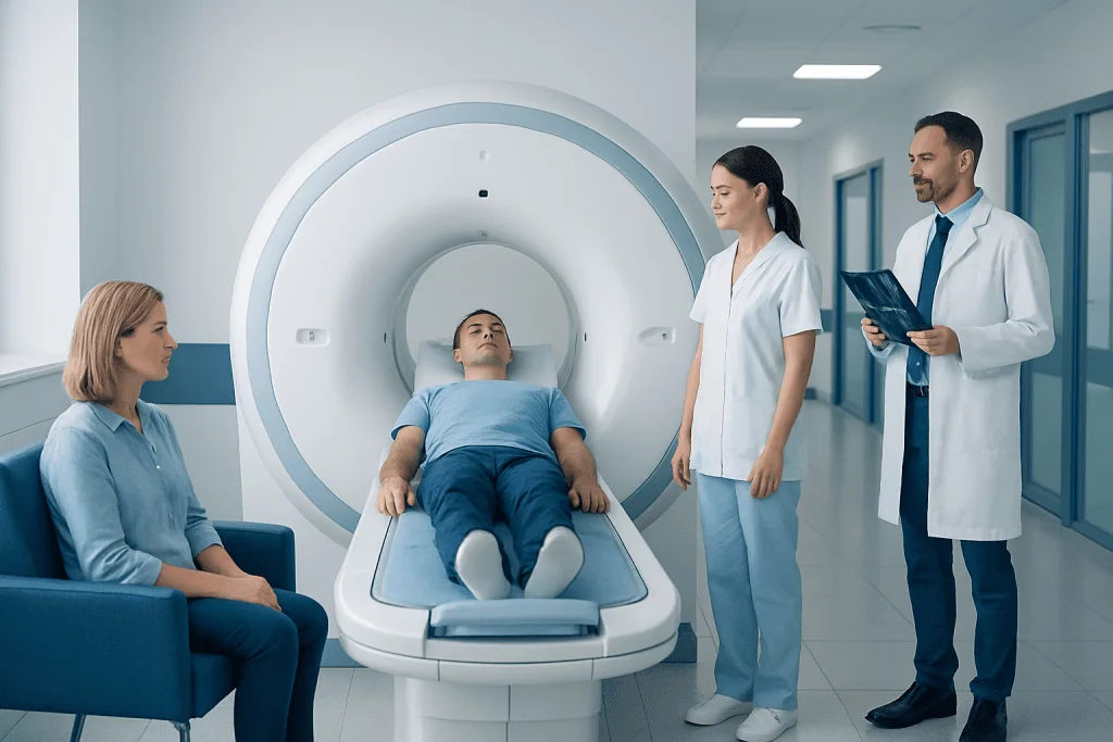
An MRI scan uses a strong magnet and radio waves to create detailed images without ionising radiation. The most realistic “after” result is a clearer explanation of a clinical concern, although some scans are normal or inconclusive.
Key Takeaways
- An MRI scan uses a strong magnet and radio waves to create detailed images without ionising radiation.
- The most realistic “after” result is a clearer explanation of a clinical concern, although some scans are normal or inconclusive.
- MRI findings must be interpreted alongside symptoms, examination findings, and medical history.
- Most people return to usual activities immediately after a routine MRI without sedation.
- Metal implants, certain devices, pregnancy considerations, kidney problems, and claustrophobia should be discussed before booking.
A private MRI scan before and after comparison is mainly about the change in available medical information: before the scan, symptoms or a clinical question may be uncertain; after it, detailed images and a radiology report can help guide next steps. An MRI is a diagnostic examination rather than a treatment, so it does not directly cure pain, injury, or disease.
Private MRI Scan Before and After: What Changes Realistically
A private MRI scan before and after experience is best understood as a diagnostic journey, not a visible transformation. Before the scan, a person may have persistent symptoms, an injury, an abnormal examination finding, or a question raised by another test. After the scan, the main change is usually that a clinician has more detailed information about structures inside the body.
MRI, or magnetic resonance imaging, can show soft tissues in high detail. Depending on the area examined, it may help assess the brain, spine, joints, muscles, nerves, blood vessels, abdominal organs, pelvis, or other structures. The images are reviewed by a radiologist, who provides a written report for the referring clinician.
A scan may identify a likely cause of symptoms, show that no significant structural abnormality is visible, confirm a known condition, or help monitor change over time. None of these outcomes automatically determines treatment. The result needs to be considered together with the person’s symptoms, physical examination, previous tests, and health history.
For an overview of arranging and preparing for the examination itself, see private MRI scan services.
How MRI Works and What It Can Show
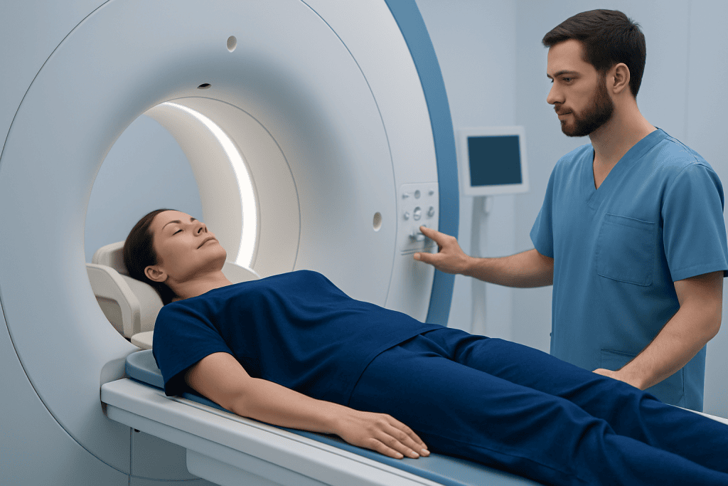
MRI uses a powerful magnetic field and radiofrequency energy to produce images of the body. Unlike X-rays and CT scans, MRI does not use ionising radiation. Different MRI sequences highlight different tissue characteristics, allowing doctors to distinguish, for example, fluid, inflammation, fat, muscle, cartilage, ligaments, bone marrow, and many types of soft-tissue changes.
Some MRI examinations use contrast material, most commonly gadolinium-based contrast given through a vein. Contrast can make certain blood vessels, areas of inflammation, tumours, scars, or active disease more visible. It is not necessary for every scan, and the imaging team decides whether it is appropriate for the clinical question.
What an MRI can show depends on the body area and the protocol requested. A knee MRI may assess ligaments, menisci, cartilage, tendons, and bone marrow. A spine MRI may show discs, nerves, the spinal cord, and narrowing around nerve pathways. A brain MRI can provide detailed information about brain tissue, blood vessels, and surrounding structures.
Importantly, MRI findings do not always explain symptoms. Common changes such as mild disc wear or joint degeneration may be present in people without pain. A radiology report is therefore a valuable part of assessment, but it is not a diagnosis in isolation.
Who May Benefit and How Suitability Is Assessed
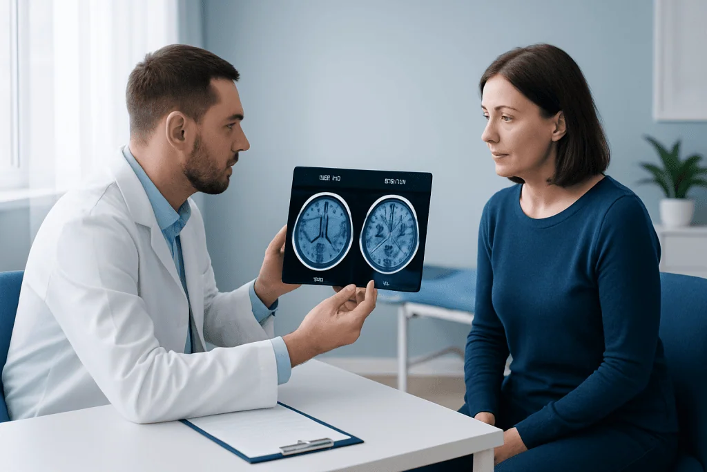
People may be referred for MRI when symptoms persist, when an examination suggests a problem involving soft tissues or the nervous system, when previous imaging needs clarification, or when a clinician needs detailed information for treatment planning. The best type of scan is determined by the clinical question rather than simply by the severity of discomfort.
Before an MRI, the imaging team reviews safety information carefully. People should disclose any pacemaker, implanted neurostimulator, cochlear implant, aneurysm clip, artificial joint, metal fragments, insulin pump, medication patch, or previous surgery. Many modern implants are MRI-compatible under specific conditions, but this must be checked in advance.
Pregnancy, breastfeeding, kidney disease, prior contrast reactions, and claustrophobia should also be discussed. MRI may still be possible in many of these situations, but the team may adjust the plan, avoid contrast, arrange additional support, or recommend another approach where appropriate.
Someone with sudden severe neurological symptoms, signs of a stroke, major trauma, or rapidly worsening illness should seek urgent assessment rather than relying on a routine outpatient scan. For conditions involving persistent back or neck symptoms, a specialist may also consider whether herniated disc or another spinal cause is clinically relevant.
What Happens During a Private MRI Scan
Preparation varies by the body area being scanned. Many MRI examinations require no special preparation, although some abdominal or pelvic studies may involve instructions about eating, drinking, or bladder preparation. The person is asked to remove jewellery, watches, hearing aids, removable dental items, and other metal-containing objects before entering the scan room.
During the examination, the person lies on a padded table that moves into the MRI scanner. The body part being scanned is positioned with a device called a coil, which helps obtain detailed images. It is important to remain still while image sequences are taken. The machine makes repetitive loud tapping or knocking sounds, so ear protection is provided.
The radiographer can see, hear, and communicate with the person throughout the scan. An alarm device is usually available if assistance is needed. A standard scan can take from roughly 20 minutes to an hour or more, depending on the area, the number of sequences, whether contrast is used, and whether more than one body region is being examined.
If contrast is required, a small cannula is placed in a vein. Images may be taken before and after contrast administration. This “before and after” wording refers to image sequences, not an immediate change in the underlying health condition.
Realistic Results: Example Outcomes and Next Steps
Results are often more useful when expectations are realistic. For example, a person with knee pain after a twisting injury may have an MRI showing a ligament or meniscal injury, which can help guide rehabilitation, activity changes, or orthopaedic review. Another person with similar symptoms may have no major tear visible, helping the clinician consider non-surgical causes and conservative management.
For back pain, an MRI may show disc changes, narrowing, or nerve compression. However, these findings need to match the location and pattern of symptoms before they are considered clinically important. A scan may support a treatment plan such as physiotherapy, medication review, targeted procedures, or surgical consultation, but it does not mean that surgery is automatically needed.
A normal MRI can also be a meaningful result. It may reduce concern about certain structural problems and allow the clinician to focus on other possible explanations. Occasionally, an MRI identifies an incidental finding unrelated to the original symptoms. These are often harmless, but they may require explanation, comparison with previous imaging, or follow-up.
Reports are commonly sent to the referring clinician, who discusses what the findings mean in the individual situation. If an MRI confirms a musculoskeletal problem, a clinician may discuss physical therapy and rehabilitation as part of a tailored recovery plan.
Recovery, Benefits, and Possible Risks
There is usually no recovery period after a routine MRI. People can normally eat, drink, work, drive, and continue usual activities immediately afterwards. If sedation has been used for severe anxiety or difficulty remaining still, recovery instructions will differ and the person will need someone to accompany them home.
The main benefit of MRI is its ability to provide detailed images without ionising radiation. This can improve diagnostic confidence, help avoid unnecessary procedures in some situations, assist with treatment planning, and provide a baseline for monitoring certain conditions over time.
MRI is generally considered safe when appropriate screening is completed. Possible difficulties include anxiety in the scanner, discomfort from lying still, noise exposure without adequate ear protection, and rarely heating or movement risks involving undisclosed metal. Contrast reactions are uncommon but can occur; people with severe kidney impairment need specific assessment before gadolinium-based contrast is used.
An MRI result can sometimes lead to further tests or referrals, especially when an unexpected abnormality is found. This does not necessarily mean a serious condition is present. A qualified doctor can explain the level of concern and whether any follow-up is needed.
When to Seek Medical Care
Medical advice should be sought for symptoms that are persistent, worsening, recurrent, or affecting daily activities. A doctor can assess whether MRI is likely to be helpful or whether another examination, laboratory test, or imaging method is more suitable. Seeking advice early may be particularly helpful after an injury or when pain is associated with weakness, numbness, or reduced function.
Urgent medical assessment is needed for sudden facial drooping, new difficulty speaking, sudden weakness or numbness on one side of the body, severe sudden headache, loss of consciousness, chest pain, severe shortness of breath, new loss of bladder or bowel control, or rapidly progressing weakness. These symptoms should not wait for a routine MRI appointment.
People should also contact their care team promptly if they develop new symptoms after contrast administration, such as breathing difficulty, widespread rash, facial swelling, or severe dizziness. Although serious reactions are rare, they require immediate assessment.
Acibadem International’s multidisciplinary specialists and JCI-accredited hospitals can assess imaging needs and coordinate MRI-based diagnosis and treatment planning for international patients.
Frequently asked questions
What does “before and after” mean for a private MRI scan?
For MRI, “before and after” usually refers to the difference in information available before the examination and after the images have been reviewed. It may also refer to imaging performed before and after contrast material is given. MRI itself does not create an immediate physical improvement because it is a diagnostic test, not a treatment.
How soon are MRI results available?
Timing varies between imaging centres and depends on the scan type, radiologist reporting processes, and whether comparison with earlier images is needed. A radiologist reviews the images and prepares a report, which is then discussed with the referring clinician. The clinician can explain what the findings mean in relation to symptoms.
Can an MRI be normal even if symptoms are real?
Yes. A normal MRI does not mean that symptoms are not real or important. Some conditions are not visible on MRI, and some symptoms can arise from functional, inflammatory, early-stage, or intermittent problems that require clinical assessment beyond imaging.
Is a private MRI scan painful?
MRI is not usually painful, but remaining still on the scanning table can be uncomfortable for some people, particularly if they have pain or limited mobility. The scanner is noisy, and some people find the enclosed space challenging. The radiography team can provide support and discuss options before the examination.
Can everyone have an MRI scan?
Many people can have MRI safely, but careful screening is essential. Some implanted medical devices, metal fragments, or certain surgical clips may make MRI unsuitable or require a modified protocol. Pregnancy, kidney disease, and previous contrast reactions should also be discussed with the imaging team.
What should a person do after receiving an abnormal MRI result?
An abnormal result should be reviewed with the clinician who requested the scan or another qualified specialist. They can explain whether the finding is likely to be related to symptoms, whether treatment is needed, and whether follow-up imaging or referral is appropriate. Avoid drawing conclusions from report wording alone without clinical context.
References
- Radiological Society of North America
- American College of Radiology
- National Health Service
- U.S. Food and Drug Administration
- World Health Organization
This article is for general information only and is not a substitute for professional medical advice. Please consult a qualified doctor about your individual situation.
Explore treatments in Turkey — costs, top hospitals & a free quote
JCI-accredited · board-certified surgeons · reply within 24h
Add us as a Preferred Source to see more of our trusted health content across Google Search, AI Overviews and Discover.

More from the Health Library



Related Specialists
Dr. G. Pınar Özeren
Gynecology & Obstetrics
Dr. Gürhan Şişman
Gastroenterology
Dr. Ateş Önal
Anesthesiology
Prof. Dr. Ahmet Ümit Güllü
Cardiovascular Surgery