Pttd Treatment: How It Works, Results and What to Expect

Early PTTD treatment commonly includes activity changes, supportive shoes, orthotics or braces, and guided rehabilitation. The best treatment depends on tendon damage, foot flexibility, alignment changes and everyday mobility needs.
Key Takeaways
- Early PTTD treatment commonly includes activity changes, supportive shoes, orthotics or braces, and guided rehabilitation.
- The best treatment depends on tendon damage, foot flexibility, alignment changes and everyday mobility needs.
- Surgery is usually reserved for persistent symptoms, substantial deformity or tendon failure despite appropriate non-surgical treatment.
- Recovery after posterior tibial tendon surgery often involves a period of immobilization followed by gradual weight-bearing and physiotherapy.
- New or worsening inner-ankle pain, loss of arch height, or difficulty walking should be assessed by a qualified clinician.
PTTD treatment aims to reduce pain, protect the posterior tibial tendon and maintain or restore the foot’s arch. Many people improve with early non-surgical care, while surgery may be considered when symptoms persist or the foot becomes increasingly flat or rigid.
Overview: how PTTD treatment works
PTTD treatment addresses problems affecting the posterior tibial tendon, a tendon that runs behind the inner ankle and helps support the arch while walking. Posterior tibial tendon dysfunction (PTTD), also called progressive collapsing foot deformity, can cause inner-ankle pain, a flattening arch and the feeling that the foot rolls inward. Treatment works by reducing strain on the tendon, supporting the foot’s alignment and restoring strength where possible.
For many people, especially when symptoms are recognized early, non-surgical care can ease pain and improve daily function. If the tendon has significantly weakened or torn, or if the foot has developed a fixed deformity, an orthopedic foot and ankle specialist may discuss reconstructive surgery. The appropriate plan is individualized rather than based on symptoms alone.
PTTD is not simply an inflammation problem. Although it may begin with tendon irritation, longstanding dysfunction can affect ligaments, joints and the overall shape of the foot. This is why timely assessment is helpful when persistent inner-ankle pain is accompanied by changes in arch height or walking ability.
PTTD stages and treatment options
Clinicians often describe PTTD in stages because the tendon and foot shape can change over time. In earlier stages, the tendon may be painful and weakened but the foot remains flexible. In later stages, the arch may collapse more noticeably, the heel may turn outward and arthritis or a rigid deformity may develop. Staging helps guide treatment, but individual symptoms and examination findings remain important.
Early-stage care commonly includes relative rest from painful impact activities, supportive footwear, an ankle-foot brace or walking boot when needed, and custom or prefabricated orthotics. Physiotherapy can then help improve calf flexibility, balance and the strength of muscles that support the foot and ankle. A clinician may also recommend short-term pain relief measures when medically appropriate.
When non-surgical measures do not provide sufficient relief, or there is progressive deformity, surgical options may include tendon cleaning or repair, tendon transfer, realignment of the heel bone, lengthening a tight calf or Achilles tendon, and procedures to stabilize the arch. In severe rigid deformity with arthritis, joint fusion may be considered. These procedures are selected to address the particular structures involved, not as a one-size-fits-all operation.
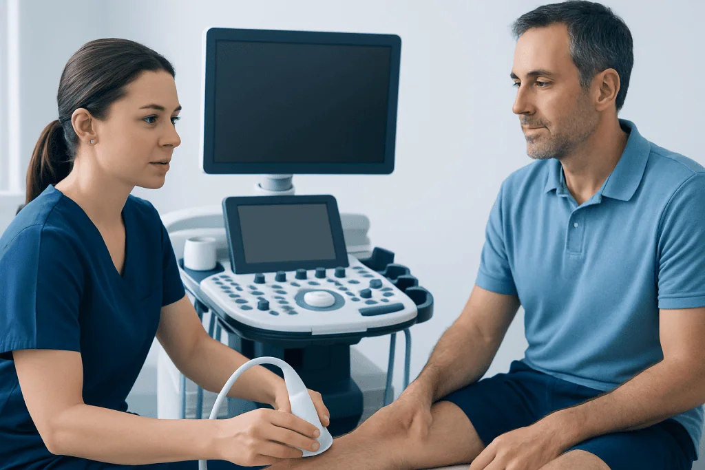
Imaging, especially weight-bearing X-rays, can show alignment changes. Ultrasound or MRI may be useful when more information is needed about the tendon, ligaments or nearby joints. An evaluation also considers medical conditions, circulation, nerve health, smoking status and personal recovery goals before deciding how to treat PTTD.
What shoes are best for PTTD?

Shoes that provide a stable heel, firm midsole and adequate arch support are generally best for PTTD. A shoe with a wide, supportive base can help limit excessive inward rolling of the foot, while a structured heel counter may improve rearfoot stability. Shoes should feel comfortable from the outset and leave enough space for an orthotic or brace if one is prescribed.
Walking shoes, supportive athletic shoes and certain stability-oriented footwear can be practical choices. The best option varies with a person’s foot shape, work demands and level of deformity, so a podiatrist, orthopedic specialist or physiotherapist may recommend trying shoes with an insole or custom device. A brace may be more useful than a shoe change alone when symptoms are more pronounced.
Very flexible, worn-out shoes and unsupportive sandals may allow more strain through the tendon and arch. High heels can also increase pressure in ways that are uncomfortable for some people. Footwear should not be expected to correct an established deformity by itself, but it can be an important part of symptom control and protection.
What is the fastest way to heal posterior tibial tendonitis?
There is no safe instant cure for posterior tibial tendonitis or PTTD. The fastest route toward recovery is usually early, accurate assessment followed by reducing the activities that aggravate the tendon, protecting the foot with appropriate support, and progressing rehabilitation at a pace the tendon can tolerate. Continuing high-impact activity through significant pain can prolong recovery.
For short periods, clinicians may recommend limiting running, jumping, long walks on uneven ground or other triggers. A walking boot or brace may be used when pain makes normal walking difficult. Once acute symptoms settle, a structured rehabilitation plan typically focuses on restoring calf flexibility, foot and ankle strength, balance and gradual loading rather than complete inactivity.
PTTD treatment at home may include wearing recommended footwear or orthotics consistently, avoiding painful activities, using cold packs briefly for comfort after activity and completing clinician-prescribed exercises. Self-treatment should not replace assessment when pain persists, the arch appears to be changing or there is weakness during walking. People with diabetes, neuropathy, poor circulation or inflammatory arthritis should seek individualized advice before starting an exercise plan.
Can I still walk with posterior tibial tendonitis?
Many people can still walk with posterior tibial tendonitis, particularly in the earlier stages, but walking through substantial pain or limping may worsen symptoms. The goal is to remain safely active within a tolerable level of discomfort while avoiding activities that repeatedly overload the tendon. Supportive shoes, orthotics or a brace can make walking more comfortable and may reduce strain.
A person should reduce walking distance and intensity if pain increases during activity, lingers afterward or causes a change in gait. Low-impact alternatives such as cycling, swimming or water-based exercise may sometimes help maintain fitness while the tendon settles, provided they are comfortable and approved by a clinician.
Difficulty standing on the affected foot to rise onto the toes, increasing ankle swelling, or a foot that is visibly flattening or turning outward warrants medical review. These signs can suggest more than a minor tendon irritation and may influence which PTTD treatment options are most suitable.
Posterior tibial tendon surgery: candidacy, steps and recovery
Posterior tibial tendon surgery is generally considered when a well-planned course of non-surgical treatment has not controlled pain or maintained function, or when deformity is progressing. Candidacy depends on whether the foot remains flexible, the condition of the tendon and joints, the degree of arthritis, overall health and the person’s activity goals. A specialist explains which procedure, if any, is appropriate and why.
The operation may combine several techniques. The surgeon may remove damaged tendon tissue, repair or reinforce the tendon, transfer a nearby functioning tendon to support the arch, and reposition the heel bone to improve alignment. If a tight calf contributes to strain, it may be lengthened. In more advanced arthritis or rigid deformity, fusion of selected joints can provide a stable, pain-reducing foot, though it limits movement at those joints.
Recovery begins with protecting the reconstruction in a cast, splint or boot. The timing of weight-bearing differs by procedure and surgeon, but it commonly advances gradually over weeks rather than days. Physical therapy is often introduced in stages to regain mobility, strength, balance and walking control. Swelling and improvement in endurance can continue for many months, so patience with rehabilitation is important.
Benefits may include less pain, better foot alignment and improved ability to walk in supportive footwear. Potential risks include infection, wound-healing problems, blood clots, nerve irritation, stiffness, persistent pain, failure of healing, recurrence of deformity and the possibility of further treatment. Smoking and poorly controlled medical conditions can raise certain surgical risks, so these should be discussed before an operation.
What is the success rate of posterior tibial tendon surgery?
A single success rate for posterior tibial tendon surgery is not reliable because the term covers different operations performed at different stages of PTTD. Outcomes are influenced by the flexibility of the deformity, the presence of arthritis, tendon quality, surgical technique, rehabilitation, body weight, smoking and other health conditions. For appropriately selected patients, reconstructive surgery often improves pain and function, but no procedure can promise a particular result.
It is useful to ask the surgeon what outcomes are realistic for the proposed procedure, including expected walking ability, footwear needs, return-to-work considerations and the chance of residual stiffness or discomfort. A surgeon can also explain how the person’s stage of disease changes the likely benefits and limitations of surgery.
Follow-up appointments and rehabilitation are central to the result. Protecting the repair early, attending physiotherapy when advised and returning gradually to activity can support recovery. New pain, increasing redness, wound drainage, calf swelling or sudden shortness of breath after surgery requires urgent medical assessment.
When to seek medical care
Medical assessment is advisable for pain or swelling along the inner ankle that lasts more than a short period, particularly if it limits walking or does not improve with rest and supportive footwear. A person should also arrange an assessment if one foot’s arch appears lower than the other, shoes suddenly fit differently, or it becomes difficult to rise onto the toes on one foot.
Urgent care is appropriate after a significant injury, with an inability to bear weight, marked redness or fever, or symptoms that could indicate a blood clot, such as new calf swelling and pain. Sudden chest pain or shortness of breath should be treated as an emergency.
Assessment by an orthopedic foot and ankle specialist can clarify whether symptoms are due to PTTD or another condition, such as arthritis, a stress injury or nerve-related pain. Acibadem International’s multidisciplinary specialists and JCI-accredited hospitals assess and treat foot and ankle conditions for international patients, with treatment planning based on the individual’s examination and imaging findings.
Frequently asked questions
What is PTTD treatment?
PTTD treatment is care for posterior tibial tendon dysfunction, a condition that can cause inner-ankle pain and a flattening foot arch. It commonly starts with supportive footwear, orthotics or bracing, activity modification and rehabilitation. Surgery may be considered for persistent symptoms or progressive structural changes.
How long does it take for posterior tibial tendonitis to heal?
Recovery time varies with the severity of tendon injury, the amount of activity modification needed and whether the foot shape has changed. Mild symptoms may improve over weeks with appropriate protection and rehabilitation, while longstanding dysfunction may require a longer course of care. Recovery after surgery generally takes months and progresses in stages.
Can orthotics help posterior tibial tendon dysfunction?
Orthotics can help support the arch and reduce strain on the posterior tibial tendon, especially in flexible early-stage PTTD. Some people benefit from prefabricated inserts, while others need a custom orthotic or ankle-foot brace. The most suitable device depends on symptoms, foot alignment and footwear.
Is surgery always needed for PTTD?
No. Many people with early PTTD improve with non-surgical treatment, particularly when treatment begins before major deformity develops. Surgery is usually discussed when symptoms continue despite appropriate conservative care or when the deformity is progressing, rigid or associated with arthritis.
What activities should be avoided with PTTD?
Activities that increase pain, such as running, jumping, long walks on uneven ground or high-impact sports, may need to be reduced temporarily. The specific restrictions depend on symptom severity and a clinician’s advice. Low-impact exercise may be possible if it does not aggravate symptoms.
Can PTTD get worse if untreated?
PTTD can progress in some people because the posterior tibial tendon supports the arch and helps control foot alignment. Ongoing tendon weakness may contribute to increasing flatfoot deformity and, in later stages, joint changes. Early evaluation can help identify supportive treatment before symptoms become more limiting.
References
- American Academy of Orthopaedic Surgeons
- American Orthopaedic Foot & Ankle Society
- National Health Service
- Mayo Clinic
This article is for general information only and is not a substitute for professional medical advice. Please consult a qualified doctor about your individual situation.
Joint and spine care in Turkey — expert assessment & treatment
JCI-accredited · board-certified surgeons · reply within 24h
Add us as a Preferred Source to see more of our trusted health content across Google Search, AI Overviews and Discover.