Pulmonary Nodules: CT Findings, Cancer Risk, and Follow-Up Decisions
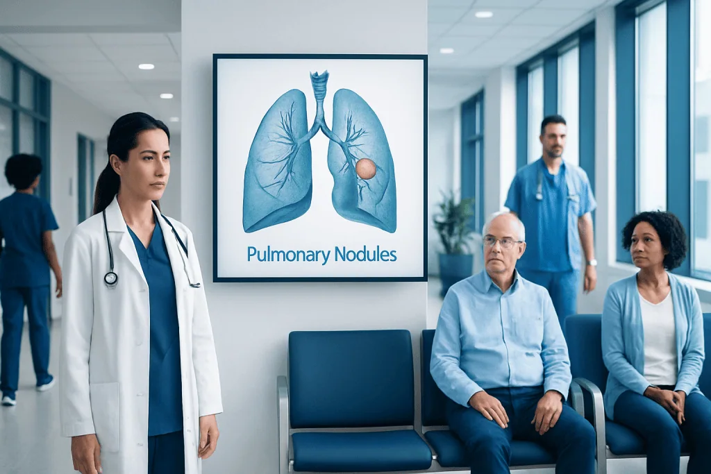
A pulmonary nodule is a small round or oval spot in the lung, usually found incidentally on a chest X-ray or CT scan. Most pulmonary nodules are benign, especially when they are small, stable over time, or related to past infection or inflammation.
Key Takeaways
- A pulmonary nodule is a small round or oval spot in the lung, usually found incidentally on a chest X-ray or CT scan.
- Most pulmonary nodules are benign, especially when they are small, stable over time, or related to past infection or inflammation.
- Cancer risk is estimated using CT features such as size, growth, shape, borders, density, and location, along with age, smoking history, and cancer history.
- Follow-up decisions are individualized; some nodules need no further testing, while others need repeat CT, PET-CT, biopsy, or surgical evaluation.
- Patients should keep prior imaging records, avoid smoking, and discuss the follow-up plan with a qualified pulmonologist, radiologist, or thoracic specialist.
Pulmonary nodules are common findings on chest CT scans, and most are not cancer. Careful review of CT features, personal risk factors, and any change over time helps doctors decide whether observation, repeat imaging, PET-CT, biopsy, or treatment is appropriate.
Overview: What Are Pulmonary Nodules?
Pulmonary nodules are small, rounded areas seen within the lung on imaging tests. They are sometimes called lung nodules, coin lesions, or solitary pulmonary nodules when only one is present. Many are found incidentally during a CT scan done for another reason, such as chest pain, infection, trauma, preoperative evaluation, or lung cancer screening.
A nodule is not a diagnosis by itself; it is a description of what appears on the image. A pulmonary nodule may represent a healed infection, a tiny scar, inflammation, a benign growth, or less commonly an early lung cancer or a spread from another cancer. The goal of medical evaluation is to identify which nodules are very low risk and which need closer attention.
CT is the most useful imaging test for evaluating pulmonary nodules because it shows the size, density, edges, and location of the nodule more clearly than a standard chest X-ray. In many cases, the safest and most accurate approach is not immediate biopsy, but careful comparison with previous scans and planned follow-up imaging.
CT Findings Doctors Look For
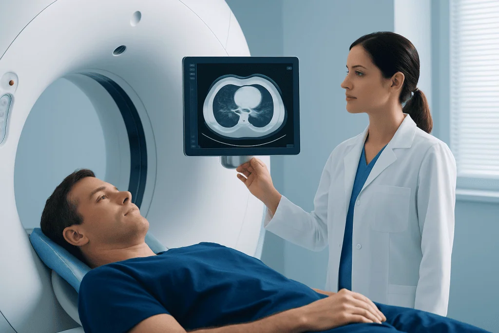
When a lung nodule is seen on CT, the radiologist evaluates several features that help estimate the likelihood of a benign or concerning cause. Size is one of the most important factors. Very small nodules are often monitored or may need no follow-up in low-risk people, while larger nodules are assessed more carefully, especially if they are new or growing.
The nodule’s density also matters. Solid nodules are completely dense on CT. Subsolid nodules include pure ground-glass nodules, which look hazy, and part-solid nodules, which contain both hazy and solid areas. Some subsolid nodules are inflammatory and disappear, while persistent part-solid nodules may require longer follow-up because certain early lung cancers can appear this way.
Other CT details include shape, borders, calcification pattern, and location. Smooth, sharply defined nodules and nodules with certain benign calcification patterns are more reassuring. Irregular or spiculated edges, upper-lobe location, associated airway changes, or growth over time may increase concern. Radiology reports may also mention whether there are multiple nodules, enlarged lymph nodes, emphysema, scarring, or signs of infection.
- Size: measured in millimeters, ideally on thin-section CT images.
- Density: solid, ground-glass, or part-solid.
- Margins: smooth, lobulated, irregular, or spiculated.
- Change over time: new, stable, shrinking, or growing.
- Context: infection, immune status, smoking history, and prior cancer history.
Cancer Risk: How Doctors Estimate Probability

Most pulmonary nodules are benign, but a structured risk assessment is important. Doctors estimate cancer probability by combining the CT appearance with patient-specific factors. Age, smoking history, exposure to certain occupational or environmental risks, family history, and a personal history of cancer can influence the level of concern. Symptoms such as coughing blood, unexplained weight loss, or persistent new cough may also affect the evaluation, although many nodules cause no symptoms at all.
Growth is one of the strongest clues. A nodule that is unchanged over a suitable period may be considered stable, depending on its type. Solid nodules that remain stable for a long enough interval are often benign. Ground-glass and part-solid nodules may grow slowly, so they sometimes require longer observation than solid nodules.
Doctors may use validated clinical models or guideline categories to classify risk as low, intermediate, or high. These tools do not replace medical judgment, but they help standardize decisions. For example, a small smooth nodule in a young person who has never smoked is usually approached differently from a larger spiculated nodule in an older person with a long smoking history.
It is also important to remember that risk is not the same as a diagnosis. A higher-risk nodule does not automatically mean cancer, and a lower-risk nodule should still be followed if the doctor recommends it. Clear communication about the reason for follow-up can reduce anxiety and prevent missed appointments.
Diagnosis and Tests Beyond the Initial CT
The first diagnostic step is often comparison with previous imaging. If an older CT scan or chest X-ray shows the same nodule without meaningful change, the finding may be far less concerning. Patients are encouraged to share prior imaging discs, digital records, and reports with their doctor, especially if scans were done at different hospitals or in different countries.
If the nodule needs more evaluation, doctors may recommend a dedicated thin-section chest CT. This can confirm the exact size and type of nodule, reduce measurement uncertainty, and look for additional findings. In some cases, contrast-enhanced CT may be helpful, but many nodule follow-up scans are performed without contrast, depending on the clinical question.
PET-CT may be used for selected nodules, especially when they are large enough for metabolic assessment and the result would change management. PET-CT can show increased activity, but inflammation and infection can also be active, and small or slow-growing cancers may not always be clearly detected. For this reason, PET-CT results are interpreted together with CT features and clinical risk.
Biopsy is considered when imaging and risk assessment suggest that tissue diagnosis is needed. Options may include bronchoscopy, navigational bronchoscopy, CT-guided needle biopsy, or surgical biopsy. The choice depends on nodule location, size, patient health, and local expertise. Because biopsy has potential risks, it is usually recommended only when the expected benefit outweighs those risks.
Follow-Up Decisions: Observation, Imaging, or Intervention
Follow-up decisions for pulmonary nodules are commonly guided by recommendations such as those from the Fleischner Society, the American College of Chest Physicians, and other professional groups. These guidelines consider nodule size, number, density, and the patient’s cancer risk. They are designed for typical incidental nodules, but they may not apply in the same way to people with known cancer, active infection, significant immune suppression, or very young patients.
Some nodules need no routine follow-up because their appearance is clearly benign or their risk is extremely low. Others may need repeat CT after a defined interval to confirm stability or resolution. If a nodule shrinks or disappears, infection or inflammation becomes more likely. If it grows, develops a new solid component, or changes shape, further testing may be advised.
For intermediate-risk nodules, options may include short-interval CT, PET-CT, or biopsy. For high-risk nodules, referral to a pulmonologist, thoracic surgeon, or multidisciplinary lung nodule board may be appropriate. When early lung cancer is suspected and the patient is fit for treatment, surgery or other local therapies may be discussed after careful staging and shared decision-making.
The best plan is individualized. A person who is older or has other serious medical conditions may choose a different approach from someone who is younger and otherwise healthy. Doctors should explain the expected benefits, limitations, and risks of surveillance versus invasive testing so the patient can participate in the decision.
Prevention and Self-Care While Being Monitored
Many pulmonary nodules cannot be prevented because they may result from past infections, scars, or environmental exposures. However, reducing lung cancer risk and protecting lung health are important for everyone. The most important step is to avoid smoking or to stop smoking with professional support. Avoiding secondhand smoke and using proper respiratory protection for occupational exposures can also help.
Patients being monitored for a lung nodule should keep all follow-up appointments and maintain a personal record of imaging reports. Missed CT follow-up can create uncertainty later, while repeated scans done too frequently may add unnecessary radiation exposure. The goal is to use the right test at the right time, not to scan more often than needed.
Healthy habits support overall respiratory wellness, although they do not replace medical follow-up. These include regular physical activity as tolerated, vaccination when recommended by a doctor, management of chronic lung diseases such as COPD or asthma, and prompt evaluation of persistent respiratory symptoms. Patients should also tell their doctor about prior tuberculosis, fungal infections, autoimmune disease, or cancer treatment, as these details may change the interpretation of the nodule.
When to See a Doctor
A person should discuss any pulmonary nodule finding with a qualified doctor, even if the radiology report says it is likely benign. The doctor can interpret the report in the context of age, symptoms, smoking history, previous imaging, and medical background. A clear written plan helps patients understand whether no action, repeat CT, specialist referral, or further testing is recommended.
Medical review is especially important if the nodule is described as growing, spiculated, part-solid, or larger; if there are enlarged lymph nodes; or if the person has a history of cancer. Patients should also seek medical care for symptoms such as coughing blood, worsening shortness of breath, persistent chest pain, unexplained weight loss, or a cough that does not improve as expected.
International patients may benefit from coordinated review when previous scans were performed in different health systems. Acibadem International’s multidisciplinary specialists and JCI-accredited hospitals diagnose and treat pulmonary nodules for international patients, including radiology review, pulmonology evaluation, and thoracic oncology input when needed. The most appropriate next step should always be based on an individual medical assessment.
Frequently asked questions
Are pulmonary nodules usually cancer?
Most pulmonary nodules are not cancer, especially when they are small, smooth, calcified in a benign pattern, or stable over time. Cancer risk depends on CT features and personal risk factors such as age, smoking history, and prior cancer. A doctor can explain whether the nodule is low, intermediate, or higher risk.
What is the difference between a solid nodule and a ground-glass nodule?
A solid nodule appears dense throughout on CT, while a ground-glass nodule looks hazy and does not fully obscure the lung structures behind it. A part-solid nodule has both ground-glass and solid components. Subsolid nodules can be inflammatory, but if they persist, they may need longer follow-up than many solid nodules.
How often should a pulmonary nodule be followed with CT?
The timing depends on the nodule's size, appearance, number, and the patient's risk factors. Some nodules require no further imaging, while others need repeat CT after a specific interval. Patients should follow the plan recommended by their doctor rather than arranging repeated scans on their own.
Does a growing lung nodule always mean cancer?
Growth can increase concern, but it does not always mean cancer. Infection, inflammation, bleeding, or other benign processes can also change over time. If growth is confirmed, the doctor may recommend additional imaging, PET-CT, biopsy, or specialist evaluation.
When is a biopsy needed for a pulmonary nodule?
Biopsy is considered when the chance of cancer is high enough that tissue diagnosis would help guide treatment, or when imaging results are uncertain. The method depends on the nodule's size and location and the patient's overall health. Not every nodule should be biopsied, because biopsy has risks and very small nodules may be difficult to sample accurately.
Can a pulmonary nodule disappear?
Yes. Nodules caused by infection or inflammation may shrink or disappear on follow-up CT. When a nodule resolves, it is usually reassuring, but the doctor should confirm whether any further monitoring is needed based on the full clinical picture.
References
- Fleischner Society
- American College of Chest Physicians
- American College of Radiology
- British Thoracic Society
- National Comprehensive Cancer Network
This article is for general information only and is not a substitute for professional medical advice. Please consult a qualified doctor about your individual situation.
Oncology care in Turkey — second opinion and treatment plan
JCI-accredited · board-certified surgeons · reply within 24h
Add us as a Preferred Source to see more of our trusted health content across Google Search, AI Overviews and Discover.