Pvl Medical Abbreviation Ultrasound: Preparation, Procedure and Results

PVL stands for periventricular leukomalacia, not a type of ultrasound examination. Cranial ultrasound is commonly used in premature infants because it is safe, painless and can be performed at the bedside.
Key Takeaways
- PVL stands for periventricular leukomalacia, not a type of ultrasound examination.
- Cranial ultrasound is commonly used in premature infants because it is safe, painless and can be performed at the bedside.
- An ultrasound can identify cystic PVL and other signs of brain injury, but subtle non-cystic changes may be clearer on MRI.
- PVL has a wide range of outcomes; some children have mild or no long-term difficulties, while others need ongoing developmental support.
- Early developmental follow-up and therapy can support a child’s mobility, learning, communication and independence.
In ultrasound reports, PVL usually means periventricular leukomalacia: injury to white matter near the brain’s fluid-filled ventricles, most often associated with premature birth. A cranial ultrasound is a safe bedside test that can help identify some forms of PVL and guide follow-up care, although MRI may provide more detail in some babies.
PVL Medical Abbreviation Ultrasound: What Does It Mean?
PVL is the medical abbreviation for periventricular leukomalacia. It describes damage to white matter, the nerve-fiber tissue around the brain’s ventricles. In newborn care, the phrase “PVL ultrasound” usually means a cranial ultrasound performed to check for signs of this injury, particularly in babies born very early.
PVL is most often linked with prematurity because the developing brain is especially vulnerable to reduced blood flow, inflammation and changes in oxygen delivery. It is not caused by anything a parent did or did not do. The finding needs to be interpreted by the neonatal team alongside the baby’s birth history, examination and other test results.
A cranial ultrasound uses sound waves rather than radiation. The scan is usually performed through the soft spot on top of a baby’s head, called the fontanelle. It helps clinicians monitor the brain after preterm birth and look for concerns such as bleeding, enlarged ventricles or changes consistent with PVL.
How Cranial Ultrasound Works and Who May Need It
During a cranial ultrasound, a trained clinician places a small probe with gel over the baby’s fontanelle. Sound waves travel through this natural opening in the skull and create images of structures inside the brain. The test does not expose a baby to X-rays, and it generally does not require sedation.
Screening cranial ultrasounds are commonly offered to babies born very prematurely, with very low birth weight, or who have had a complicated course in a neonatal intensive care unit. A scan may also be recommended when there has been severe illness, infection, breathing difficulty, low blood pressure, seizures or another concern for brain injury.
The timing and number of scans depend on gestational age, health needs and local neonatal protocols. Early scans may look for bleeding, while later scans can assess evolving white-matter changes. Repeat imaging can be helpful because some features of PVL become more visible over time.
- Benefits: bedside access, no ionizing radiation, no injections and the ability to repeat the examination safely.
- Limitations: ultrasound may not show every mild or diffuse white-matter change; MRI can sometimes provide a more detailed view.
Step-by-Step Procedure, Preparation and Recovery
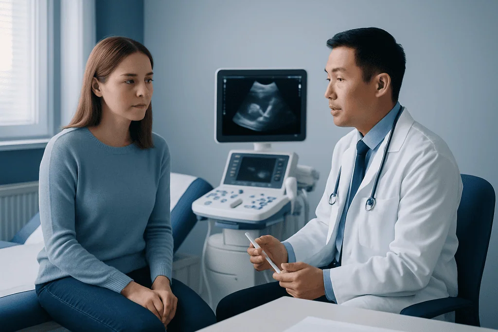
Families usually do not need to prepare a baby for a cranial ultrasound. Feeding and routine medicines can generally continue unless the neonatal team provides different instructions. Keeping the baby warm, settled and comfortable is the main practical consideration.
For the examination, the baby lies in an incubator, cot or caregiver’s arms when appropriate. The sonographer or doctor applies a small amount of clear gel to the probe and gently moves it over the fontanelle from different angles. Images are recorded for a radiologist, neonatologist or pediatric specialist to review. The scan commonly takes only a short time.
There is no recovery period. The gel is wiped away, and the baby can return to normal feeding, sleep and neonatal care immediately. Most babies tolerate the procedure well; some may briefly fuss because of handling or the cool gel. The meaningful “next step” is discussion of the report, including whether another ultrasound, MRI or developmental assessment is recommended.
Can Periventricular Leukomalacia Be Seen on an Ultrasound?
Yes. Periventricular leukomalacia can sometimes be seen on a cranial ultrasound, especially when it causes cysts in the white matter near the ventricles. This is often called cystic PVL. Early ultrasound findings can include brighter-than-expected areas around the ventricles, although these changes are not always specific to PVL and may need repeat imaging for clarification.
Ultrasound is very useful for regular monitoring in premature babies, but it has limits. Non-cystic or diffuse white-matter injury can be subtle and may not be fully apparent on ultrasound. If the care team needs a more detailed assessment, they may recommend brain MRI at a clinically appropriate time, often nearer to the baby’s expected due date.
An abnormal ultrasound does not predict one exact outcome for an individual child. Specialists consider the location and extent of imaging changes, the baby’s overall medical history and developmental progress over time. Families should ask the neonatal team to explain the wording of the specific report and the plan for follow-up.
How Serious Is Periventricular Leukomalacia?
PVL can be serious because white matter supports communication between different areas of the brain. The possible effects vary widely. Some children with mild imaging changes develop normally or have only subtle challenges, while others may develop movement, learning, vision, hearing, speech or behavioral differences that require support.
PVL is associated with a higher likelihood of cerebral palsy, particularly spastic diplegia, which affects muscle tone and movement in both legs more than the arms. It may also be associated with developmental delay, coordination difficulties, attention concerns or learning differences. However, a diagnosis of PVL does not by itself determine which challenges a child will have or how severe they will be.
Early follow-up is important because support can begin as soon as needs are identified. This may include physiotherapy, occupational therapy, speech and language therapy, vision or hearing assessment, and developmental pediatric care. Ongoing monitoring allows the care plan to change as the child grows.
What Is the Average Life Expectancy for Someone With Periventricular Leukomalacia (PVL)?
There is no single average life expectancy for a person with PVL. PVL is a brain injury pattern rather than a progressive disease, and life expectancy depends mainly on the person’s overall health and the severity of associated conditions, such as cerebral palsy, epilepsy, swallowing problems or serious breathing difficulties.
Many people with PVL, including those with mild to moderate developmental or motor differences, can have a typical or near-typical lifespan. Children with more complex disabilities may need specialized, lifelong medical and practical support. Their outlook is influenced by individual factors rather than the imaging label alone.
It is understandable for families to want clear predictions, but newborn imaging cannot provide a precise life-expectancy estimate. A pediatric neurologist, developmental specialist and the child’s wider care team can give more individualized guidance as the child’s health and development become clearer.
Is PVL Considered a Disability?
PVL itself is a medical diagnosis and imaging finding; it is not automatically a disability. Whether a child meets the definition of disability depends on whether they develop functional limitations in areas such as walking, hand use, communication, learning, vision, hearing or everyday activities.
Some children with PVL may qualify for disability services because they have cerebral palsy, developmental delay or other ongoing needs. Others may need temporary early-intervention support without having a long-term disability. Eligibility criteria for educational, social and financial support vary by country and local system.
Families do not need to wait for a final long-term label before seeking help. Developmental surveillance, early-intervention referral and regular pediatric review can identify needs early and help children access appropriate therapies and school-based accommodations when needed.
When to Seek Medical Care and What Follow-Up Involves
Parents should contact their child’s doctor promptly if a baby or child with a history of prematurity or PVL has seizures, repeated unusual jerking or staring episodes, trouble feeding or swallowing, breathing concerns, loss of skills already gained, or a sudden change in alertness. Urgent symptoms should be assessed without delay according to local emergency guidance.
Less urgent but important reasons to arrange a review include persistent stiffness or floppiness, using one hand much more than the other, delayed sitting or walking, difficulty seeing or hearing, feeding problems, speech delay, or concerns about learning and behavior. These signs do not always mean PVL is the cause, but they deserve professional assessment.
Follow-up commonly involves a neonatologist or pediatrician, with referral to pediatric neurology, developmental pediatrics, rehabilitation and therapy services when appropriate. Acibadem International’s multidisciplinary specialists and JCI-accredited hospitals support diagnosis, imaging review and individualized care planning for international patients with pediatric neurological needs.
Frequently asked questions
What does PVL mean on a baby ultrasound report?
PVL means periventricular leukomalacia, a form of injury affecting white matter near the brain’s ventricles. A report may describe suspected, evolving or confirmed features, and the neonatal team may repeat imaging because findings can change over time.
Does a PVL ultrasound hurt a baby?
No. Cranial ultrasound is noninvasive and uses sound waves, not radiation. A baby may be briefly unsettled by handling or gel on the head, but the procedure is generally painless and does not require recovery time.
Can an ultrasound diagnose all cases of PVL?
No. Ultrasound can identify many important findings, particularly cystic PVL, but mild or diffuse white-matter injury may be difficult to see. MRI may be used when more detail is needed or when ultrasound results are uncertain.
Can PVL improve over time?
The original white-matter injury does not simply disappear, but a child’s abilities and function can improve substantially with development, learning and appropriate support. The developing brain can adapt, and early therapies can help children build skills and independence.
Does every child with PVL develop cerebral palsy?
No. PVL increases the risk of cerebral palsy, but not every child with PVL develops it. Outcomes depend on the extent of injury and many individual health and developmental factors.
What follow-up is needed after suspected PVL?
Follow-up may include repeat cranial ultrasound, MRI when appropriate, pediatric examinations and developmental assessments. Depending on the child’s needs, the care team may recommend physiotherapy, occupational therapy, speech and language therapy, and hearing or vision evaluation.
References
- American Academy of Pediatrics
- National Institute of Neurological Disorders and Stroke
- Centers for Disease Control and Prevention
- MedlinePlus
- World Health Organization
This article is for general information only and is not a substitute for professional medical advice. Please consult a qualified doctor about your individual situation.
Ultrasound in Turkey — costs, top hospitals & a free quote
JCI-accredited · board-certified surgeons · reply within 24h
Add us as a Preferred Source to see more of our trusted health content across Google Search, AI Overviews and Discover.
Check your numbers in seconds
BMI, calories, due date, blood pressure and 30+ more clinical calculators — free, instant, doctor-reviewed ranges.

Check-up & Preventive Medicine
Comprehensive health screenings designed for early detection and prevention.
3 specialists in this unitMore from the Health Library


Related Specialists
Dr. Orkun Cem Akyazı
Emergency Service
Prof. Dr. Simay A. Kara
Interventional Radiology
Assoc. Prof. Dr. Selen Kanar
Ophthalmology
Dr. Şafak Çakmakçı
Ophthalmology