Pvns MRI: Preparation, Procedure and Results
MRI is the preferred imaging test for evaluating suspected PVNS because it shows synovial tissue, fluid, cartilage and nearby bone in detail. PVNS commonly affects one large joint, especially the knee, and may cause persistent swelling, pain, stiffness or repeated fluid buildup.
Key Takeaways
- MRI is the preferred imaging test for evaluating suspected PVNS because it shows synovial tissue, fluid, cartilage and nearby bone in detail.
- PVNS commonly affects one large joint, especially the knee, and may cause persistent swelling, pain, stiffness or repeated fluid buildup.
- Characteristic low-signal areas on MRI can reflect hemosiderin, an iron-containing pigment from prior bleeding in the joint lining.
- MRI findings are interpreted alongside symptoms, examination and sometimes tissue sampling to confirm the diagnosis.
- Treatment may include monitoring, surgery, rehabilitation and, in selected cases, medicines or other specialist therapies.
- PVNS can recur after treatment, so follow-up imaging and orthopedic review may be recommended.
A PVNS MRI is the main imaging examination used to assess pigmented villonodular synovitis, now often called tenosynovial giant cell tumor. It can show the extent of abnormal synovial tissue within a joint, particularly the knee, and helps clinicians plan appropriate care.
PVNS MRI: what it is and why it is used
A PVNS MRI is a magnetic resonance imaging scan performed to investigate pigmented villonodular synovitis (PVNS), a rare disorder in which the synovium—the tissue lining a joint or tendon sheath—grows abnormally. The condition is also commonly described as tenosynovial giant cell tumor (TGCT). MRI does not use ionizing radiation and provides detailed images of soft tissues that are not seen as clearly on standard X-rays.
The scan helps determine whether abnormal synovial tissue is present, where it is located and how extensively it involves the joint. It can also show joint fluid, cartilage changes, bone erosion and involvement of structures around the joint. This information is especially useful when planning care with orthopedic, musculoskeletal radiology and rehabilitation teams.
PVNS may be localized, meaning it forms a more defined nodule, or diffuse, meaning it involves a broader area of synovium. MRI helps distinguish these patterns, although the final diagnosis may occasionally require a biopsy or examination of tissue removed during surgery.
How PVNS MRI works and who may need it
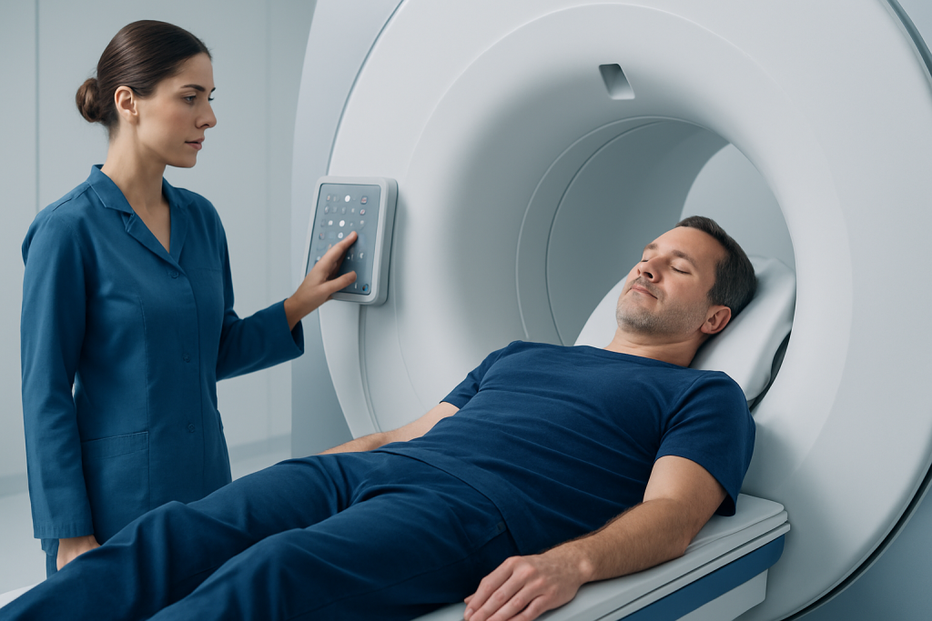
MRI uses a strong magnetic field and radio waves to create detailed cross-sectional images. During a PVNS MRI, the person lies still on a movable examination table while the body part being examined is positioned in or near the scanner. The machine makes loud tapping or knocking sounds while images are acquired, but the scan itself should not be painful.
A clinician may request MRI when a person has unexplained joint swelling, recurring collections of fluid, stiffness, reduced range of motion or ongoing pain that has not been explained by more common joint conditions. The knee is the most frequent site, but PVNS can also affect the hip, ankle, shoulder, elbow, wrist or smaller joints.
Not everyone can have MRI without additional planning. People should tell the imaging team about implanted medical devices, metal fragments, prior surgery, kidney disease, pregnancy, allergies and severe claustrophobia. Many modern implants are MRI-compatible, but the team must check device details before scanning. A contrast agent may be used in some cases to clarify the extent of inflamed or abnormal tissue.
Preparing for a PVNS MRI and what happens during the scan

Most people can eat, drink and take regular medicines as usual before a non-contrast PVNS MRI. If contrast is planned or sedation is being considered, the imaging center may provide specific instructions. Comfortable clothing without metal fasteners can be helpful, although patients are often asked to change into a gown and remove jewelry, watches, hearing aids, removable dental items and other metal objects.
Before the scan, a radiographer reviews safety questions and confirms the body area to be examined. If contrast is needed, it is usually given through a small intravenous line. The person is positioned carefully, sometimes with supports to help keep the joint comfortable and still. Ear protection is provided because the scanner is noisy.
The imaging portion commonly takes about 30 to 60 minutes, depending on the joint and whether contrast sequences are required. It is important to remain as still as possible because movement can blur the images. The radiographer can communicate throughout the examination, and an emergency call device is typically available.
After a standard MRI, normal activities can usually resume immediately. If sedation was used, the person will need recovery time and should arrange for someone else to drive them home. The images are reviewed by a radiologist, who sends a report to the referring clinician for discussion in the context of the person’s symptoms and examination.
What are the MRI findings of PVNS?
PVNS has MRI features that can be strongly suggestive of the diagnosis. The scan may show thickened, irregular or nodular synovium, often with joint effusion (extra fluid). In diffuse disease, the abnormal tissue can extend throughout the joint lining; in localized disease, it may appear as a more discrete mass within the joint or tendon sheath.
A characteristic feature is low signal intensity in parts of the synovium on particular MRI sequences. This is often due to hemosiderin, an iron-containing pigment left after repeated small amounts of bleeding within the tissue. Specialized sequences may make this susceptibility effect more conspicuous and help radiologists recognize the pattern.
MRI can also assess the effects of PVNS on surrounding structures. Depending on the severity and duration of disease, findings may include cartilage damage, bone erosions, cyst-like changes, ligament or tendon involvement, and swelling in nearby soft tissues. These details help distinguish PVNS from other causes of synovitis, such as inflammatory arthritis, infection or another type of joint tumor.
Although MRI is highly informative, it is not the only part of diagnosis. A clinician may recommend a biopsy when imaging is not definitive or when confirmation is needed before treatment. The report should be reviewed with an orthopedic specialist familiar with joint and soft-tissue conditions.
Is PVNS in the knee painful?
PVNS in the knee can be painful, but symptoms vary between individuals. Some people experience a dull ache that gradually worsens, while others mainly notice swelling, stiffness, a feeling of fullness in the joint or difficulty bending and straightening the knee. Pain may be more noticeable with walking, climbing stairs, squatting or other weight-bearing activity.
Repeated swelling or fluid accumulation is a common reason people seek assessment. In some cases, the knee may feel unstable, catch or lock because thickened synovial tissue interferes with joint movement. Symptoms may develop slowly, which can delay diagnosis if they resemble a sports injury, osteoarthritis or another common knee problem.
Persistent knee pain and swelling deserve medical assessment, particularly when there is no clear injury or symptoms keep returning after rest or routine treatment. MRI can help identify the cause and assess whether the synovium has features compatible with PVNS.
Treatment planning, benefits and possible risks
The benefit of PVNS MRI is that it provides a detailed map of the condition before treatment. It can help determine whether the disease is localized or diffuse, whether it involves difficult-to-reach portions of the joint and whether there is damage to cartilage or bone. This supports individualized planning and provides a baseline for future comparison.
For many symptomatic cases, surgery to remove abnormal synovial tissue is considered. This may be performed arthroscopically through small incisions, through open surgery, or with a combined approach depending on the location and extent of disease. Treatment decisions take account of symptoms, joint function, imaging findings, the likelihood of complete removal and the person’s overall health.
Some people with mild symptoms may be monitored with clinical review and repeat imaging. For diffuse, recurrent or difficult-to-operate disease, a multidisciplinary team may discuss additional options, including medicines that target pathways involved in tenosynovial giant cell tumor. Physical therapy is often important for restoring movement, strength and confidence after treatment.
MRI itself is generally very safe. Potential issues include anxiety in confined spaces, discomfort from staying still, and rare reactions to contrast material when it is used. Staff screen for contraindications and take precautions for people with kidney impairment or implanted devices. Acibadem International’s multidisciplinary specialists and JCI-accredited hospitals support diagnosis and treatment planning for international patients with complex joint conditions.
How long does it take to recover from PVNS knee surgery?
Recovery after PVNS knee surgery depends on the procedure performed, how much synovial tissue is removed, whether cartilage or other joint structures need treatment, and the person’s health and activity needs. After a straightforward arthroscopic procedure, many people begin gentle movement and rehabilitation relatively soon, but full recovery commonly takes several weeks to a few months.
Recovery may take longer after open surgery or combined procedures, especially in diffuse PVNS. Swelling, stiffness and reduced muscle strength can persist while tissues heal. A rehabilitation program may include exercises to restore knee range of motion, strengthen the thigh and hip muscles, improve walking mechanics and gradually return the person to work, exercise or sport.
The surgical team provides individualized guidance about weight-bearing, wound care, driving, activity restrictions and follow-up appointments. Follow-up is important because symptoms and imaging may need to be monitored for recurrence. People should avoid comparing their recovery directly with another patient’s, as disease extent and procedures can differ substantially.
What is the prognosis for PVNS?
The outlook for PVNS is often favorable in terms of controlling symptoms and improving joint function, particularly when the condition is identified and managed before major joint damage develops. PVNS does not usually spread to distant organs in the way malignant cancers do. However, it can be locally aggressive and may gradually damage cartilage and bone if ongoing inflammation and synovial overgrowth are not controlled.
Recurrence is possible after treatment, particularly with diffuse PVNS because abnormal tissue can be difficult to remove completely from all parts of the joint. Regular clinical follow-up, and sometimes repeat MRI, can help identify recurrent disease early. Persistent or returning pain, swelling or stiffness should be discussed with the treating team rather than assumed to be a normal part of recovery.
Long-term outcomes depend on the affected joint, whether disease is localized or diffuse, pre-existing cartilage damage, completeness of treatment and response to rehabilitation. A coordinated approach involving orthopedic surgery, radiology, pathology, rehabilitation and, where appropriate, medical oncology can help address both disease control and joint preservation.
When to seek medical care
A person should arrange medical assessment for joint swelling, pain or stiffness that persists for several weeks, repeatedly returns or limits normal activities. Evaluation is particularly important if a knee or another joint repeatedly fills with fluid without an obvious injury, or if symptoms are steadily worsening.
Urgent medical care is appropriate for a hot, very swollen joint with fever or feeling unwell, sudden inability to bear weight, severe pain after injury, new numbness or weakness, or symptoms suggesting infection. These symptoms are not specific to PVNS, but they require prompt assessment to rule out conditions that need immediate treatment.
For suspected or confirmed PVNS, an orthopedic specialist can explain the role of MRI, whether tissue confirmation is needed and which treatment approach best fits the imaging findings and personal goals. Keeping copies of imaging reports and prior scans can be useful when seeking a specialist opinion.
Frequently asked questions
Does an MRI confirm PVNS?
MRI can show features that are highly suggestive of PVNS, including thickened synovium and hemosiderin-related signal changes. However, clinicians interpret the scan together with symptoms, examination findings and other tests. In some situations, a biopsy or surgical tissue sample is needed to confirm the diagnosis.
Do I need contrast for a PVNS MRI?
Contrast is not always required, but it may help define the active synovium and the full extent of disease. The radiologist or referring clinician decides based on the joint being examined and the diagnostic question. Patients should tell the team about kidney disease, past contrast reactions or pregnancy.
Can a PVNS MRI show cartilage damage?
Yes. MRI can assess cartilage, bone beneath the cartilage, joint fluid, ligaments, tendons and soft tissues around the joint. This helps clinicians understand whether PVNS has affected the joint beyond the synovial lining.
Is PVNS in the knee painful?
PVNS in the knee may cause aching pain, swelling, stiffness and reduced movement. Symptoms can be mild initially or gradually become more disruptive with activity. Recurrent unexplained knee swelling should be assessed by a clinician.
How long does it take to recover from PVNS knee surgery?
Recovery varies according to whether surgery is arthroscopic, open or combined, and whether the disease is localized or diffuse. Gentle movement and rehabilitation often begin early, while fuller recovery may take weeks to months. The surgical and rehabilitation teams provide personalized milestones and activity advice.
What is the prognosis for PVNS?
Many people achieve improved symptoms and function with appropriate treatment and follow-up. PVNS can recur, particularly when it is diffuse, and untreated disease may contribute to joint damage over time. Ongoing review helps clinicians monitor symptoms and decide whether further imaging or treatment is needed.
References
- American Academy of Orthopaedic Surgeons
- National Organization for Rare Disorders
- Orphanet
- Radiological Society of North America
- National Cancer Institute
This article is for general information only and is not a substitute for professional medical advice. Please consult a qualified doctor about your individual situation.
Joint and spine care in Turkey — expert assessment & treatment
JCI-accredited · board-certified surgeons · reply within 24h
Add us as a Preferred Source to see more of our trusted health content across Google Search, AI Overviews and Discover.

More from the Health Library


Related Specialists
Assoc. Prof. Dr. Mehmet Fatih Karakaş
Cardiology
Dyt. Beyza Erdoğan Aksan
Nutrition & Diet
Assoc. Prof. Dr. Semiha Urvay
Medical Oncology
Prof. Dr. Mehmet Serdar Binnet
Orthopedic Surgery & Traumatology