Renal Abscess Ultrasound: Preparation, Procedure and Results
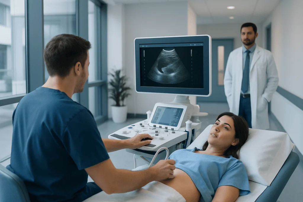
Ultrasound is radiation-free and usually takes about 20 to 40 minutes. Preparation varies, but patients may be asked to drink water and arrive with a comfortably full bladder.
Key Takeaways
- Ultrasound is radiation-free and usually takes about 20 to 40 minutes.
- Preparation varies, but patients may be asked to drink water and arrive with a comfortably full bladder.
- A renal abscess may appear as a fluid-containing or complex area in the kidney, but ultrasound does not confirm every case.
- CT imaging may be recommended when ultrasound results are unclear or an abscess is strongly suspected.
- Kidney abscesses need timely medical assessment because treatment may include antibiotics and, in some cases, drainage.
A renal abscess ultrasound is a painless imaging test that uses sound waves to examine the kidneys for a possible pocket of infection. It can help clinicians identify an abnormal collection, assess the kidneys and bladder, and decide whether further imaging, antibiotics or drainage may be needed.
Renal Abscess Ultrasound: What It Shows
A renal abscess ultrasound is an imaging examination used when a clinician suspects a collection of pus within or around a kidney. This can occur when a urinary tract infection reaches the kidney or when bacteria spread through the bloodstream. The test uses high-frequency sound waves rather than radiation, making it a safe way to examine the kidneys, nearby tissues and often the bladder.
The scan can identify kidney enlargement, blockage of urine flow, stones, fluid collections and other changes that may support a diagnosis. A renal abscess ultrasound appearance may be a rounded area with fluid, internal echoes or an irregular wall. However, appearances vary, and small or deeply located abscesses can be difficult to see.
Ultrasound is often an early imaging option because it is accessible and noninvasive. If the findings are uncertain, or if symptoms suggest a complicated infection, a clinician may recommend contrast-enhanced CT or MRI for more detailed assessment. The scan result is always interpreted alongside symptoms, examination findings, urine testing and blood tests.
How the Ultrasound Works and Who May Need It
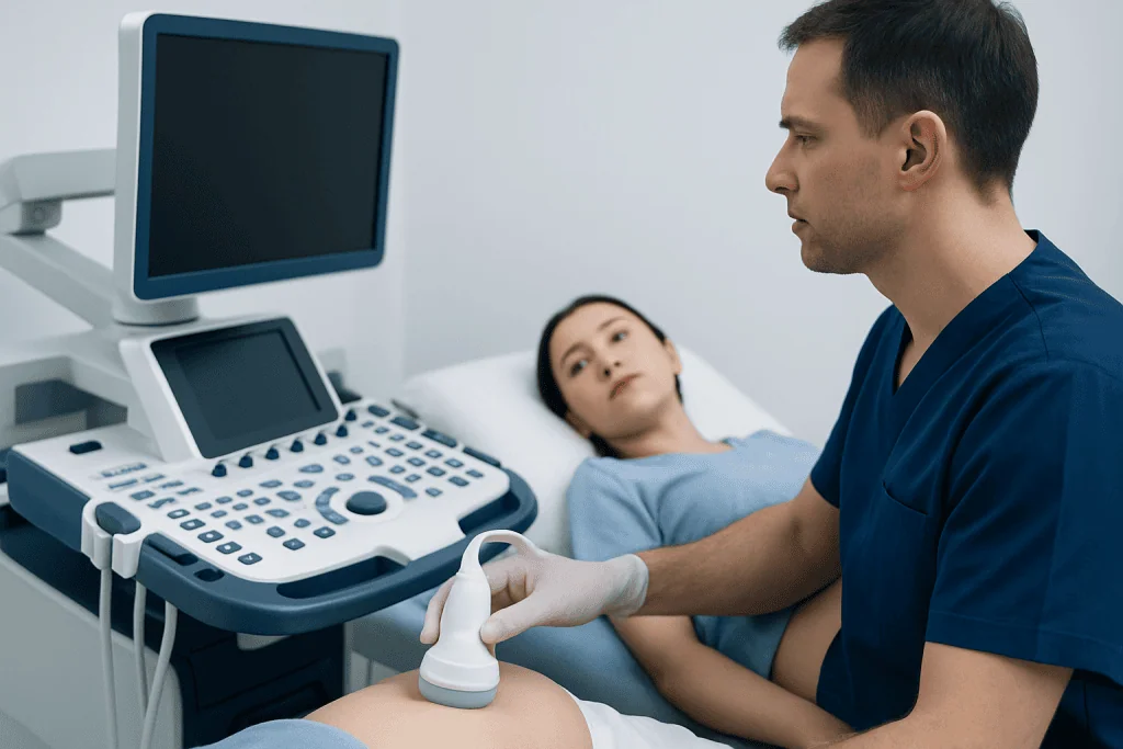
During ultrasound, a trained sonographer places a handheld device called a transducer on the skin. The transducer sends sound waves into the body and receives returning echoes. A computer converts these echoes into real-time images that a radiologist or treating clinician reviews.
A renal ultrasound may be requested for fever with urinary symptoms, pain in the side or back, repeated urinary infections, blood in the urine, abnormal kidney blood tests, or a suspected obstruction. It may also be used to monitor a known kidney infection or evaluate recovery after treatment.
People with diabetes, kidney stones, structural urinary tract conditions, reduced immune function or a recent urinary infection may have a higher likelihood of complicated infection. Nevertheless, symptoms alone cannot determine whether an abscess is present. Imaging helps the clinical team choose the safest next step.
Because no ionizing radiation is used, ultrasound can be appropriate in many situations, including pregnancy when imaging is needed. The care team will select the most suitable test based on the person’s symptoms, medical history and urgency of care.
What Is the Prep for a Renal Ultrasound?

Renal ultrasound prep instructions differ between imaging centers, so patients should follow the appointment-specific guidance they receive. In many cases, no special diet is required. Some centers ask patients to avoid a large meal for several hours beforehand because bowel gas can make it harder to obtain clear images.
Patients may be asked to drink water before arriving and not urinate until after the examination. A comfortably full bladder can improve views of the bladder and the lower portions of the urinary tract. The instructions commonly specify a volume and timing, but patients should use the exact directions supplied by their imaging department.
Regular medicines can usually be taken as prescribed unless a doctor provides different instructions. Patients should tell the team about pregnancy, recent surgery, an inability to hold urine, severe pain, mobility needs or an active urinary catheter. These details help the staff adapt the examination safely and comfortably.
It is helpful to bring relevant prior imaging reports, a medication list and information about recent infections or antibiotics. If a person feels too unwell to wait for a scheduled outpatient examination, they should contact their clinician or seek urgent medical advice instead of relying on preparation instructions alone.
Why Do I Have to Drink Water Before a Renal Ultrasound?
Drinking water before a renal ultrasound fills the bladder. A full bladder can create a clearer acoustic window for ultrasound waves and may help the clinician assess the bladder wall, urine retention and the parts of the urinary tract near the bladder.
This instruction does not mean that every renal scan requires a full bladder. When the main concern is the kidneys alone, preparation may be different. In suspected infection, the imaging team may want images of both the kidneys and bladder, so arriving with a comfortably full bladder can add useful information.
Patients should not force large amounts of water if this causes pain, nausea or worsening symptoms. Anyone with heart failure, severe kidney disease or a fluid restriction should ask their clinician or imaging center how to prepare. The goal is useful imaging, not discomfort.
Do I Have to Undress for a Renal Ultrasound?
For a renal ultrasound, patients usually need to expose the abdomen, sides and sometimes the lower back so the sonographer can position the transducer over the kidneys. Most people can wear loose, comfortable clothing and may only need to lift a top or change into a gown, depending on the facility’s protocol.
Privacy is maintained during the examination. The area being scanned is covered with warm, water-based gel, and the rest of the body should remain covered as much as possible. Patients can ask for a chaperone, interpreter or additional privacy support if needed.
Jewelry or belts around the abdomen may need to be removed. The sonographer may ask the patient to lie on their back and turn slightly onto either side, as the kidneys are viewed from several angles. If movement is difficult or painful, patients should let the team know.
Step-by-Step Procedure, Results and Recovery
After check-in, the patient lies on an examination table. The sonographer applies gel to the skin and moves the transducer over the abdomen, sides and back. Gentle pressure may be used to obtain images, which can be briefly uncomfortable if the area is tender but should not be painful. The patient may be asked to hold their breath or change position.
The examination commonly takes 20 to 40 minutes, although timing varies. There are no injections, needles or radiation in a standard ultrasound. Once images have been collected, the gel is wiped away and the patient can usually return to usual activities immediately, including eating, drinking and driving.
A radiologist interprets the images and sends a report to the requesting clinician. Results may describe a fluid collection, kidney swelling, obstruction, stones, bladder findings or no clear abnormality. An ultrasound can raise or lower concern for an abscess, but a normal or inconclusive scan does not always exclude one.
If the results suggest a renal abscess, the care team may arrange further imaging and start or adjust treatment promptly. Management can include intravenous or oral antibiotics, treatment of an underlying blockage, and image-guided drainage for selected larger or persistent collections. Treatment for kidney stones may also be relevant when a stone is contributing to urinary blockage or recurrent infection.
How Serious Is an Abscess on Your Kidney?
An abscess on the kidney is a potentially serious infection because it can damage kidney tissue, spread beyond the kidney or, in severe cases, lead to infection throughout the body. With timely diagnosis and appropriate treatment, many people recover well. The level of concern depends on the size and location of the abscess, the person’s overall health, kidney function and whether urine flow is blocked.
Antibiotics are central to treatment, but some abscesses do not resolve fully with medicine alone. If the collection is large, does not improve, or is difficult for antibiotics to penetrate, a specialist may recommend drainage through the skin using imaging guidance. Surgery is less commonly needed but can be considered in complex cases.
People should not try to manage suspected kidney infection symptoms only with fluids or over-the-counter pain relief. Fever, chills, worsening side or back pain, vomiting, confusion, reduced urine output or marked weakness require urgent medical assessment. People at higher risk of complications should contact a clinician early when urinary symptoms occur.
Acibadem International’s multidisciplinary specialists and JCI-accredited hospitals diagnose and treat kidney and urinary tract conditions for international patients, coordinating imaging, urology, infectious disease and interventional care when appropriate.
When to Seek Medical Care
Medical advice should be sought promptly for fever with burning urination, frequent urination, blood in the urine, new pain in the side or back, or symptoms that persist despite treatment for a urinary infection. A clinician can determine whether testing, antibiotics or imaging such as renal ultrasound is appropriate.
Urgent assessment is important for high fever, shaking chills, severe or rapidly worsening pain, repeated vomiting, fainting, confusion, breathing difficulty, very little urine, or an inability to keep fluids or medicines down. These symptoms can indicate a serious infection or another condition requiring prompt treatment.
Pregnant people, children, older adults and people with diabetes, a weakened immune system, known kidney disease, a urinary catheter or a history of urinary obstruction should contact a healthcare professional early if infection is suspected. Follow-up is also important if symptoms return after antibiotics or do not improve as expected.
Frequently asked questions
Can an ultrasound diagnose a renal abscess?
Ultrasound can identify findings that suggest a renal abscess, such as a complex fluid collection within or near the kidney. However, it may miss small or deep abscesses, and CT imaging is often used when the diagnosis remains uncertain or detailed treatment planning is needed.
What does a renal abscess look like on ultrasound?
A renal abscess may appear as a rounded or irregular area that is darker than surrounding kidney tissue because it contains fluid. It can also have internal debris, partitions or a thick wall. The appearance is not identical in every person, so a radiologist interprets it in clinical context.
Is a renal ultrasound painful?
A renal ultrasound is usually painless because it does not involve needles or radiation. Some pressure from the transducer may be uncomfortable, particularly if the kidney area is already sore. Patients should tell the sonographer if pressure is painful.
How long does it take to get renal ultrasound results?
The images are often reviewed after the examination by a radiologist, who prepares a report for the requesting clinician. Timing varies by facility and clinical urgency. If there is concern for a serious infection, the healthcare team may discuss preliminary findings and next steps quickly.
Can I eat before a renal ultrasound?
Many renal ultrasounds do not require fasting, but some imaging departments ask patients to avoid food for a period before the scan to reduce bowel gas. Patients should follow the specific instructions from their appointment center. If instructions are unclear, contacting the imaging department is the best approach.
What happens if the ultrasound finds a kidney abscess?
The treating clinician will assess the imaging result together with symptoms, urine tests and blood tests. Treatment commonly includes antibiotics, and further CT imaging may be needed to define the abscess. Some collections require drainage, especially if they are large, persistent or associated with obstruction.
References
- National Institute of Diabetes and Digestive and Kidney Diseases
- American College of Radiology
- National Health Service
- Merck Manual Professional Edition
This article is for general information only and is not a substitute for professional medical advice. Please consult a qualified doctor about your individual situation.
Ultrasound in Turkey — costs, top hospitals & a free quote
JCI-accredited · board-certified surgeons · reply within 24h
Add us as a Preferred Source to see more of our trusted health content across Google Search, AI Overviews and Discover.

Check-up & Preventive Medicine
Comprehensive health screenings designed for early detection and prevention.
3 specialists in this unit