Screening Rop Guidelines: How It Works, Results and What to Expect

ROP screening is designed for babies born very early or with very low birth weight, with local protocols defining the exact eligibility criteria. The examination is performed by a trained eye specialist using dilating drops and a retinal viewing device.
Key Takeaways
- ROP screening is designed for babies born very early or with very low birth weight, with local protocols defining the exact eligibility criteria.
- The examination is performed by a trained eye specialist using dilating drops and a retinal viewing device.
- Many mild cases of ROP, including most stage 1 and stage 2 cases, improve without treatment but still need scheduled monitoring.
- ROP can progress quickly in some infants, so attending every recommended eye appointment is important.
- Treatment is reserved for higher-risk disease and may include anti-VEGF eye injections or laser treatment.
- Parents should seek urgent medical advice if they miss follow-up or have concerns about a baby’s eye care plan after neonatal discharge.
Screening ROP guidelines help neonatal teams identify premature babies who need retinal examinations for retinopathy of prematurity (ROP), an eye condition caused by abnormal blood-vessel development. Regular screening and follow-up allow most concerning changes to be found early, when treatment can be most effective if it is needed.
Overview: what screening ROP guidelines mean
Screening ROP guidelines outline which premature babies should have eye examinations, when the first examination should occur, and how often follow-up is needed. Their purpose is to detect retinopathy of prematurity (ROP) before it threatens vision. ROP develops when the blood vessels of the retina, the light-sensitive tissue at the back of the eye, grow abnormally after an early birth.
Screening is not a test of whether a baby can see. It is a detailed assessment of the developing retina by an ophthalmologist or retinal specialist with experience in newborn care. Most babies who are screened will not develop severe ROP, and many babies with early changes recover without treatment. However, careful surveillance is essential because a smaller number can develop changes that require prompt treatment.
Protocols vary slightly between countries, neonatal units, and professional organizations. The neonatal team uses the local guideline together with the baby’s gestational age, birth weight, medical course, and retinal findings to make an individualized screening plan.
What is the ROP screening test and how is it performed?
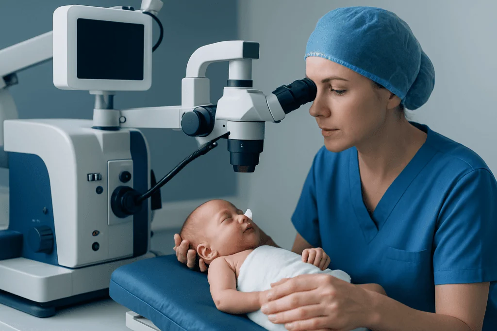
The ROP screening test is a dilated retinal examination. Before the examination, staff place eye drops to enlarge the pupils. The eye specialist then examines the retina using a light and specialized lens, often with a small instrument that gently keeps the eyelids open. Some services also use wide-field digital retinal imaging to document findings and support follow-up.
The examination is usually performed in the neonatal intensive care unit or a pediatric eye clinic. Babies are kept warm and monitored during the process. Comfort measures may include swaddling, a pacifier, and small amounts of soothing oral solution where appropriate. The examination can be briefly uncomfortable and may cause temporary changes in heart rate, breathing, or oxygen levels, so neonatal staff observe the baby closely.
Parents may be told that the eyes look normal, that immature retinal vessels need repeat checks, or that ROP is present. The report describes the location of the changes, the stage, and whether features associated with faster progression are seen. The specialist then sets the next examination date, which may range from days to several weeks depending on the result.
What are the criteria for ROP screening?

ROP screening criteria are based mainly on prematurity and low birth weight. In many widely used guidelines, screening includes babies born at or before 30 weeks of gestation and/or with a birth weight of 1,500 grams or less. Some neonatal units also screen larger or more mature babies whose clinical course suggests increased risk, based on the judgment of the neonatal team.
Factors that may increase the likelihood of ROP include a very early birth, very low birth weight, prolonged need for oxygen or respiratory support, severe infection, anemia or blood transfusions, and poor postnatal growth. These factors do not mean that an individual baby will develop sight-threatening ROP. They help clinicians recognize babies who may benefit from careful observation.
The timing of the first examination is calculated from both gestational age at birth and the baby’s age after birth. In many protocols, it occurs at approximately 4 weeks after birth or around 31 weeks postmenstrual age, whichever is later, although exact timing differs by guideline. The neonatal team should provide parents with the specific schedule used for their baby.
- Screening continues until the retina is sufficiently developed or ROP has clearly regressed.
- Babies discharged before screening is complete need a confirmed outpatient appointment.
- Missing follow-up appointments can delay detection of progression, even if the baby appears well.
What are the 5 stages of ROP?
ROP is classified from stage 1 to stage 5 according to how far abnormal retinal changes have progressed. The stages describe anatomy, not a prediction of vision by themselves. Specialists also assess the retinal zone involved and look for “plus disease,” meaning unusually enlarged and twisted blood vessels that can indicate more active, higher-risk disease.
Stage 1 is a faint line between vascularized and nonvascularized retina. Stage 2 is a raised ridge at this boundary. Stage 3 involves abnormal blood-vessel growth extending from the ridge into the eye. In stage 4, partial retinal detachment has occurred. Stage 5 is total retinal detachment, the most advanced stage.
Early stages often improve as the retina matures. By contrast, certain stage 3 findings, especially when combined with plus disease or involvement of the central retina, may need treatment to lower the risk of retinal detachment. Classification and treatment decisions should always be made by the examining pediatric retinal specialist.
What is the prognosis for ROP stage 2?
The prognosis for ROP stage 2 is generally favorable. Many babies with stage 2 ROP do not need treatment because the ridge can gradually flatten as normal retinal blood-vessel growth continues. Nonetheless, stage 2 requires repeat examinations because a minority of cases can progress, particularly when disease occurs in a more central retinal zone or plus disease is present.
The eye specialist will determine how often the baby should be reviewed. Follow-up may be more frequent when the findings are closer to the central retina, change rapidly, or are accompanied by concerning blood-vessel changes. Parents should not interpret a stage number alone as a complete prognosis; the location, activity, and overall health of the infant all matter.
Even after ROP has resolved, children born prematurely may have a higher chance of refractive errors such as nearsightedness, eye misalignment, or reduced vision in one eye. Routine pediatric eye care supports early recognition of these issues, which can often be managed more effectively when found early.
How screening works: preparation, results and follow-up
Before the examination, the neonatal team confirms the baby’s medical stability and administers pupil-dilating drops. The specialist records the retinal findings in each eye. The process itself is short, but dilation and monitoring mean the overall visit may take longer. Temporary sensitivity to light or mild eyelid redness can occur after the examination and usually settles quickly.
Results generally fall into three broad groups: retina still immature and requiring routine recheck; mild ROP that needs closer surveillance; or treatment-requiring ROP. The timing of the next review is one of the most important parts of the result. Parents should ask for the date, location, and purpose of the next eye examination before discharge from the neonatal unit.
The main benefit of screening is early detection of disease before retinal detachment develops. Risks of the examination are usually brief and manageable, but may include discomfort, temporary irritation from drops, and short-lived changes in breathing or heart rate in medically fragile infants. These risks are weighed against the substantial benefit of identifying serious ROP early.
Treatment options and recovery timeline
Most screened babies do not need ROP treatment. When treatment is advised, the aim is to stop abnormal blood-vessel growth and reduce the chance of retinal detachment. The two main options are laser treatment to the peripheral retina and injections of medicines that block vascular endothelial growth factor (anti-VEGF). The preferred approach depends on the retinal findings, the baby’s overall condition, and the specialist’s assessment.
Laser treatment is generally performed under anesthesia or carefully managed sedation in an appropriate hospital setting. Anti-VEGF treatment is given as an injection into the eye under sterile conditions and requires prolonged follow-up because late reactivation can occur. In advanced disease with retinal detachment, surgery may be considered, although screening aims to identify high-risk changes before this is necessary.
Recovery depends on the treatment and the baby’s neonatal needs. After laser or injection treatment, the eye specialist performs repeat retinal checks within a defined interval and continues monitoring until the retina is safely mature or disease has regressed. Parents should follow instructions on appointments, medications if prescribed, and warning signs. Acibadem International’s multidisciplinary specialists and JCI-accredited hospitals support diagnosis and treatment planning for international patients requiring pediatric eye care.
When to seek medical care
Parents should contact the neonatal team or pediatric ophthalmology service promptly if a scheduled ROP examination is missed, if they are unsure where follow-up will occur after discharge, or if they have not received clear screening instructions. ROP usually cannot be recognized at home by looking at a baby’s eyes, so planned examinations are the safest way to monitor it.
Urgent medical assessment is also appropriate if a baby appears unwell after an eye procedure, has persistent eye redness or swelling, discharge, or any symptom that concerns the parent. These symptoms do not necessarily indicate an ROP-related problem, but they should be assessed by a qualified clinician.
For children with a history of prematurity or ROP, routine pediatric follow-up and recommended eye examinations remain important as they grow. A pediatrician or eye specialist can advise on visual development, refractive testing, and whether additional support is needed.
Frequently asked questions
Is ROP screening painful for a baby?
The examination can be uncomfortable for a short time because pupils are dilated and the eyelids are gently held open. Neonatal staff use comfort measures and monitor the baby throughout. Any temporary changes in breathing, heart rate, or oxygen levels are managed by the clinical team.
When does ROP screening stop?
Screening stops when the retina has developed sufficiently, when retinal blood vessels have matured into the peripheral retina, or when ROP has regressed and the specialist considers further examinations unnecessary. The exact endpoint varies between babies. Parents should confirm the follow-up plan with the eye specialist before leaving neonatal care.
Can a full-term baby develop ROP?
ROP is primarily associated with premature birth because retinal blood-vessel development is incomplete at birth. It is very uncommon in full-term babies. Other eye conditions can affect full-term infants, so concerns about a baby’s vision should still be discussed with a pediatrician.
Does stage 2 ROP always need treatment?
No. Many cases of stage 2 ROP improve without treatment as the retina matures. The need for treatment depends on the retinal zone, whether plus disease is present, the speed of progression, and other examination findings.
Can ROP cause problems later in childhood?
Some children with a history of prematurity or ROP may be more likely to develop nearsightedness, strabismus, or other visual-development concerns. This does not mean such problems will occur. Regular recommended eye checks help detect and manage issues early.
What should parents bring to an outpatient ROP appointment?
Parents should bring the baby’s discharge summary, previous eye examination records if available, and details of current medicines or medical care. It is also helpful to bring the next appointment information and a list of questions. Because pupil-dilating drops may be used, parents may wish to plan for a longer visit and protect the baby from bright light afterward.
References
- American Academy of Pediatrics
- American Academy of Ophthalmology
- International Classification of Retinopathy of Prematurity
- National Eye Institute
- Royal College of Ophthalmologists
This article is for general information only and is not a substitute for professional medical advice. Please consult a qualified doctor about your individual situation.
Explore treatments in Turkey — costs, top hospitals & a free quote
JCI-accredited · board-certified surgeons · reply within 24h
Add us as a Preferred Source to see more of our trusted health content across Google Search, AI Overviews and Discover.
Check your numbers in seconds
BMI, calories, due date, blood pressure and 30+ more clinical calculators — free, instant, doctor-reviewed ranges.

More from the Health Library



Related Specialists

Prof. Dr. Mehmet Ali Şehitoğlu
Otorhinolaryngology
Dr. Cihan Göktaş
Inpatient Clinic Physicians Clinical Service
Assoc. Prof. Dr. Yetkin Zeki Yılmaz
Ear Nose & Throat
Asst. Prof. Dr. Murat Tuğrul Eren
Urology