Signs of Endometrial Cancer on Ultrasound: Preparation, Procedure and Results

Transvaginal ultrasound is commonly used to assess the uterine lining in people with abnormal bleeding. A thickened endometrium can have non-cancerous causes, including polyps, fibroids, hormone changes, and some medicines.
Key Takeaways
- Transvaginal ultrasound is commonly used to assess the uterine lining in people with abnormal bleeding.
- A thickened endometrium can have non-cancerous causes, including polyps, fibroids, hormone changes, and some medicines.
- Postmenopausal bleeding should always be assessed by a qualified clinician, even if it occurs only once.
- Endometrial biopsy or hysteroscopy with tissue sampling is needed to diagnose or rule out endometrial cancer.
- Most transvaginal ultrasounds are quick, do not require recovery time, and have very low risk.
Signs of endometrial cancer on ultrasound may include an unusually thick, irregular, or poorly defined endometrial lining, sometimes with a mass or increased blood flow. Ultrasound can identify findings that need further assessment, but it cannot confirm cancer; tissue testing is required.
Overview: What ultrasound can and cannot show
Signs of endometrial cancer on ultrasound can include a thickened uterine lining, an irregular or uneven endometrium, a focal growth within the uterine cavity, or changes in the way blood flows through abnormal tissue. These findings can raise concern and guide the next steps, but they do not diagnose cancer by themselves.
The endometrium is the lining inside the uterus. It naturally changes thickness during the menstrual cycle before menopause, which is why an ultrasound result must be interpreted in the context of age, symptoms, menstrual status, medicines, and individual risk factors. After menopause, the lining is normally thin, so a thickened lining in a person with bleeding often needs additional evaluation.
A pelvic ultrasound may be performed through the abdomen, through the vagina, or both. Transvaginal ultrasound generally provides the clearest view of the endometrium because the probe is placed close to the uterus. If imaging suggests an abnormality, a clinician may recommend an endometrial biopsy or hysteroscopy to obtain tissue for laboratory examination.
Would endometrial cancer show up on an ultrasound?
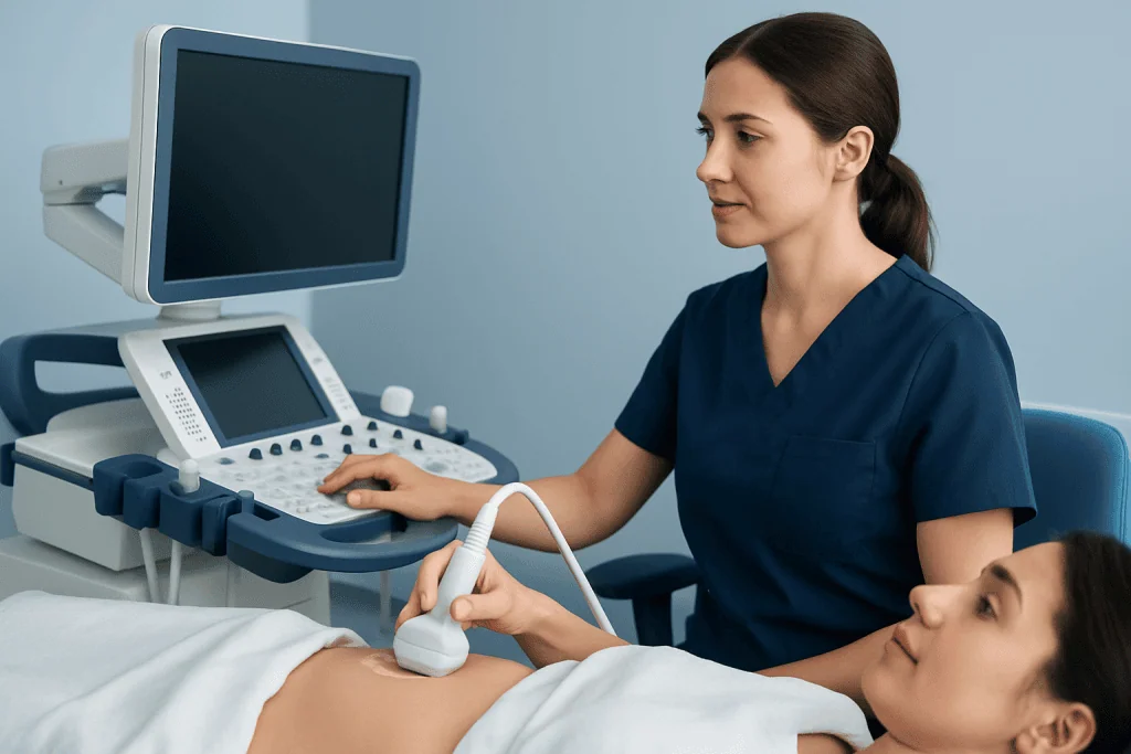
Endometrial cancer may show up on ultrasound as an abnormal appearance of the endometrial lining, but some cancers are not clearly visible and ultrasound cannot determine whether abnormal tissue is cancerous. The scan is best viewed as an important assessment tool rather than a final diagnostic test.
In a postmenopausal person with vaginal bleeding, transvaginal ultrasound often measures endometrial thickness. A thin lining makes endometrial cancer less likely in many situations, while a thicker lining, irregularity, or inability to clearly see the lining may prompt tissue sampling. The appropriate next step depends on the full clinical picture, including whether bleeding continues or returns.
Before menopause, thickness alone is less informative because normal hormonal cycling affects the endometrium. Ultrasound may still identify polyps, fibroids, ovarian concerns, or other structural causes of bleeding, but a biopsy may be advised when bleeding is persistent, unusual, or accompanied by risk factors.
What does cancer look like on uterine ultrasound?
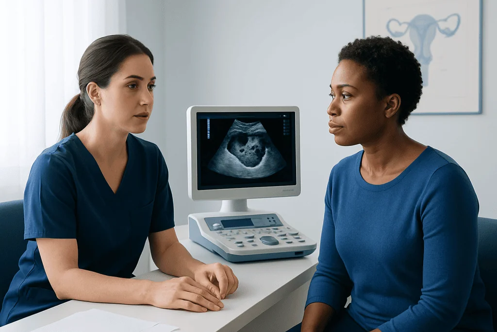
There is no single ultrasound appearance that proves endometrial cancer. Radiologists and gynecologists look for patterns such as a diffusely thickened lining, uneven thickness, a heterogeneous or mixed internal texture, an irregular border between the endometrium and uterine muscle, or a localized mass within the uterine cavity.
Color Doppler ultrasound, which displays blood flow, may show more blood vessels within an abnormal area. However, benign conditions can also produce irregular-looking tissue or increased blood flow. Endometrial polyps, fibroids that extend into the cavity, infection, hormone therapy, and endometrial hyperplasia are examples of conditions that can overlap with cancer on imaging.
For this reason, clinicians do not diagnose endometrial cancer from an image alone. Ultrasound findings are combined with symptoms, examination findings, and—when indicated—sampling of the endometrium. This approach helps avoid unnecessary treatment while ensuring concerning symptoms are investigated appropriately.
What is one of the most common signs of endometrial cancer?
One of the most common signs of endometrial cancer is abnormal vaginal bleeding. In people who have gone through menopause, any vaginal bleeding, spotting, or pink or brown discharge should be discussed with a clinician. Although bleeding often has a non-cancerous cause, prompt assessment is important.
Before menopause, possible warning signs include bleeding between periods, unusually heavy or prolonged periods, or a noticeable change from the person’s usual cycle. Pelvic pressure or pain, watery or blood-tinged discharge, and unexplained weight loss can occur, but they are less specific and may have many causes.
Symptoms do not establish the cause, and many people with abnormal bleeding do not have cancer. Still, early evaluation allows clinicians to identify and treat common benign conditions and, when necessary, detect endometrial cancer at an earlier stage.
How a transvaginal ultrasound works: candidacy and preparation
Transvaginal ultrasound uses high-frequency sound waves to create images of the uterus, endometrium, ovaries, and nearby pelvic structures. It does not use radiation. It is commonly considered for postmenopausal bleeding, abnormal uterine bleeding, an abnormal pelvic examination, follow-up of a known uterine condition, or evaluation of a previously identified thickened endometrium.
People who are pregnant, have active pelvic infection symptoms, have recently had pelvic surgery, or feel unable to tolerate an internal examination should tell their care team. Alternatives or modifications may be possible. A transabdominal pelvic ultrasound may be used as well, although it may not show the endometrial lining as clearly.
Preparation is usually simple. For a transvaginal scan, patients may be asked to empty their bladder beforehand. If a transabdominal scan is planned, they may instead be asked to arrive with a comfortably full bladder. The imaging center will provide instructions based on the planned examination.
Step-by-step procedure, benefits, risks and recovery
During the examination, the patient usually lies on an examination table with privacy measures in place. A trained sonographer covers a slim ultrasound probe with a protective sheath and gel, then gently inserts it a short distance into the vagina. The patient can ask for the examination to pause or stop at any time.
The scan often takes about 15 to 30 minutes, although timing can vary. Images are reviewed by a radiologist or another qualified clinician, who prepares a report for the referring healthcare professional. Results may describe endometrial thickness, whether the lining appears regular, and any fibroids, polyps, fluid, or masses seen in the pelvis.
The benefits include detailed pelvic imaging without radiation and no need for anesthesia in most cases. Risks are very low; some people experience temporary pressure, mild discomfort, or light spotting if the vaginal tissues are sensitive. There is no routine recovery period, and most people can return to usual activities immediately after the scan.
If a scan is concerning or symptoms persist, the next step may be endometrial biopsy or hysteroscopy. These procedures allow tissue to be examined by a pathologist and can also help identify benign problems such as polyps or hyperplasia.
What is the gold standard for diagnosing endometrial cancer?
The gold standard for diagnosing endometrial cancer is examination of an endometrial tissue sample by a pathologist. This is usually obtained with an office-based endometrial biopsy. If the sample is insufficient, symptoms continue, or a focal abnormality is suspected, hysteroscopy with directed biopsy or dilation and curettage may be recommended.
During an endometrial biopsy, a clinician passes a narrow instrument through the cervix into the uterus to collect a small tissue sample. Cramping and brief bleeding may occur afterward. Hysteroscopy uses a thin camera to look directly inside the uterus and can help clinicians target a polyp, mass, or other localized area for sampling.
If cancer is confirmed, further testing helps determine its type, grade, and extent. Management often involves gynecologic oncology specialists and may include surgery, radiation therapy, chemotherapy, hormone therapy, or targeted treatments depending on the individual diagnosis. Gynecologic oncology care coordinates diagnosis, treatment planning, and follow-up.
When to seek medical care
Medical assessment is recommended for any vaginal bleeding after menopause, even when it is light, happens only once, or seems to stop. A clinician should also evaluate bleeding between periods, new heavy or prolonged periods, recurrent watery or blood-stained discharge, or pelvic symptoms that do not settle.
Urgent care may be appropriate for very heavy bleeding, dizziness, fainting, severe pelvic pain, fever, or symptoms that suggest significant blood loss or infection. These symptoms are not specific to endometrial cancer, but they should not be managed without medical advice.
For patients seeking evaluation internationally, Acibadem International’s multidisciplinary specialists and JCI-accredited hospitals provide assessment and treatment planning for gynecologic conditions. A clinician can explain whether ultrasound, biopsy, hysteroscopy, or further imaging is appropriate based on the patient’s symptoms and medical history.
Frequently asked questions
Can a normal ultrasound rule out endometrial cancer?
A normal ultrasound can be reassuring, especially in some postmenopausal patients with bleeding and a thin endometrial lining. However, it does not rule out every case. Persistent or recurrent bleeding should be reviewed by a clinician, who may recommend biopsy despite a reassuring scan.
What endometrial thickness is concerning after menopause?
In a postmenopausal person with bleeding, an endometrial lining measuring 4 mm or less on transvaginal ultrasound is generally associated with a low likelihood of endometrial cancer. A thicker lining may require further assessment, but it does not mean cancer is present. Individual decisions also depend on bleeding pattern, risk factors, image quality, and prior treatment.
Can a thickened endometrium be benign?
Yes. A thickened endometrium can result from normal hormonal changes before menopause, endometrial polyps, fibroids, hormone therapy, tamoxifen use, or endometrial hyperplasia. A clinician may recommend follow-up imaging or tissue sampling to determine the cause.
Is transvaginal ultrasound painful?
Most people find transvaginal ultrasound uncomfortable rather than painful. The probe is narrow and inserted only a short distance into the vagina, and no anesthesia is usually needed. Patients should tell the sonographer if they have pain, anxiety, previous pelvic trauma, or difficulty tolerating internal examinations.
How long does it take to get ultrasound results?
The images are typically obtained during the appointment, but the formal report may be available the same day or within several days depending on the facility. The referring clinician explains what the findings mean in relation to symptoms and medical history. If an abnormality is found, they will discuss whether additional testing is needed.
Does an endometrial biopsy always find cancer if it is present?
Endometrial biopsy is highly useful, but occasionally the sample is too small or does not capture a focal abnormality such as a polyp or localized tumor. If bleeding persists, the biopsy is inconclusive, or ultrasound suggests a focal lesion, hysteroscopy with directed sampling may be recommended. Follow-up is important even after a non-diagnostic result.
References
- American College of Obstetricians and Gynecologists
- National Cancer Institute
- American Cancer Society
- Society of Gynecologic Oncology
- World Health Organization
This article is for general information only and is not a substitute for professional medical advice. Please consult a qualified doctor about your individual situation.
Endometrial Cancer in Turkey — costs, top hospitals & a free quote
JCI-accredited · board-certified surgeons · reply within 24h