Skeletal Dysplasia Ultrasound: Preparation, Procedure and Results
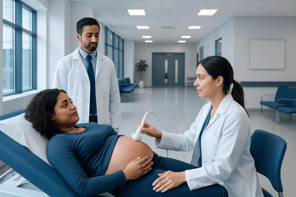
Skeletal dysplasia ultrasound is a noninvasive prenatal examination that uses sound waves, not radiation. The scan measures long bones and evaluates bone shape, mineralization, the chest, spine, skull, hands and feet.
Key Takeaways
- Skeletal dysplasia ultrasound is a noninvasive prenatal examination that uses sound waves, not radiation.
- The scan measures long bones and evaluates bone shape, mineralization, the chest, spine, skull, hands and feet.
- Ultrasound can raise or lower suspicion for a skeletal dysplasia, but it does not confirm every type.
- Follow-up scans and genetic counselling may help clarify the diagnosis and expected outlook.
- Many skeletal dysplasias have different levels of severity, so individualized specialist counselling is important.
A skeletal dysplasia ultrasound is a detailed prenatal scan that assesses a baby's bones, growth pattern, chest and other features when a skeletal condition is suspected. It can identify important clues, but additional imaging, genetic testing and specialist assessment may be needed to confirm a specific diagnosis.
Overview: what is a skeletal dysplasia ultrasound?
A skeletal dysplasia ultrasound is a detailed prenatal ultrasound examination performed when routine screening suggests that a baby’s bones may be growing differently or have an unusual shape. Skeletal dysplasia is a broad term for genetic conditions that affect bone and cartilage development. These conditions vary widely, from forms associated mainly with short stature to forms that can seriously affect breathing and survival around birth.
The examination uses high-frequency sound waves to create images of the developing baby. It does not use ionizing radiation and is generally considered safe when performed for an appropriate medical reason by trained professionals. The purpose is not only to measure bone length, but also to examine the pattern of findings across the skeleton and assess features that help clinicians plan the next steps.
Results are interpreted in the context of gestational age, family history, previous pregnancies, parental height and other ultrasound findings. A concerning scan result can be emotionally difficult, but it is important to remember that a single measurement or isolated short bone does not automatically mean a baby has skeletal dysplasia.
Can skeletal dysplasia be diagnosed through ultrasound?
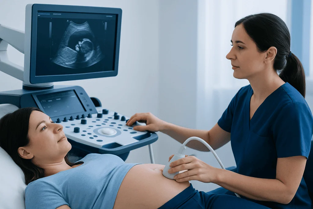
Ultrasound can detect findings that strongly suggest fetal skeletal dysplasia, especially when several bones are markedly short, unusually shaped, poorly mineralized or associated with a small chest. However, ultrasound alone often cannot identify the exact condition. Some skeletal dysplasias have overlapping appearances before birth, while others are subtle or become more apparent later in pregnancy.
A specialist may recommend serial ultrasounds to follow growth and bone development over time. Depending on the findings, further evaluation may include genetic counselling, diagnostic testing such as chorionic villus sampling or amniocentesis, and genetic analysis of fetal cells. In selected situations, fetal MRI or a postnatal skeletal survey may also provide helpful information.
Diagnostic testing is a personal decision. A maternal-fetal medicine specialist and genetic counsellor can explain what each test can and cannot answer, its timing, and any potential risks. The aim is to provide the family with the clearest possible information for pregnancy care, birth planning and newborn support.
How the examination works and who may need it
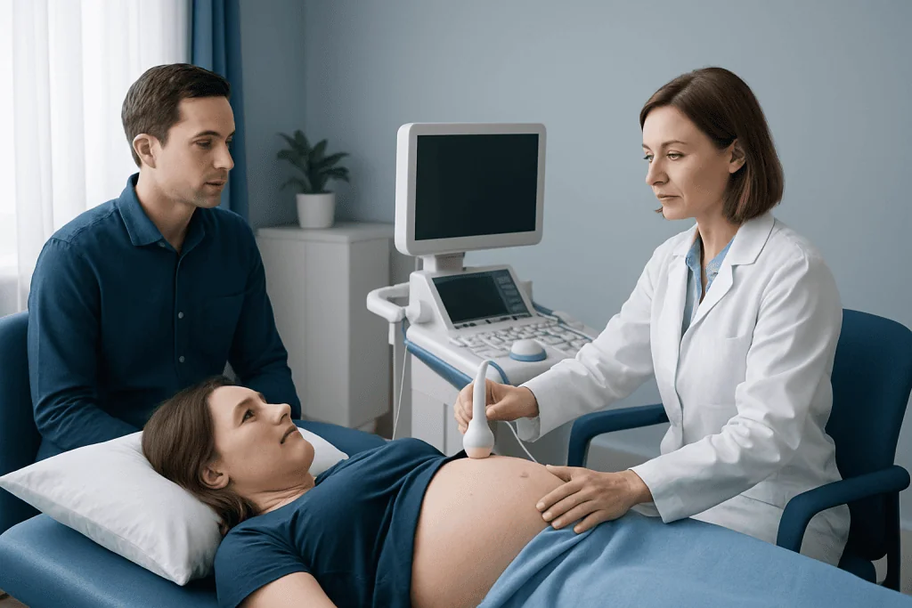
The ultrasound transducer sends sound waves through the abdomen and receives returning echoes from fetal tissues. A computer converts these echoes into real-time images. During a targeted skeletal assessment, the sonographer and fetal medicine specialist compare measurements with expected ranges for the stage of pregnancy and look closely at the structure of individual bones.
A detailed skeletal assessment may be advised after a routine scan shows shortened long bones, an unusual bone contour, a small chest, fractures, reduced bone brightness or abnormal hands, feet, spine or skull shape. It may also be recommended when there is a known family history of skeletal dysplasia, a previous affected pregnancy, or a genetic test result suggesting a bone-development condition.
Not every baby with shorter-than-average bones has a dysplasia. Some babies are constitutionally small, have inherited familial short stature, or have a growth pattern related to another pregnancy condition. This is why clinicians evaluate the whole picture rather than relying on one measurement.
- Preparation is usually simple: normal food and fluids are generally allowed unless the care team gives different instructions.
- Wear comfortable clothing that allows access to the abdomen.
- Bring prior ultrasound reports, genetic test results and relevant family medical information if available.
What happens during the procedure and afterward?
The appointment commonly begins with a review of the pregnancy history and the reason for referral. The pregnant person lies comfortably on an examination couch while gel is applied to the abdomen. The sonographer moves a handheld probe across the skin to obtain images. Pressure may be felt, but the scan should not be painful.
The assessment may include measurements of the femur, humerus, tibia, fibula, radius and ulna; evaluation of the ribs, chest circumference, spine, pelvis, skull, jaw, fingers and toes; and review of bone mineralization and possible fractures or bowing. The specialist also checks overall fetal growth, amniotic fluid, placenta and other organs, because associated findings can influence interpretation.
Scan duration varies with fetal position, gestational age and the complexity of the findings. There is no physical recovery period after an ultrasound: people can normally return to usual activities immediately. If invasive genetic testing is discussed, its preparation, procedure and aftercare are separate from the ultrasound and should be reviewed carefully with the treating team.
The benefits of the scan include noninvasive assessment, earlier specialist involvement and more informed pregnancy and birth planning. Its limitations include imperfect visibility, variation in fetal position and the fact that not all conditions can be confirmed prenatally. Ultrasound itself has no known harmful effects when used appropriately, but it can sometimes lead to uncertain findings that require additional follow-up.
What are the radiological findings of fetal skeletal dysplasia?
On prenatal ultrasound, clinicians look for a pattern of skeletal findings rather than one sign alone. Important observations include whether limb shortening affects the upper arms and thighs, the forearms and lower legs, or the hands and feet. They also assess whether the shortening is symmetrical and whether growth changes over time.
Potential findings include very short long bones, curved or bowed bones, fractures, widened metaphyses, poor bone mineralization, a narrow or bell-shaped chest, short ribs, abnormal vertebral development, a large head compared with body size, unusual skull shape, or differences in the hands and feet. The ratio of chest size to abdominal size and the relationship between femur length and abdominal circumference can help estimate whether the chest may be too small to support normal lung development.
These findings are not specific to one diagnosis. For example, shortened long bones can occur in several skeletal dysplasias and may also be seen with fetal growth restriction or normal familial variation. Specialists may use standardized measurements, growth charts and repeat imaging to identify the most likely explanation.
Following birth, radiological assessment may include a skeletal survey, which is a set of carefully selected X-rays used to examine bone shape and development. Postnatal examination and genetic testing frequently provide the most definitive classification.
What does a baby with skeletal dysplasia look like?
There is no single appearance for a baby with skeletal dysplasia. The term includes many conditions, and each may affect the body differently. Some babies have proportionate or disproportionate short limbs, while others have a comparatively larger head, a smaller chest, bowed limbs, differences in hand or foot shape, or changes in spinal curvature.
Some differences may be visible on ultrasound, but prenatal images cannot always predict a baby’s appearance or function precisely. In many cases, appearance becomes clearer after birth through a newborn examination, measurements, imaging and genetic assessment. The care team should avoid making assumptions based on appearance alone.
Families may benefit from speaking with specialists who understand both the medical and practical aspects of skeletal conditions. This can include fetal medicine, clinical genetics, neonatology, paediatric orthopedics, pulmonology and other professionals based on the suspected diagnosis and the baby’s needs.
Can a baby survive with skeletal dysplasia?
Many babies with skeletal dysplasia survive and can lead full lives, although they may need monitoring or treatment for issues such as growth, spine alignment, limb differences, hearing, sleep or breathing. The outlook depends on the exact type of dysplasia and the severity of its effects, not simply on the presence of short bones.
Some forms are life-limiting before or shortly after birth, most often because the chest is too small for the lungs to develop adequately or because of severe bone mineralization problems. When ultrasound raises concern for a potentially severe form, a multidisciplinary team can assess the findings, discuss uncertainty honestly and help the family prepare for delivery and newborn care.
Accurate diagnosis matters because it supports appropriate planning. This may include choosing a delivery hospital with neonatal intensive care, arranging specialist reviews after birth and discussing comfort-focused care when a condition is expected to be life-limiting. Families should be offered compassionate counselling and ongoing emotional support throughout decision-making.
Follow-up, self-care and when to seek medical care
There is no proven self-care measure that can change a fetal skeletal dysplasia during pregnancy. The most helpful steps are attending recommended prenatal appointments, taking prescribed pregnancy supplements, avoiding tobacco, alcohol and non-prescribed substances, and discussing all medicines with the obstetric care team. Parents should ask for clear explanations of results and may request genetic counselling or a second specialist opinion when needed.
Follow-up may involve repeat ultrasound scans, diagnostic genetic testing and coordinated delivery planning. Acibadem International’s multidisciplinary specialists and JCI-accredited hospitals support international patients needing fetal medicine, genetics, neonatal and paediatric care for complex prenatal findings.
When to seek medical care: Pregnant people should contact their maternity care team promptly for vaginal bleeding, fluid leakage, regular painful contractions before term, severe abdominal pain, fever, severe headache with visual changes, or a noticeable reduction in fetal movements after movements have become established. These symptoms are not specific to skeletal dysplasia, but they need timely assessment during any pregnancy.
After a scan suggests possible skeletal dysplasia, families should arrange the recommended specialist review rather than waiting for the next routine appointment. Urgent medical evaluation is also appropriate if the pregnancy care team advises immediate assessment based on ultrasound findings or maternal symptoms.
Frequently asked questions
Is skeletal dysplasia ultrasound painful?
No. The examination is performed over the abdomen using a handheld ultrasound probe and is usually painless. Some pressure may be felt while images are being obtained, particularly if the baby's position makes imaging more difficult.
When can skeletal dysplasia be seen on ultrasound?
Some severe skeletal conditions may be suspected in the first or early second trimester, but many are recognized during the detailed anatomy scan or later growth scans. The timing depends on the condition, how it affects bone development and the quality of imaging.
Does a short femur on ultrasound always mean skeletal dysplasia?
No. An isolated short femur can reflect normal familial body proportions, an inaccurate gestational age estimate, fetal growth restriction or other causes. Clinicians consider all measurements, growth over time and any additional ultrasound findings before drawing conclusions.
Will I need genetic testing after an abnormal skeletal ultrasound?
Genetic testing may be offered when the scan findings raise concern for a genetic skeletal condition, but the decision is individual. A genetic counsellor or fetal medicine specialist can explain the possible benefits, limitations and choices for testing.
How accurate is ultrasound for fetal skeletal dysplasia?
Ultrasound is valuable for detecting patterns that suggest skeletal dysplasia and for identifying features linked with greater severity. However, it may not determine the exact diagnosis, and confirmation may require genetic testing and assessment after birth.
What happens after a suspected fetal skeletal dysplasia is found?
The care team may recommend a detailed review by maternal-fetal medicine and clinical genetics, follow-up imaging and discussion of diagnostic testing. If appropriate, they also help plan delivery and newborn care with relevant paediatric specialists.
References
- American College of Obstetricians and Gynecologists
- International Society of Ultrasound in Obstetrics and Gynecology
- American College of Medical Genetics and Genomics
- National Institutes of Health
- World Health Organization
This article is for general information only and is not a substitute for professional medical advice. Please consult a qualified doctor about your individual situation.
Ultrasound in Turkey — costs, top hospitals & a free quote
JCI-accredited · board-certified surgeons · reply within 24h
Add us as a Preferred Source to see more of our trusted health content across Google Search, AI Overviews and Discover.

Check-up & Preventive Medicine
Comprehensive health screenings designed for early detection and prevention.
3 specialists in this unit